. 2004 Oct;78(19):10310-9.
doi: 10.1128/JVI.78.19.10310-10319.2004.

Np9 protein of human endogenous retrovirus K interacts with ligand of numb protein X

Affiliations

Np9 protein of human endogenous retrovirus K interacts with ligand of numb protein X

Vivienne Armbruester et al. J Virol. 2004 Oct.

Abstract

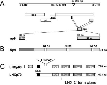
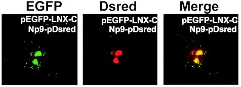
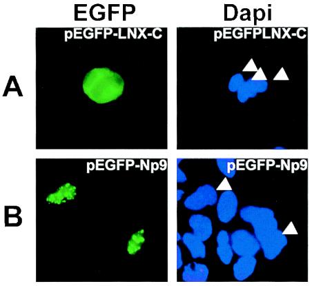
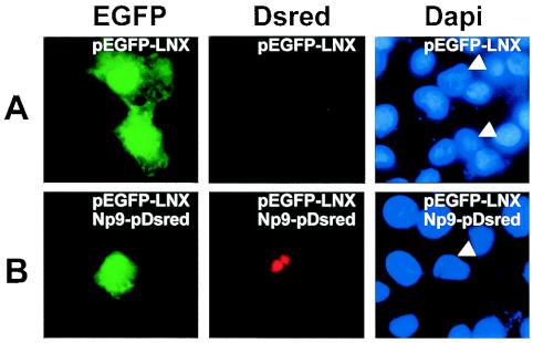
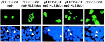
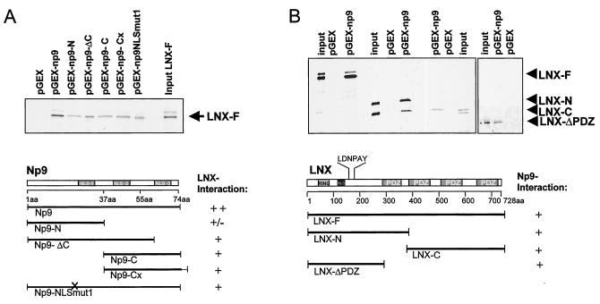
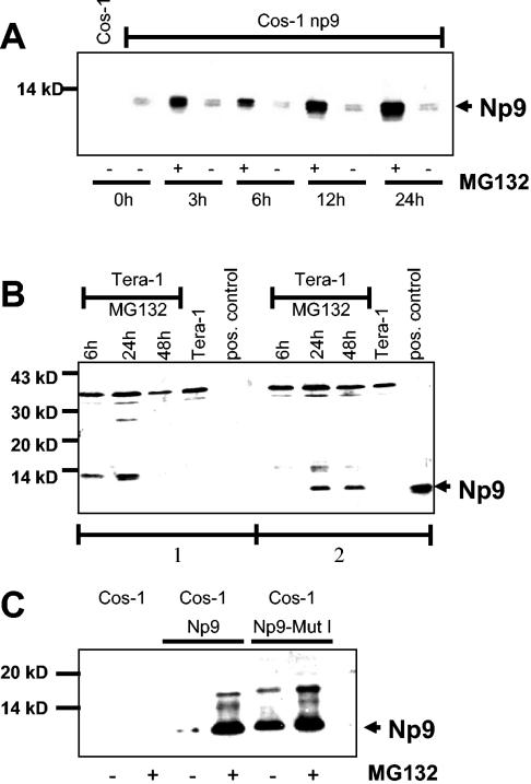
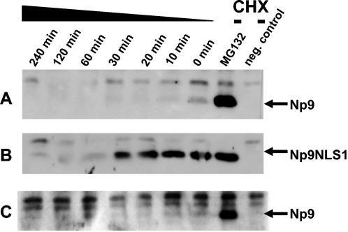
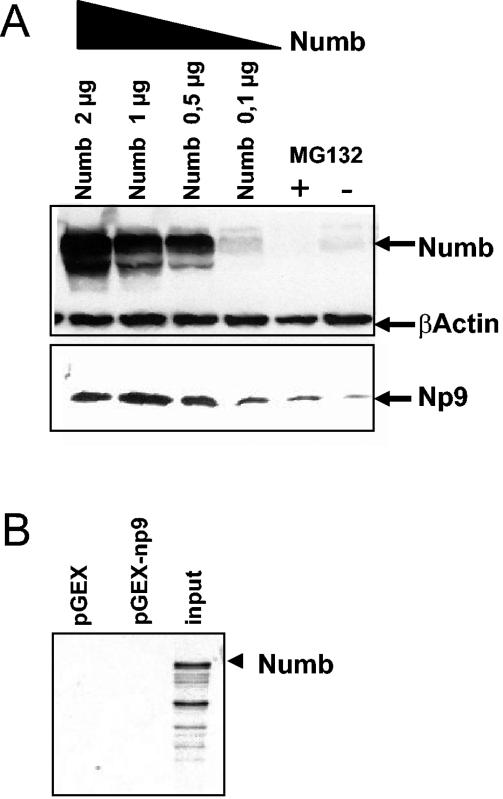
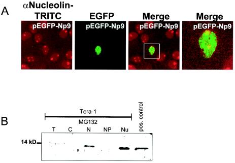
We have recently identified Np9 as a novel nuclear protein produced by the human endogenous retrovirus K and were able to document the exclusive presence of np9 transcript in tumors and transformed cells. With the aim of studying whether Np9 has a role in tumorigenesis, a systematic search for interacting proteins was performed. Here, we identify the RING-type E3 ubiquitin ligase LNX (ligand of Numb protein X) as an Np9-interacting partner. We furthermore show that the interaction involves N- and C-terminal domains of both proteins and can affect the subcellular localization of LNX. LNX has been reported to target the cell fate determinant and Notch antagonist Numb for proteasome-dependent degradation, thereby causing an increase in transactivational activity of Notch. We document that LNX-interacting Np9, like Numb, is unstable and degraded via the proteasome pathway and that ectopic Numb can stabilize recombinant Np9. Combined, these findings point to the possibility that Np9 affects tumorigenesis through the LNX/Numb/Notch pathway.


Figures

FIG. 1.
FIG. 1.
(A) Schematic representation of the HERV-K101 provirus. The open reading frames encoding the viral proteins Gag, Prot, and Pol are indicated. The env reading frame encodes Np9. The two exons of the np9 gene are highlighted. The shown provirus type fails to code for Rec, due to the type 1-specific deletion of 292 bp depicted. (B) Np9 protein. The positions of the three putative NLSs are depicted. (C) Schematic representation of the two human LNX splice variants, LNXp80 and LNXp70. The RING finger domain and the four PDZ domains are designated. The depicted LNX-C-term clone represents the Np9-interacting domain. A putative NLS and the N-terminal-binding site for Numb protein (LDNPAY), are indicated.
FIG. 2.
FIG. 2.
Fluorescence of Cos-1 cells transiently cotransfected to produce the fusion proteins EGFP-LNX-C (green fluorescence) and Np9Dsred (red fluorescence). Images were obtained by confocal laser-scanning microscopy. The righthand panel shows the colocalization sites in the merged images.
FIG. 3.
FIG. 3.
Fluorescence of Cos-1 cells transfected with pEGFP-LNX-C (A) or pEGFP-Np9 (B). Cell nuclei were stained with DAPI as a control. DAPI-stained relevant nuclei are marked by arrowheads.
FIG. 4.
FIG. 4.
(A) Localization of full-length LNX in Cos-1 cells transfected with pEGFP-LNX. (B) Colocalization of LNX and Np9. Cos-1 cells were cotransfected with pEGFP-LNX and Np9pDsred constructs. The relevant cell nucleus is marked by arrowheads in the DAPI-stained control.
FIG. 5.
FIG. 5.
Intracellular localization of EGFP-GST-Np9, EGFP-GST-Np9 NLS mutants (EGFP-Np9NLS1Mut, EGFP-Np9NLS2Mut, and EGFP-Np9NLS3Mut), and EGFP-GST alone in transiently transfected 293gp cells. Cell nuclei were stained with DAPI as a control, and relevant nuclei are marked by arrowheads.
FIG. 6.
FIG. 6.
Mapping of the Np9-LNX interaction domains by GST pulldown assays. (A) Full-length Np9 and variants with the indicated N- and C-terminal truncation, additional C-terminal amino acid residues (Cx), or a mutated NLS1 (NLSmut1) were employed as GST fusion products and incubated with in vitro-translated radiolabeled full-length LNX (LNX-F). A GST-only construct (pGEX) served as a control. (B) GST-full-length Np9 was incubated with the indicated variants of LNX. A putative NLS and the N-terminal-binding site for Numb (LDNPAY) are shown.
FIG. 7.
FIG. 7.
(A) Western blot analysis of protein extracts prepared from Cos-1 cells transiently transfected with pSG5-Np9. Twenty-four hours posttransfection, cells were treated with 5 μM MG132 (MG132 +), and extracts were harvested at the designated time points posttreatment with MG132. Untreated transiently transfected cells (MG132 −) and Cos-1 cells served as controls. Extracts were separated on a SDS-9.5 to 20% PAGE gradient and immunoblotted. Np9 was detected with the anti-Np9 polyclonal antiserum K82. (B) Western blot analysis of Tera-1 cell extracts partly treated with 5 μM MG132 for the times shown. A transiently pSG5-Np9-transfected Cos-1 cell extract served as a positive control. The total cellular protein extracts were separated on a SDS-9.5 to 20% PAGE gradient. Panel 1 shows a blot stained with anti-Np9 serum K82 preincubated with bacterially expressed Np9 protein. Panel 2 shows a blot stained with anti-Np9 serum K82 preincubated with the corresponding bacterial vector control. Np9-specific signals are marked with an arrowhead. (C) Western blot analysis of a cytolplasmic Np9 mutant (pSG5-Np9NLS1) in transiently transfected Cos-1 cells. Parallel cultures of Cos-1 cells were transiently transfected with pSG5-Np9 or a pSG5-Np9NLS1 mutant. One culture was treated 24 h posttransfection with 5 μM MG132 for an additional 24 h (MG132 +); the other culture remained untreated (MG132 −). Untransfected Cos-1 cells, with and without MG132 treatment, served as controls. Total cellular protein extracts were separated on a SDS-9.5 to 20% PAGE gradient. The arrowhead marks Np9-specific signals detected with the anti-Np9 polyclonal antiserum K82.
FIG. 8.
FIG. 8.
(A) Half-life of ectopically expressed Np9 in Tera-1 cells. Tera-1 cultures were transfected for 24 h with pSG5-Np9. The cultures were then treated either with MG132 (5 μM) for another 24 h or with CHX (25 μg/ml) for the indicated times at 48 h after transfection. Np9 protein was subjected to immunoprecipitation with K82 antiserum and analyzed by immunoblotting. Untreated Tera-1 extracts served as negative controls. (B) Half-life of transfected cytoplasmic Np9NLS1 in Tera-1 cells. Tera-1 cultures were treated and analyzed as described in panel A but were transfected with plasmid pSG5-Np9NLS1. (C) Stability of transfected Np9 in the presence of transfected p65 Numb. Tera-1 cultures were transfected and analyzed as outlined in panel A, except that pSG5-Np9 was cotransfected with Numb-pSG5-HA. Transfection efficiency was controlled by Western blotting (results not shown).
FIG. 9.
FIG. 9.
(A) Changes in steady-state levels of Np9 in dependence of Numb. Cos-1 cells were transiently transfected with pSG5-Np9 plus Numb-pSG5-HA at increasing quantities (0.1 to 2 μg) or with pSG5-Np9 and empty vector only. MG132 (5 μM) was added to the latter, and protein extracts were prepared at 48 h after transfection. Numb was detected with anti-Numb polyclonal antibody at a 1:500 dilution; β-actin was detected with anti-β-actin monoclonal antibody at a 1:1,000 dilution, and Np9 was detected with anti-Np9 polyclonal antibody at a 1:100 dilution. (B) Results of a GST pulldown assay documenting that Numb fails to directly interact with Np9.
FIG. 10.
FIG. 10.
(A) Combined immunofluorescence and autofluorescence analysis of nucleolin and EGFP-Np9. HeLa cells were transiently transfected with EGFP-Np9 to express Np9 hybrid protein. At 48 h posttransfection, cell nucleoli were immunostained with an anti-nucleolin polyclonal antibody and visualized with tetramethyl rhodamine isocyanate (TRITC) anti-rabbit secondary antibody. Images were produced by confocal laser-scanning microscopy. Colocalization of Np9 and nucleolin within the nucleoli is shown in the merged image. (B) Western immunoblot identifying Np9 in subcellular fractions. Subcellular fractions of exponentially growing Tera-1 cells were subjected to SDS-PAGE and were analyzed with the rabbit anti-Np9 polyclonal antibody K82 at a 1:100 dilution. T, total cell protein; C, free cytoplasmic protein; N, nuclear protein; NP, nucleoplasm without nucleoli fraction; Nu, nucleoli fraction; pos. control, total protein from Np9-transfected Cos-1 cells.

References

    1. Armbruester, V., M. Sauter, E. Krautkraemer, E. Meese, A. Kleiman, B. Best, K. Roemer, and N. Mueller-Lantzsch. 2002. A novel gene from the human endogenous retrovirus K expressed in transformed cells. Clin. Cancer Res. 8:1800-1807. - PubMed
    1. Barbulescu, M., G. Turner, M. I. Seaman, A. S. Deinhard, K. K. Kidd, and J. Lenz. 1999. Many human endogenous retrovirus K (HERV-K) proviruses are unique to humans. Curr. Biol. 9:861-868. - PubMed
    1. Beverly, L. J., and A. J. Capobianco. 2003. Perturbation of Ikaros isoform selection by MLV integration is a cooperative event in Notch(IC)-induced T cell leukemogenesis. Cancer Cell 3:551-564. - PubMed
    1. Bezprozvanny, I., and A. Maximov. 2001. PDZ domains: more than just a glue. Proc. Natl. Acad. Sci. USA 98:787-789. - PMC - PubMed
    1. Blond, J. L., D. Lavillette, V. Cheynet, O. Bouton, G. Oriol, S. Chapel-Fernandes, B. Mandrand, F. Mallet, and F. L. Cosset. 2000. An envelope glycoprotein of the human endogenous retrovirus HERV-W is expressed in the human placenta and fuses cells expressing the type D mammalian retrovirus receptor. J. Virol. 74:3321-3329. - PMC - PubMed

Publication types

MeSH terms

LinkOut - more resources