Case Reports
. 2004 Oct;78(19):10320-7.
doi: 10.1128/JVI.78.19.10320-10327.2004.

Direct evidence for a chronic CD8+-T-cell-mediated immune reaction to tax within the muscle of a human T-cell leukemia/lymphoma virus type 1-infected patient with sporadic inclusion body myositis

Affiliations
Case Reports

Direct evidence for a chronic CD8+-T-cell-mediated immune reaction to tax within the muscle of a human T-cell leukemia/lymphoma virus type 1-infected patient with sporadic inclusion body myositis

Simona Ozden et al. J Virol. 2004 Oct.

Abstract

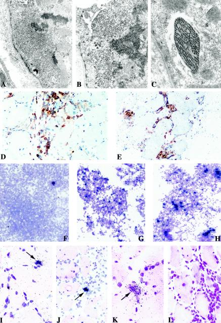
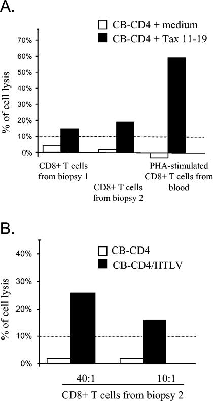
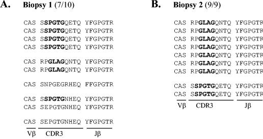
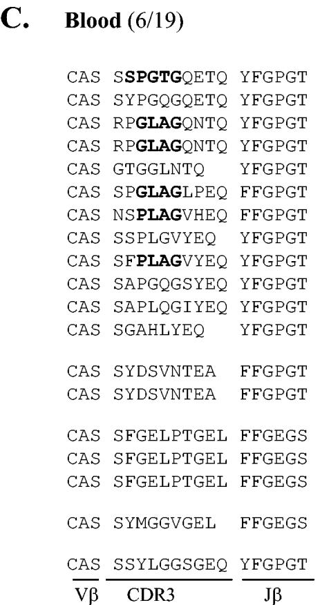
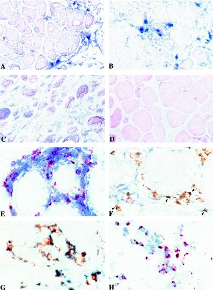
Human T-cell leukemia/lymphoma virus type 1 (HTLV-1) infection can lead to the development of HTLV-1-associated myelopathy/tropical spastic paraparesis (HAM/TSP), concomitantly with or without other inflammatory disorders such as myositis. These pathologies are considered immune-mediated diseases, and it is assumed that migration within tissues of both HTLV-1-infected CD4(+) T cells and anti-HTLV-1 cytotoxic T cells represents a pivotal event. However, although HTLV-1-infected T cells were found in inflamed lesions, the antigenic specificity of coinfiltrated CD8(+) T cells remains to be determined. In this study, we performed both ex vivo and in situ analyses using muscle biopsies obtained from an HTLV-1-infected patient with HAM/TSP and sporadic inclusion body myositis. We found that both HTLV-1-infected CD4(+) T cells and CD8(+) T cells directed to the dominant Tax antigen can be amplified from muscle cell cultures. Moreover, we were able to detect in two successive muscle biopsies both tax mRNA-positive mononuclear cells and T cells recognized by the Tax11-19/HLA-A*02 tetramer and positive for perforin. These findings provide the first direct demonstration that anti-Tax cytotoxic T cells are chronically recruited within inflamed tissues of an HTLV-1 infected patient, which validates the cytotoxic immune reaction model for the pathogenesis of HTLV-1-associated inflammatory disease.


Figures

FIG.1.
FIG.1.
Histological and virological features of muscle tissues and cultures. Electron micrographs of deltoid muscle sections from patient CB showing intracytoplasmic accumulation of abnormal filaments (magnification, ×9,800) (A) and clusters of straight (B) and paired helical (C) filaments seen at higher magnification (×14,000 and ×18,000). Immunochemical detection of CD4+ (D) and CD8+ (E) T cells on muscle sections counterstained with Harris hematoxylin. Detection of tax mRNA-positive cells among fresh PBMCs (F) and CD4+ T lymphocytes sorted from muscle culture (G) compared to PHA-stimulated CD4+ T cells from blood (H) (counterstained with Giemsa; exposure time, 8 days). In situ detection of tax mRNA with an antisense probe on muscle sections from the first (I and J) and the second (K) biopsies; arrows show focal positivity for HTLV-1 in mononuclear infiltrated cells and not in muscle fibers. Negative control analyses were performed with a tissue section from the second biopsy with a sense probe (L). Sections were counterstained with hematoxylin-eosin, and the exposure time was 21 days.
FIG. 2.
FIG. 2.
Cytotoxic activities of CD8+ T cells expanded from muscle cultures. (A) Chromium release assay to measure the cytotoxic activity directed to the immunodominant Tax 11-19 epitope of CD8+ T cells sorted from muscle cultures (performed in absence of mitogen), compared to PHA-stimulated CD8+ T cells from blood. For 4 days, PHA-stimulated CD4+ T lymphocytes from blood (in which HTLV-1 antigen production was still undetectable) incubated with or without the immunodominant Tax 11-19 epitope (10 μM) were used as targets cells (effector/target ratio, 40:1). (B) Chromium release assay to detect the ability of CD8+ T cells sorted after culture of biopsy 2 to kill chronically infected cells (CB-CD4/HTLV) at two effector/target ratios. Nonproducing autologous PHA-stimulated CD4+ cells were also used as a negative control. A percentage of cell lysis up to 10% was considered significant.
FIG. 3.
FIG. 3.
Molecular characterization of CD8+ T cells expanded from muscle cultures. Determination of amino acid composition of the TCR β-chain CDR3 sequences of 9 (biopsy 1), 10 (biopsy 2), and 19 (blood) DNA products obtained from Vβ13.1/Cβ-specific PCRs. Amino acid sequences corresponding to the conserved P/GLA/RG and SPGTG motifs previously found in anti-Tax 11-19 CD8+ T clonotypes (2, 29) are in boldface. The number of clones containing the characteristic motifs among the total number of sequenced clones is indicated between brackets.
FIG. 3.
FIG. 3.
Molecular characterization of CD8+ T cells expanded from muscle cultures. Determination of amino acid composition of the TCR β-chain CDR3 sequences of 9 (biopsy 1), 10 (biopsy 2), and 19 (blood) DNA products obtained from Vβ13.1/Cβ-specific PCRs. Amino acid sequences corresponding to the conserved P/GLA/RG and SPGTG motifs previously found in anti-Tax 11-19 CD8+ T clonotypes (2, 29) are in boldface. The number of clones containing the characteristic motifs among the total number of sequenced clones is indicated between brackets.
FIG. 4.
FIG. 4.
Immunochemical analyses of inflammatory lesions within deltoid muscle. Serial 5-μm frozen sections were fixed in acetone, processed for antigen detection, and counterstained with Harris hematoxylin. Staining was performed with the specific PE-conjugated HLA-A*02 Tax 11-19 tetramer on sections from the first (A) and the second (B) biopsies or on a control section from an HLA-A*02 uninfected patient with sIBM (C). Staining with a control HLA-A*02 EBV tetramer was also carried out on the second biospy sample from patient CB (D). Sections from the first biopsy were stained with anti-perforin antibody (E), with both anti-perforin antibody and PE-conjugated HLA-A*02 Tax 11-19 (F) (arrowheads indicate double-stained cells), and with both anti-TNF-α and CD68+ antibodies (G). Detection of apoptotic mononuclear cells by the terminal deoxynucleotidyltransferase-mediated dUTP-biotin nick end labeling method (H) was also performed.

References

    1. Bieganowska, K., P. Hollsberg, G. J. Buckle, D. G. Lim, T. F. Greten, J. Schneck, J. D. Altman, S. Jacobson, S. L. Ledis, B. Hanchard, J. Chin, O. Morgan, P. A. Roth, and D. A. Hafler. 1999. Direct analysis of viral-specific CD8+ T cells with soluble HLA-A2/Tax11-19 tetramer complexes in patients with human T cell lymphotropic virus-associated myelopathy. J. Immunol. 162:1765-1771. - PubMed
    1. Bourcier, K. D., D. G. Lim, Y. H. Ding, K. J. Smith, K. Wucherpfennig, and D. A. Hafler. 2001. Conserved CDR3 regions in T-cell receptor (TCR) CD8+ T cells that recognize the Tax11-19/HLA-A*0201 complex in a subject infected with human T-cell leukemia virus type 1: relationship of T-cell fine specificity and major histocompatibility complex/peptide/TCR crystal structure. J. Virol. 75:9836-9843. - PMC - PubMed
    1. Cavrois, M., A. Gessain, O. Gout, S. Wain-Hobson, and E. Wattel. 2000. Common human T cell leukemia virus type 1 (HTLV-1) integration sites in cerebrospinal fluid and blood lymphocytes of patients with HTLV-1-associated myelopathy/tropical spastic paraparesis indicate that HTLV-1 crosses the blood-brain barrier via clonal HTLV-1-infected cells. J. Infect. Dis. 182:1044-1050. - PubMed
    1. Cupler, E. J., M. Leon-Monzon, J. Miller, C. Semino-Mora, T. L. Anderson, and M. C. Dalakas. 1996. Inclusion body myositis in HIV-1 and HTLV-1 infected patients. Brain 119:1887-1893. - PubMed
    1. Dalakas, M. C., and R. Hohlfeld. 2003. Polymyositis and dermatomyositis muscle biopsy findings in inflammatory myopathies. Understanding the immunopathogenesis of inclusion-body myositis: present and future prospects. Lancet 362:971-982.

Publication types

MeSH terms