. 2004 Oct;78(19):10378-89.
doi: 10.1128/JVI.78.19.10378-10389.2004.

Human cytomegalovirus inhibits cytokine-induced macrophage differentiation

Affiliations

Human cytomegalovirus inhibits cytokine-induced macrophage differentiation

Sara Gredmark et al. J Virol. 2004 Oct.

Abstract

Human cytomegalovirus (HCMV) infection in immunocompromised patients is associated with impaired immunological function. Blood monocytes, which differentiate into macrophage effector cells, are of central importance for immune reactivity. Here, we demonstrate that HCMV transiently blocks cytokine-induced differentiation of monocytes into functionally active phagocytic macrophages. In HCMV-treated cultures, the cells had classical macrophage markers but lacked the classical morphological appearance of macrophages and had impairments in migration and phagocytosis. Even at very low multiplicities of infection, macrophage differentiation was almost completely inhibited. The inhibition appeared to be mediated by a soluble factor released upon viral treatment of monocytes. Human immunodeficiency virus or measles virus had no such effects. These findings suggest that HCMV impairs immune function by blocking certain aspects of cytokine-induced differentiation of monocytes and demonstrate an efficient pathway for this virus to evade immune recognition that may have clinical implications for the generalized immunosuppression often observed in HCMV-infected patients.


Figures
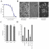
FIG. 1.
FIG. 1.
HCMV inhibits the differentiation of monocytes into macrophages. (A) Dose-dependent effects of infection with AD169 (MOI, 1 to 0.01) on macrophage differentiation. Values shown are means ± SEM for six separate experiments performed in duplicate. Esterase-negative cells were subtracted for the data presentation. (B) Phase-contrast microscopy photograph of mock-treated, HCMV-treated (AD169), and unstimulated monocytes at 7 days. Original magnification, ×40. (C) Effects of HCMV infection with AD169, Towne, or the clinical isolate PO at an MOI of 1. Values are means ± SEM for 10 separate experiments performed in duplicate. (D) Differentiation of monocytes stimulated with LPS, PMA, or IFN-γ plus TNF-α in the presence or absence of HCMV (AD169). Values are means ± SEM for three separate experiments performed in duplicate. The differentiation was defined by esterase staining as well as by morphology.
FIG. 2.
FIG. 2.
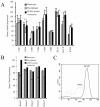
HCMV-treated monocytes retain high levels of CD14 expression. (A) Expression of cellular markers by uninfected monocytes, macrophages, and HCMV-treated monocytes after 7 days in culture as determined by flow cytometric analysis. The data represent mean channel fluorescence values ± SEM for five separate experiments. ***, P < 0.001 (two-tailed, paired t test; fresh monocytes versus HCMV-treated monocytes). (B) CD14 expression of donors 1 to 5, comparing the mean channel fluorescence values for fresh monocytes, macrophages, and HCMV-treated monocytes after 7 days in culture. (C) Representative histogram of CD14 expression by HCMV-treated monocytes and macrophages, assessed by flow cytometry.
FIG. 3.
FIG. 3.
HCMV's ability to inhibit macrophage differentiation declines with time after stimulation and is reversible. (A) Monocyte-enriched cultures were stimulated with allo-cytokines; treated with HCMV 0, 3, 6, 12, 24, 48, or 72 h after stimulation; and cultured for 7 days. Values are the means ± SEM for six separate experiments performed in duplicate. Esterase-negative cells were subtracted for the data presentation. (B) Monocyte-enriched cultures were stimulated with allo-cytokines, treated with HCMV, and restimulated once for 24 h 1 to 18 days after infection. Phase-contrast microscopy photographs are of mock-treated monocyte-derived macrophages, HCMV-treated monocytes, HCMV-treated monocytes restimulated 6 days after infection, and HCMV-treated monocytes restimulated 18 days after infection. The photographs were taken 7 days after restimulation. Original magnification, ×40. The differentiation was defined by esterase staining as well as by morphology.
FIG. 3.
FIG. 3.
HCMV's ability to inhibit macrophage differentiation declines with time after stimulation and is reversible. (A) Monocyte-enriched cultures were stimulated with allo-cytokines; treated with HCMV 0, 3, 6, 12, 24, 48, or 72 h after stimulation; and cultured for 7 days. Values are the means ± SEM for six separate experiments performed in duplicate. Esterase-negative cells were subtracted for the data presentation. (B) Monocyte-enriched cultures were stimulated with allo-cytokines, treated with HCMV, and restimulated once for 24 h 1 to 18 days after infection. Phase-contrast microscopy photographs are of mock-treated monocyte-derived macrophages, HCMV-treated monocytes, HCMV-treated monocytes restimulated 6 days after infection, and HCMV-treated monocytes restimulated 18 days after infection. The photographs were taken 7 days after restimulation. Original magnification, ×40. The differentiation was defined by esterase staining as well as by morphology.
FIG. 4.
FIG. 4.
HCMV-treated monocytes exhibit decreased migration in response to chemoattractants. (A) Cells were mock treated or treated with HCMV and cultured for 7 days, and migration in response to RANTES, MIP-1α, and MCP-1 was assessed with a transwell culture system. Cells that migrated were counted by light microscopy in four representative fields at ×40 magnification. Values are the means ± SEM for three separate experiments performed in duplicate. (B) Phase-contrast microscopy photographs of mock-treated macrophages or HCMV-treated monocytes that migrated through the filter pores. Original magnification, ×40. Arrows are pointing at cells to exemplify migrated cells. (C) Cells were mock treated, treated with HCMV, or restimulated 1 day postinfection after HCMV treatment. Migration in response to RANTES was assessed after 7 days in a transwell culture system. Values are the means ± SEM for three separate experiments performed in duplicate.
FIG. 5.
FIG. 5.
HCMV-treated monocytes exhibit impaired phagocytosis. (A) After 7 days in culture, mock-treated and HCMV-treated monocytes were tested for their ability to phagocytose FITC-labeled yeast particles. Cells that had phagocytosed yeast particles were quantified after 10 and 60 min. Values are means ± SEM for six separate experiments performed in duplicate. (B) Fluorescence microscopy photographs showing phagocytosed yeast particles in mock-treated and HCMV-treated monocytes at 30 min. Original magnification, ×40. Arrows are pointing at cells to exemplify phagocytosed FITC-labeled yeast particles. (C) Cells were mock treated, treated with HCMV, or restimulated 1 day postinfection after HCMV treatment. Phagocytosis of FITC-labeled yeast particles was assessed after 7 days. Cells that had phagocytosed yeast particles were quantified after 30 min. Values are the means ± SEM for three separate experiments performed in duplicate.
FIG. 6.
FIG. 6.
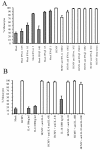
Inhibition of macrophage differentiation is not a general event after virus exposure and is mediated by a structural component of HCMV. Macrophage differentiation after treatment of monocytic cultures with HIV or measles virus (MV) (A), ultracentrifuged virus-free supernatants (HCMV-sup), concentrated virus particles (UC-HCMV), or supernatants from mock-infected cultures (HL-sup) (B), or HCMV inactivated by treatment with UV or IVIG (HCMV UV and HCMV IVIG) (C). Values are means ± SEM for three separate experiments performed in duplicate. The differentiation was defined by esterase staining as well as by morphology.
FIG. 7.
FIG. 7.
HCMV-treated monocytes release a soluble factor that blocks macrophage differentiation. (A) Effects of supernatants on macrophage differentiation of monocytic cultures stimulated with allo-cytokines. Supernatants from HCMV-treated and mock-treated monocytic cultures were collected 6, 12, and 24 h after HCMV treatment, cleared by centrifugation, filtered to remove infectious virus, and diluted 1:10. (B) Effects of supernatants on macrophage differentiation of monocytic cultures stimulated with allo-cytokines. Supernatants from HCMV-treated and mock-treated monocytic cultures were collected 24 h after HCMV treatment, cleared by centrifugation, filtered to remove infectious virus, and diluted 1:10 and 1:100. Values are means ± SEM for three separate experiments performed in duplicate. The differentiation was defined by esterase staining as well as by morphology. (C to E) Levels of IFN-α (undetectable IFN-α levels in mock-treated cultures) (C), IL-6 (D), and IL-10 (E) as detected by ELISA in the supernatants obtained from mock- and HCMV-infected cultures, respectively.
FIG. 8.
FIG. 8.
IFN-α affects macrophage differentiation. (A) Recombinant IFN-α and IFN-β were added to monocytic cultures at the same time as cytokine stimulation (IFN concentrations, 0.05 to 5 ng/ml). Antibodies against IFN-α (10 to 1,000 U) and IFN-β (10 to 2,500 U) and a control sheep serum were added to the cultures at the same time point as HCMV treatment. (B) Recombinant IL-6 (100 to 1,000 ng/ml) or recombinant IL-10 (1,000 ng/ml) was added to the cultures at the same time as cytokine stimulation. Antibodies against IL-6 and/or IL-6 receptor or antibodies against IL-10 and/or the IL-10-receptor were added at the same time point as HCMV treatment. The differentiation was defined by esterase staining as well as by morphology.

Similar articles

Cited by

References

    1. Allavena, P., L. Piemonti, D. Longoni, S. Bernasconi, A. Stoppacciaro, L. Ruco, and A. Mantovani. 1998. IL-10 prevents the differentiation of monocytes to dendritic cells but promotes their maturation to macrophages. Eur. J. Immunol. 28:359-369. - PubMed
    1. Asadullah, K., S. Prosch, H. Audring, I. Buttnerova, H. D. Volk, W. Sterry, and W. D. Docke. 1999. A high prevalence of cytomegalovirus antigenaemia in patients with moderate to severe chronic plaque psoriasis: an association with systemic tumour necrosis factor alpha overexpression. Br. J. Dermatol. 141:94-102. - PubMed
    1. Biondi, A., T. H. Rossing, J. Bennett, and R. F. Todd III. 1984. Surface membrane heterogeneity among human mononuclear phagocytes. J. Immunol. 132:1237-1243. - PubMed
    1. Bolovan-Fritts, C. A., E. S. Mocarski, and J. A. Wiedeman. 1999. Peripheral blood CD14(+) cells from healthy subjects carry a circular conformation of latent cytomegalovirus genome. Blood 93:394-398. - PubMed
    1. Britt, W. J., and C. A. Alford. 1996. Cytomegalovirus, p. 2493-2523. In B. N. Fields, D. M. Knipe, and P. M. Howley (ed.), Fields virology, vol. 2, 3rd ed. Lippincott-Raven Publishers, Philadelphia, Pa.

Publication types

MeSH terms

LinkOut - more resources