. 2004 Oct;78(19):10410-9.
doi: 10.1128/JVI.78.19.10410-10419.2004.

Murine coronavirus nonstructural protein p28 arrests cell cycle in G0/G1 phase

Affiliations

Murine coronavirus nonstructural protein p28 arrests cell cycle in G0/G1 phase

Chun-Jen Chen et al. J Virol. 2004 Oct.

Abstract

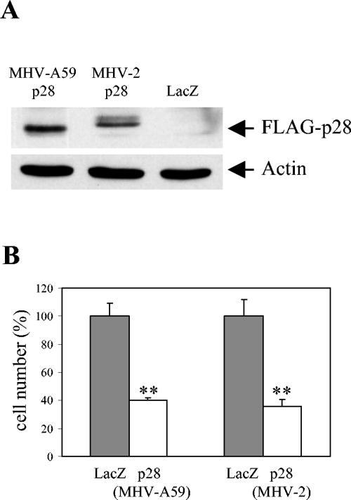
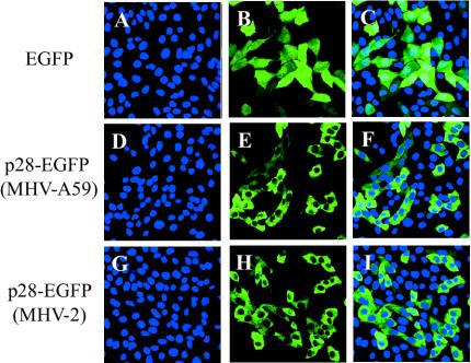
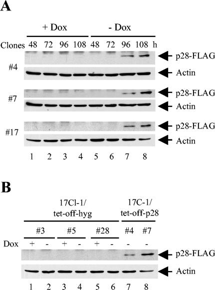
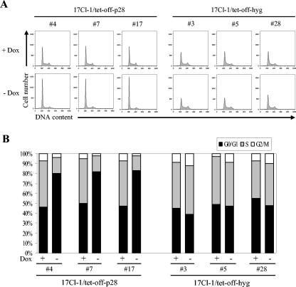
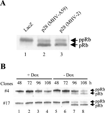
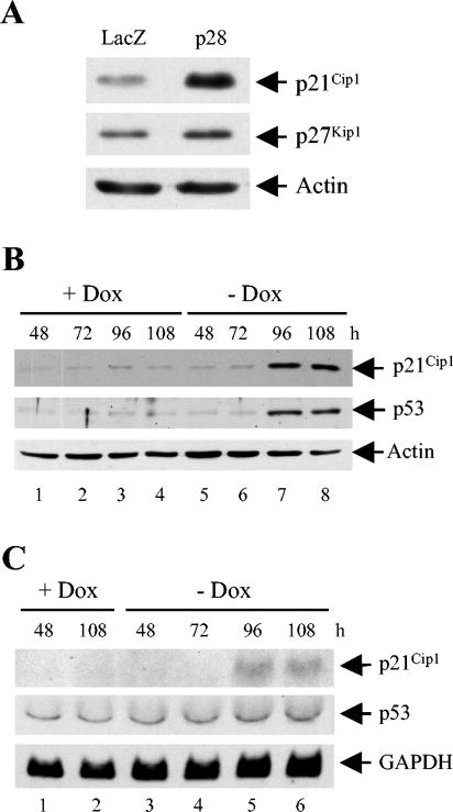
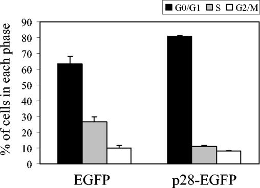
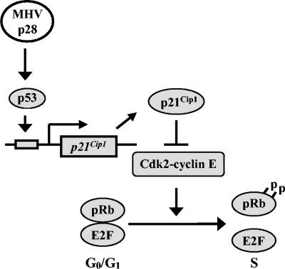
Murine coronavirus mouse hepatitis virus (MHV) gene 1 encodes several nonstructural proteins. The functions are unknown for most of these nonstructural proteins, including p28, which is encoded at the 5' end of the MHV genome. Transient expression of cloned p28 in several different cultured cells inhibited cell growth, indicating that p28 expression suppressed cell proliferation. Expressed p28 was exclusively localized in the cytoplasm. Cell cycle analysis by flow cytometry demonstrated that p28 expression induced G(0)/G(1) cell cycle arrest. Characterization of various cellular proteins that are involved in regulating cell cycle progression demonstrated that p28 expression resulted in an accumulation of hypophosphorylated retinoblastoma protein (pRb), tumor suppressor p53, and cyclin-dependent kinase (Cdk) inhibitor p21(Cip1). Expression of p28 did not alter the amount of p53 transcripts yet increased the amount of p21(Cip1) transcripts, suggesting that p28 expression increased p53 stability and that p21(Cip1) was transcriptionally activated in a p53-dependent manner. Our present data suggest the following model of p28-induced G(0)/G(1) cell cycle arrest. Expressed cytoplasmic p28 induces the stabilization of p53, and accumulated p53 causes transcriptional upregulation of p21(Cip1). The increased amount of p21(Cip1) suppresses cyclin E/Cdk2 activity, resulting in the inhibition of pRb hyperphosphorylation. Accumulation of hypophosphorylated pRb thus prevents cell cycle progression from G(0)/G(1) to S phase.


Figures

FIG. 1.
FIG. 1.
Transient expression of p28-FLAG inhibited cell proliferation. 17Cl-1 cells cultured in six-well plates were transfected with 1 μg of pA59-p28-FLAG, pV2-p28-FLAG, or pcDNA3.1/HisB/LacZ. (A) At 24 h after transfection, cell lysates were prepared and subjected to Western blot analysis with anti-FLAG M2 and antiactin as the primary antibodies. (B) At 68 h after transfection, the numbers of live cells in each sample were determined by using trypan blue exclusion. The results are presented as the mean and standard error of cell numbers for six independent experiments, and the cell numbers in p28-transfected samples were normalized to the cell numbers in LacZ-transfected samples (100%). **, P < 0.001 (Student's t test) versus LacZ-transfected samples.
FIG. 2.
FIG. 2.
Subcellular localization of p28-EGFP. 17Cl-1 cells cultured on chambered coverglasses were transfected with pEGFP-N1 (A to C), pEGFP-pA59-p28 (D to F), or pEGFP-pV2-p28 (G to I). At 24 h after transfection, Hoechst 33342 was added to the culture medium, and live cells were observed under a laser confocal microscope with appropriate filters for Hoechst 33342 (blue) (A, D, and G) and EGFP (green) (B, E, and H). Confocal images in the left two columns were superimposed (C, F, and I) to show the cytoplasmic distribution of p28-EGFP.
FIG. 3.
FIG. 3.
Tetracycline-regulated p28 expression. (A) 17Cl-1/tet-off-p28 cells were cultured in the presence (+ Dox) or absence (− Dox) of 1 μg of doxycyline per ml. At indicated times after doxycyline withdrawal, cell lysates were collected and subjected to Western blot analysis for FLAG-p28 and actin. (B) Lanes 1 to 6, 17Cl-1/tet-off-hyg cells were cultured in the presence or absence of 1 μg of doxycyline per ml for 108 h, and cell lysates were collected and used in Western blot analysis for FLAG-p28 and actin. Lanes 7 and 8, same samples as in lane 8 of panel A served as positive controls.
FIG. 4.
FIG. 4.
Cell cycle profiles of p28-expressing cell. (A) 17Cl-1/tet-off-p28 cells and 17Cl-1/tet-off-hyg cells were cultured in the presence (+ Dox) or absence (− Dox) of 1 μg of doxycyline per ml. At 120 h after doxycyline withdrawal, cells were collected and subjected to cell cycle analysis as described in Materials and Methods. (B) The percentage of cells in each phase of the cell cycle was computed by using the ModFit LT program. The data represent results from one of three independent experiments; the other two experiments showed similar results.
FIG. 5.
FIG. 5.
Phosphorylation status of pRb in p28-expressing cells. (A) 17Cl-1 cells were transfected with 1 μg of pA59-p28-FLAG, pV2-p28-FLAG, or pcDNA3.1/HisB/LacZ. At 36 h after transfection, cells were lysed with SDS-polyacrylamide gel electrophoresis sample buffer, and whole-cell lysates were subjected to Western blot analysis for pRb. The slower-migrating band and fast-migrating band are hyperphosphorylated forms of pRb (ppRb) and hypo- and unphosphosphorylated forms of pRb (pRb), respectively. (B) 17Cl-1/tet-off-p28 cells were cultured in the presence (+ Dox) or absence (− Dox) of 1 μg of doxycyline per ml. At the indicated times after doxycyline withdrawal, whole-cell lysates were collected and analyzed as described for panel A.
FIG. 6.
FIG. 6.
Accumulation of p21Cip1 and p53 proteins in p28-expressing cells. (A) 17Cl-1 cells were transfected with 1 μg of pA59-p28-FLAG or pcDNA3.1/HisB/LacZ. At 36 h after transfection, cell lysates were collected and subjected to Western blot analysis for p21Cip1, p27Kip1, and actin. (B and C) 17Cl-1/tet-off-p28 clone 17 cells were cultured in the presence (+ Dox) or absence (− Dox) of 1 μg of doxycyline per ml. At the indicated times after doxycyline withdrawal, cell lysates were collected and subjected to Western blot analysis for p21Cip1, p53, and actin (B), or total cellular RNA was extracted and equal amounts (10 μg) of RNA from each sample were subjected to Northern blot analysis and probed for p21Cip1, p53, and GAPDH (C).
FIG. 7.
FIG. 7.
Cell cycle profiles of p28-expressing LU cells. LU cells were infected with pseudotype retroviruses encoding EGFP or MHV-A59 p28-EGFP fusion protein. At 96 h p.i., cells were collected and subjected to cell cycle analysis by flow cytometry. The percentage of cells in each phase of the cell cycle was computed by using the ModFit LT program. The results are presented as means and standard errors for three independent experiments.
FIG. 8.
FIG. 8.
A model for MHV p28-induced cell cycle arrest in G0/G1 phase. p28 expression induces stabilization and accumulation of p53, which in turn transactivates p21Cip1. The upregulated p21Cip1 suppresses cyclin E-Cdk2 activity, resulting in the inhibition of pRb hyperphosphorylation and the prevention of cell cycle progression from G0/G1 to S phase.

References

    1. Adams, P. D., W. R. Sellers, S. K. Sharma, A. D. Wu, C. M. Nalin, and W. G. Kaelin, Jr. 1996. Identification of a cyclin-cdk2 recognition motif present in substrates and p21-like cyclin-dependent kinase inhibitors. Mol. Cell. Biol. 16:6623-6633. - PMC - PubMed
    1. Akagi, T., T. Shishido, K. Murata, and H. Hanafusa. 2000. v-Crk activates the phosphoinositide 3-kinase/AKT pathway in transformation. Proc. Natl. Acad. Sci. USA 97:7290-7295. - PMC - PubMed
    1. Albrecht, T., and T. H. Weller. 1980. Heterogeneous morphologic features of plaques induced by five strains of human cytomegalovirus. Am. J. Clin. Pathol. 73:648-654. - PubMed
    1. Baker, S. C., C. K. Shieh, L. H. Soe, M. F. Chang, D. M. Vannier, and M. M. Lai. 1989. Identification of a domain required for autoproteolytic cleavage of murine coronavirus gene A polyprotein. J. Virol. 63:3693-3699. - PMC - PubMed
    1. Balint, E. E., and K. H. Vousden. 2001. Activation and activities of the p53 tumour suppressor protein. Br. J. Cancer 85:1813-1823. - PMC - PubMed

Publication types

MeSH terms