. 2004 Oct;78(19):10433-41.
doi: 10.1128/JVI.78.19.10433-10441.2004.

Low pH is required for avian sarcoma and leukosis virus Env-dependent viral penetration into the cytosol and not for viral uncoating

Affiliations

Low pH is required for avian sarcoma and leukosis virus Env-dependent viral penetration into the cytosol and not for viral uncoating

Richard J O Barnard et al. J Virol. 2004 Oct.

Abstract

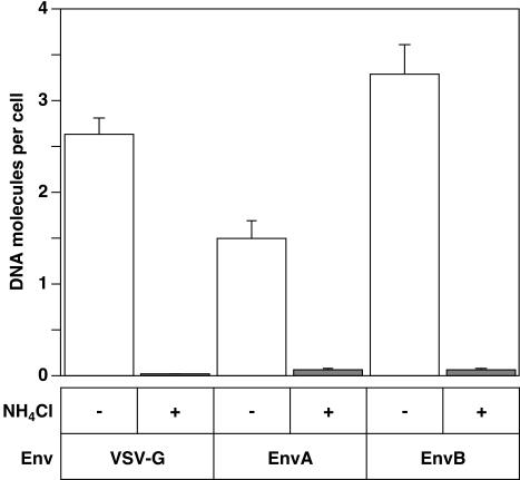
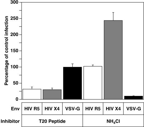
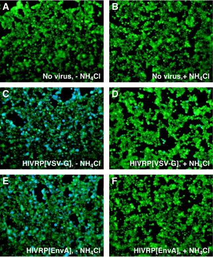
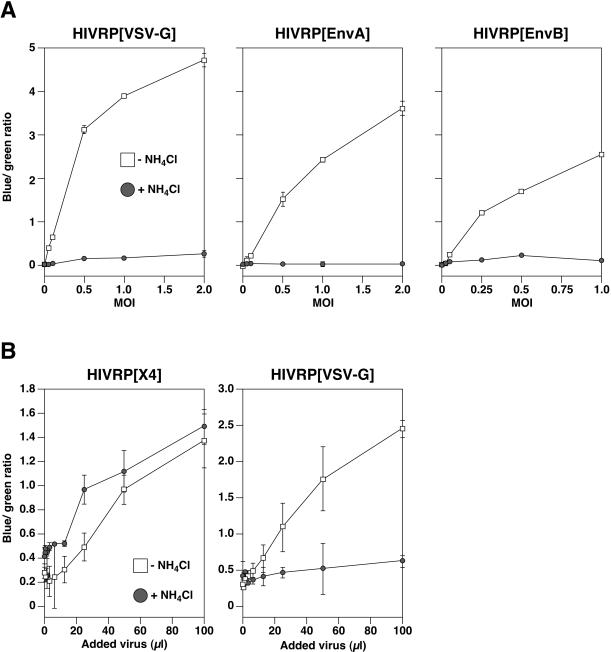
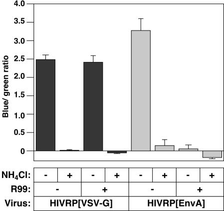
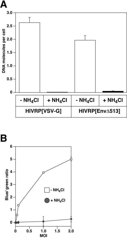
A novel entry mechanism has been proposed for the avian sarcoma and leukosis virus (ASLV), whereby interaction with specific cell surface receptors activates or primes the viral envelope glycoprotein (Env), rendering it sensitive to subsequent low-pH-dependent fusion triggering in acidic intracellular organelles. However, ASLV fusion seems to proceed to a lipid mixing stage at neutral pH, leading to the suggestion that low pH might instead be required for a later stage of viral entry such as uncoating (L. J. Earp, S. E. Delos, R. C. Netter, P. Bates, and J. M. White. J. Virol. 77:3058-3066, 2003). To address this possibility, hybrid virus particles were generated with the core of human immunodeficiency virus type 1 (HIV-1), a known pH-independent virus, and with subgroups A or B ASLV Env proteins. Infection of cells by these pseudotyped virions was blocked by lysosomotropic agents, as judged by inhibition of HIV-1 DNA synthesis. Furthermore, by using HIV-1 cores that contain a Vpr-beta-lactamase fusion protein (Vpr-BlaM) to monitor viral penetration into the cytosol, we demonstrated that virions bearing ASLV Env, but not HIV-1 Env, enter the cytosol in a low-pH-dependent manner. This effect was independent of the presence of the cytoplasmic tail of ASLV Env. These studies provide strong support for the model, indicating that low pH is required for ASLV Env-dependent viral penetration into the cytosol and not for viral uncoating.


Figures

FIG. 1.
FIG. 1.
Infection of HIV particles bearing ASLV fusion proteins is pH dependent. For the present study we quantified the HIVRP virus titer in terms of VTU/milliliter (see Materials and Methods). HIVRP[VSV-G]-, HIVRP[EnvA]-, or HIVRP[EnvB]-pseudotyped particles were bound to 293 cells stably expressing ASLV receptors at an MOI of 1 (VTU) for 1 h at 4°C and then extensively washed to remove unbound virus. Cells were then warmed to 37°C for 6 h in the presence or absence of 30 mM NH4Cl, and DNA from these cells was prepared as described earlier (31). The number of early HIV reverse transcription products was quantitated by real-time PCR with an ABI 7000 machine (Applied Biosystems). HIV plasmid DNA was used to construct a standard curve for enumeration. The results shown are representative of two independent experiments performed in triplicate. Error bars represent the standard deviation.
FIG. 2.
FIG. 2.
The pH dependence of Vpr-BlaM delivery is dictated by the viral fusion protein. HIV X4 Env-, HIV R5 Env-, or HIV[VSV-G]-pseudotyped particles were bound to cells for 1 h at room temperature in the presence or absence of either 30 mM NH4Cl or 1 μM T20 inhibitory peptide and then warmed to 37°C for 4 h in the presence or absence of inhibitors. Cells were then loaded with CCF4/AM for 18 h at 25°C, and their fluorescence was then quantitated with a BMG FluoStar plate reader with excitation at 410/12 nm, and emission was detected by using band-pass filters at 460/12 nm and 530/12 nm. The ratio of fluorescence at 460 and 530 nm was calculated, and the results were normalized to the amount of Vpr-BlaM delivery observed for each virus in the absence of inhibitors. The results shown are mean values obtained from two independent experiments. Error bars represent the standard deviation.
FIG. 3.
FIG. 3.
ASLV Env-dependent viral penetration is pH dependent. HIVRP-pseudotyped particles were bound to 293 cells stably expressing cognate ASLV receptors at an MOI of 1 (VTU) for 1 h at 4°C and then extensively washed to remove unbound virus. Cells were warmed to 37°C in the presence (B, D, and F) or absence (A, C, and E) of 30 mM NH4Cl for 6 h and then loaded with CCF2/AM for 8 to 12 h at 25°C. Cells were visualized by using an Axiovert 25 epifluorescence microscope (Carl Zeiss) equipped with a 20× objective using a β-lactamase filter set (part number 41031; Chroma Technologies). Images were captured with an AxioCam HRc camera (Carl Zeiss) and processed by using Openlab software (version 3.0.7) from Improvision, Lexington, Mass.
FIG. 4.
FIG. 4.
Quantitation of HIVRP[EnvA] and HIVRP[EnvB] viral penetration. (A) HIVRP-pseudotyped particles were bound to 293 cells stably expressing ASLV receptors at different MOIs (VTU) for 1 h at 4°C and then extensively washed to remove unbound virus. Cells were warmed to 37°C for 6 h in the presence or absence of 30 mM NH4Cl and then loaded with CCF2/AM for 8 to 12 h at 25°C. The ratio of fluorescence at 447 and 518 nm was calculated as described in Fig. 2 and was plotted against the MOI (VTU). The experiments shown are representative of three independent experiments performed in quadruplet. Error bars represent the standard deviation. (B) HIV Env-dependent viral penetration is pH independent in 293 cells. Different dilutions of a stock of HIVRPs bearing either VSV-G- or X4-tropic HIV fusion proteins were bound to 293 cells stably expressing TVA800, CXCR4, and CD4 for 1 h at room temperature in the presence or absence of either 30 mM NH4Cl and then warmed to 37°C for 4 h in the presence or absence of inhibitor. Cells were then loaded with CCF4/AM for 18 h at 25°C, their fluorescence was quantitated with a BMG FluoStar plate reader, and the blue/green ratio was calculated and plotted against the volume of virus added. The results shown are representative of data obtained in two independent experiments. Error bars represent the standard deviation.
FIG. 5.
FIG. 5.
Fusion of HIV[EnvA] is required for Vpr-BlaM delivery. HIVRP[EnvA]- or HIVRP[VSV-G]-pseudotyped particles were bound to 293 cells stably expressing TVA at an MOI of 1 (VTU) for 1 h at 4°C. Cells were then extensively washed to remove unbound virus and warmed to 37°C for 6 h in the presence or absence of 30 mM NH4Cl or 3.4 μM R99 peptide. Cells were then loaded with CCF2/AM for 12 h at 25°C. The ratio of fluorescence at 447 and 518 nm was calculated, and the blue/green ratio of uninfected control cells was subtracted from each datum point. The experiments shown are representative of two independent experiments performed in quadruplet. Error bars represent the standard deviation.
FIG. 6.
FIG. 6.
HIVRP[EnvAΔ513] enters cells by a pH-dependent mechanism. HIV[EnvAΔ513] virus-cell complexes were formed at 4°C at an MOI of 1 (VTU/cell) for 1 h, and cells were washed to remove unbound virus. The cells were warmed to 37°C for 6 h in the presence or absence of NH4Cl, and the amount of early HIV DNA products (A) and Vpr-BlaM delivery (B) was quantitated as described in Fig. 1 and 4, respectively.

References

    1. Adkins, H. B., J. Brojatsch, and J. A. T. Young. 2000. Identification and characterization of a shared TNFR-related receptor for subgroup B, D, and E avian leukosis viruses reveal cysteine residues required specifically for subgroup E entry. J. Virol. 74:3572-3578. - PMC - PubMed
    1. Barnard, R. J. O., and J. A. T. Young. 2003. Alpharetroviral receptor-envelope interactions. Curr. Top. Immunol. Microbiol. 281:107-136. - PubMed
    1. Boerger, A. L., S. Snitkovsky, and J. A. T. Young. 1999. Retroviral vectors preloaded with a viral receptor-ligand bridge protein are targeted to specific cell types. Proc. Natl. Acad. Sci. USA 96:9867-9872. - PMC - PubMed
    1. Brojatsch, J., J. Naughton, M. M. Rolls, K. Zingler, and J. A. T. Young. 1996. CAR1, a TNFR-related protein, is a cellular receptor for cytopathic avian leukosis-sarcoma viruses and mediates apoptosis. Cell 87:845-855. - PubMed
    1. Cavrios, M., C. de Noronha, and W. C. Greene. 2002. A sensitive and specific enzyme-based assay detecting HIV-1 virion fusion in primary T lymphocytes. Nat. Biotechnol. 20:1151-1154. - PubMed

Publication types

MeSH terms

LinkOut - more resources