. 2004 Oct;78(19):10685-94.
doi: 10.1128/JVI.78.19.10685-10694.2004.

Nuclear export of the nonenveloped parvovirus virion is directed by an unordered protein signal exposed on the capsid surface

Affiliations

Nuclear export of the nonenveloped parvovirus virion is directed by an unordered protein signal exposed on the capsid surface

Beatriz Maroto et al. J Virol. 2004 Oct.

Abstract

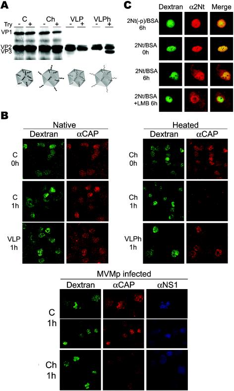
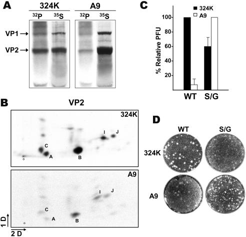
It is uncertain whether nonenveloped karyophilic virus particles may actively traffic from the nucleus outward. The unordered amino-terminal domain of the VP2 major structural protein (2Nt) of the icosahedral parvovirus minute virus of mice (MVM) is internal in empty capsids, but it is exposed outside of the shell through the fivefold axis of symmetry in virions with an encapsidated single-stranded DNA genome, as well as in empty capsids subjected to a heat-induced structural transition. In productive infections of transformed and normal fibroblasts, mature MVM virions were found to efficiently exit from the nucleus prior to cell lysis, in contrast to the extended nuclear accumulation of empty capsids. Newly formed mutant viruses lacking the three phosphorylated serine residues of 2Nt were hampered in their exit from the human transformed NB324K nucleus, in correspondence with the capacity of 2Nt to drive microinjected phosphorylated heated capsids out of the nucleus. However, in normal mouse A9 fibroblasts, in which the MVM capsid was phosphorylated at similar sites but with a much lower rate, the nuclear exit of virions and microinjected capsids harboring exposed 2Nt required the infection process and was highly sensitive to inhibition of the exportin CRM1 in the absence of a demonstrable interaction. Thus, the MVM virion exits the nucleus by accessing nonconventional export pathways relying on cell physiology that can be intensified by infection but in which the exposure of 2Nt remains essential for transport. The flexible 2Nt nuclear transport signal may illustrate a common structural solution used by nonenveloped spherical viruses to propagate in undamaged host tissues.


Figures

FIG. 1.
FIG. 1.
Exposure of 2Nt in MVM capsid and virus. (A) Transport sequences in MVM structural proteins. Shown are the NLM (residues 670 to 680 of VP1) in the common sequence of the three polypeptides and the NLS of VP1 (residues 6 to 10 and 87 to 90) (33). The phosphoserine residues at positions 2, 6, and 10 of VP2 (black balls) within 2Nt and the approximate VP2 processing site to produce VP3 (arrow) are indicated. The entire 2Nt amino acid sequence of the prototype MVM strain is shown below. (B) Immunological analysis of 2Nt exposure. 35S-labeled empty capsids (C) and DNA-filled virus (V) were purified from 324K cultures early (22 hpi) and late (48 hpi) in the infection and subsequently were immunoprecipitated as either native or denatured (boiled for 5 min in 0.2% SDS) proteins with the polyclonal α-CAP antiserum, the α-2Nt antiserum, or a control preimmune serum (cs). The positions of the virus structural proteins in the SDS-10% PAGE gels are indicated to the left.
FIG. 2.
FIG. 2.
2Nt phosphorylation is important for nuclear exit of infectious virus in transformed cells. (A) Subcellular localization of MVMp particles in a single round of infection of transformed 324K cells synchronized by growth to confluence. Shown are representative fields of cells visualized by confocal microscopy and stained with the MVM-capsid MAb and the α-2Nt antibody specific for DNA-filled virions. +LMB, wt particles at 24 hpi in cells treated with LMB (100 ng/ml) from 14 hpi. Insets, intranuclear punctate phenotype of S/G particles. (B) Subcellular distribution of newly formed MVM particles. Infected synchronous cells were labeled with 35S since 6 hpi, and wt viral particles harvested from fractionated cells at the indicated times were sedimented through a sucrose cushion and centrifuged to equilibrium in a CsCl gradient. A representative result of 35S cpm distribution in the banding positions of the empty capsids (c) and DNA-filled virions (v) is shown. (C) The S/G phosphorylation mutant shows a defect in nuclear egress. (Left) Infectious units (IU) of wt and S/G viruses in the culture medium (extra) and inside the cells (intra) at the indicated postinfection times for synchronized 324K cells. DL, detection limit of the assay. (Right) Percentage of subcellular distribution of infectious viruses in fractionated cells. Data are means and standard errors from three independent experiments.
FIG. 3.
FIG. 3.
Nuclear export activity of 2Nt in transformed cells. (A) Heat-triggered exposure of 2Nt in MVM particles. Purified native empty capsids (C) and VLPs or the respective particles heated at 50°C for 10 min (Ch and VLPh) were tested for 2Nt exposure by trypsin (Try) digestion (23), and the cleavage of VP2 to form VP3 was visualized by immunoblotting with an α-VP antiserum. The four types of microinjected particles, with phosphorylated serine residues of 2Nt in natural capsids (black balls) or no phosphorylation in VLPs, are illustrated at the bottom. (B) Subcellular localization of MVM particles injected inside the 324K nucleus. Viral particles were injected with dextran-FITC in noninfected (upper panels) or infected (lower panels) cells and analyzed either immediately (0 h) or at 1 h postinjection by confocal laser microscopy after staining with the α-CAP serum and the α-NS1 MAb antibody where indicated. Shown are representative fields at different magnifications with clusters of injected cells and FITC-dextran marking intact nuclear membranes. (C) Confocal analysis of 2Nt(−p)-BSA and 2Nt-BSA protein conjugates microinjected into the 324K nucleus. Cells were fixed at the indicated times postinjection and stained with the α-2Nt antibody. Where indicated (+LMB), LMB (100 ng/ml) was added to the cultures after injection.
FIG. 4.
FIG. 4.
Cell-type-dependent 2Nt phosphorylation and MVM infection. (A) Quantitative analysis of MVM capsid phosphorylation in permissive cells. The respective cultures (106 cells) were infected with MVMp (5 PFU/cell) and labeled in parallel at 2 hpi with [35S]Met-Cys (35S) or [32P]orthophosphate (32P). Capsid proteins were immunoprecipitated at 20 hpi with the α-VP antiserum from identical amounts of boiled homogenates and were resolved by SDS-10% PAGE. (B) Two-dimensional phosphopeptide map of VP2 capsid subunits. The VP2 protein purified from the gels was digested with trypsin and subjected to two-dimensional TLC analysis. The plates were exposed to autoradiography for 5 days with an intensifying screen at −70°C. Peptide B, corresponding to 2Nt, and other main phosphopeptides are designated as described previously (37). 1D, first dimension; 2D, second dimension; O, origin. (C) Plaque-forming capacity of wt and S/G viruses. Identical numbers (IU) of gradient-purified viruses were added to the respective cell monolayers, and the infectivity was scored as the percentage of PFU with respect to the most permissive cell for each virus. The figure outlines the averages and standard errors from three independent determinations. (D) Representative result illustrating the ratio and morphology of virus plaques in the reference cell lines.
FIG. 5.
FIG. 5.
Nuclear export of MVM virions in A9 mouse fibroblasts. (A) Involvement of CRM1 in MVM exit from the mouse fibroblast nucleus. The panels of cells visualized by confocal microscopy show the subcellular distribution of wt and S/G empty capsids (capsid MAb staining) and DNA-filled virions (α-2Nt staining) that were newly synthesized during the MVM infection cycle in synchronized A9 cells in the presence (+) or absence (−) of LMB. (B) Distribution of infectious virions. Synchronized cells were fractionated at the indicated times postinfection in the absence (−) or presence (+) of LMB, and the percentages of wt and S/G infectious viruses localized inside or outside (cytoplasm plus extracellular medium) the nucleus were determined by a plaque assay.
FIG. 6.
FIG. 6.
Analysis of 2Nt export activity in A9 mouse fibroblasts. (A) Transport of MVM particles. The native and heated MVM particles illustrated in Fig. 3A were injected into the nuclei of either growing or synchronized and MVMp-infected (1 PFU/cell) mouse fibroblasts at 6 hpi and then were stained with the α-CAP antiserum. The infection onset in some cells was demonstrated by staining with the α-NS1 antiserum. Representative fields of injected cells (two panels for infection studies) denoting intact nuclear membranes are shown. (B) Transport activity of 2Nt for a heterologous protein. The localization of phosphorylated (2Nt) and unphosphorylated [2Nt(−p)] peptides coupled to BSA and microinjected into the nuclei of uninfected (upper panels) or MVMp-infected (lower panels; injection at 14 hpi) A9 mouse fibroblasts is shown. The subcellular distribution of the conjugates was examined by confocal microscopy with the α-BSA and α-2Nt antisera. Shown are representative cells for each experiment.
FIG. 7.
FIG. 7.
Signals regulating trafficking of the MVM virus across the nuclear membrane. Cytoplasmic entry events are as follows: cleavage of 2Nt and externalization of the NLS contained in the VP1 N terminus contribute to the docking of the incoming virus to the NPC. Nuclear export events are as follows: the viral structural proteins translocated to the nucleus by import sequences (Fig. 1) assemble first into empty capsids that likely act as intermediates of viral maturation. Genome encapsidation triggers the externalization of 2Nt of some VP2 subunits outside the capsid shell, driving the active export of mature virus. In transformed cells, a high level of capsid phosphorylation endows 2Nt with NES activity (route 1), whereas in normal fibroblasts the lower 2Nt phosphorylation restricts virus nuclear export to a CRM1-dependent mechanism requiring the infection process (route 2).

Similar articles

Cited by

References

    1. Agbandje-McKenna, M., A. LLamas-Saiz, F. Wang, P. Tattersall, and M. G. Rossmann. 1998. Functional implications of the structure of the murine parvovirus minute virus of mice. Structure 6:1369-1381. - PubMed
    1. Boyle, W. J., P. van der Geer, and T. Hunter. 1991. Phosphopeptide mapping and phosphoamino acid analysis by two-dimensional separation on thin-layer cellulose plates. Methods Enzymol. 201:110-149. - PubMed
    1. Brown, V. M., E. Y. Krynetski, N. F. Krynetskaia, D. Grieger, S. T. Mukatira, K. G. Murti, C. A. Slaughter, H.-W. Park, and W. E. Evans. 2004. A novel crm1-mediated nuclear export signal governs nuclear accumulation of glyceraldehyde-3-phosphate dehydrogenase following genotoxic stress. J. Biol. Chem. 279:5984-5992. - PubMed
    1. Brownstein, D. G., A. L. Smith, R. O. Jacoby, E. A. Johnson, G. Hansen, and P. Tattersall. 1991. Pathogenesis of infection with a virulent allotropic variant of minute virus of mice and regulation by host genotype. Lab. Investig. 65:357-363. - PubMed
    1. Caillet-Fauquet, P., M. Perros, A. Brandenburger, P. Spegelaere, and J. Rommelaere. 1990. Programmed killing of human cells by means of an inducible clone of parvoviral genes encoding non-structural proteins. EMBO J. 9:2989-2995. - PMC - PubMed

Publication types

LinkOut - more resources