. 2004 Oct;78(19):10706-14.
doi: 10.1128/JVI.78.19.10706-10714.2004.

Forced selection of a human immunodeficiency virus type 1 variant that uses a non-self tRNA primer for reverse transcription: involvement of viral RNA sequences and the reverse transcriptase enzyme

Affiliations

Forced selection of a human immunodeficiency virus type 1 variant that uses a non-self tRNA primer for reverse transcription: involvement of viral RNA sequences and the reverse transcriptase enzyme

Truus E M Abbink et al. J Virol. 2004 Oct.

Abstract

Human immunodeficiency virus type 1 uses the tRNA(3)(Lys) molecule as a selective primer for reverse transcription. This primer specificity is imposed by sequence complementarity between the tRNA primer and two motifs in the viral RNA genome: the primer-binding site (PBS) and the primer activation signal (PAS). In addition, there may be specific interactions between the tRNA primer and viral proteins, such as the reverse transcriptase (RT) enzyme. We constructed viruses with mutations in the PAS and PBS that were designed to employ the nonself primer tRNA(Pro) or tRNA(1,2)(Lys). These mutants exhibited a severe replication defect, indicating that additional adaptation of the mutant virus is required to accommodate the new tRNA primer. Multiple independent virus evolution experiments were performed to select for fast-replicating variants. Reversion to the wild-type PBS-lys3 sequence was the most frequent escape route. However, we identified one culture in which the virus gained replication capacity without reversion of the PBS. This revertant virus eventually optimized the PAS motif for interaction with the nonself primer. Interestingly, earlier evolution samples revealed a single amino acid change of an otherwise well-conserved residue in the RNase H domain of the RT enzyme, implicating this domain in selective primer usage. We demonstrate that both the PAS and RT mutations improve the replication capacity of the tRNA(1,2)(Lys)-using virus.


Figures

FIG. 1.
FIG. 1.
HIV-1 genome and the PAS and PBS motifs that specify tRNA primer usage. The HIV-1 DNA genome is shown at the top. The 5′ long terminal repeat (LTR) is divided into three segments (U3, R, and U5). Transcription starts at the U3-R border (arrow). A close-up of the untranslated leader of the vRNA is shown (from the transcription start site +1 to the Gag start codon AUG). The PAS and PBS are indicated. The cloverleaf structures of the self tRNA3Lys primer and the nonself primers tRNA1,2Lys and tRNAPro are shown below. Base modifications in the tRNA molecules are indicated according to standard nomenclature (59). R29 in tRNA1,2Lys indicates that this position is G in tRNA1Lys and A in tRNA2Lys, and Y41 is C in tRNA1Lys and U in tRNA2Lys The anti-PAS and anti-PBS motifs are boxed. We mutated the HIV-1 PAS and PBS motifs to complement the nonself primers. Details of these mutations are shown in Fig. 3 and 4.
FIG. 2.
FIG. 2.
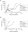
Replication of wt HIV-1 LAI and PAS and PBS mutated viruses. SupT1 cells were transfected with 10 μg (A) or 5 μg (B) of the proviral constructs. CA-p24 production was measured in the culture medium at several days posttransfection.
FIG. 3.
FIG. 3.
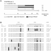
Evolution of PAS/PBS-pro mutant. SupT1 cells were transfected with 5 μg of the molecular clones. Breakthrough replication was observed in some cultures, and viruses could eventually be passaged onto fresh cells. The identity of the PBS motif is indicated as a function of the evolution time (A). The input mutant PBS is shown with open boxes, and wt revertants are shown with black boxes. (B) Culture number, day of harvest, and sequence of proviral DNA isolated from infected cells. The mutated PAS and PBS nucleotides are depicted in bold and are underlined. Nucleotide changes acquired during evolution are shown in white surrounded by a black box (“N” indicates a mixed sequence).
FIG. 4.
FIG. 4.
Evolution of PAS/PBS-lys1,2 mutant. See the legend for Fig. 3 for details. The two mutated PAS and PBS nucleotides are indicated in bold and are underlined. Mutations in the region just upstream of the PAS that were observed in some cultures may reflect G-to-A hypermutations. These transitions have been described previously for other leader revertant viruses and were therefore not analyzed further (8).
FIG. 5.
FIG. 5.
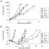
Replication of wt, PAS/PBS-lys1,2 mutant, and cloned revertant viruses. SupT1 cells were transfected with 2 μg of the proviral constructs (A) or were infected with equal amounts of viruses (1 ng of CA-p24 per 5 ml of culture) (B). CA-p24 production was measured in the culture medium for several days. M indicates the original PAS/PBS-lys1,2 mutant, R1 indicates the U126C reversion in the PAS motif, and R2 indicates the G490E reversion in the RNase H domain of RT.
FIG. 6.
FIG. 6.
Schematic representation of the gain in relative viral fitness during forced evolution of the PAS/PBS-lys1,2 mutant (M). Arrows indicate reversion events. The thickness of the arrows indicates the chance of reversion. The slope of the arrow indicates the gain of fitness. M indicates the original PAS/PBS-lys1,2 mutant, which predominantly reverts to a wt, tRNA3Lys-using virus. In the L4 culture (Fig. 4), M acquired an R2 reversion in the RT gene (G3600A). The M-R2 virus is fitter, but it can still revert back to the wt. Viral fitness increases most significantly upon acquisition of the R1 mutation in the PAS element. The M-R12 double revertant is stable in prolonged cultures and does not revert back to the wt. From competition experiments, we concluded that M-R1 replicates slightly more efficiently than M-R12. Consistent with this, the M-R12 virus lost the R2 mutation in a single culture. M-R1 stably maintained the PBS-lys1,2 sequence in all cultures, highlighting the importance of the PAS motif in tRNA1,2Lys usage.
FIG. 7.
FIG. 7.
Model for reverse transcription initiation on wt, PAS/PBS-lys1,2 mutant, and R1 revertant templates. (A) The secondary structures of the PBS region of the HIV-1 RNA genome and tRNA primers are shown schematically (black and orange lines, respectively) AC, anticodon loop; D, D loop. The tRNA primer anneals with its 3′-terminal 18 nucleotides to the PBS (the PBS and anti-PBS sequences are shown in green). An additional interaction between PAS and anti-PAS (orange) is required to activate the initiation of reverse transcription. These interactions are indicated for the wt leader with tRNA3Lys and for the PAS/PBS-lys1,2 mutant (M) and the M-R1 revertant with tRNA1,2Lys (B). The sequence differences between PAS and PBS sequences of wt, M, and R1 leader RNAs and the anti-PAS and anti-PBS sequences of tRNA3Lys and tRNA1,2Lys are indicated in bold and marked with dots. The R1 reversion (U126C; indicated by an arrowhead) stabilizes the PAS-anti-PAS interaction (replacement of a weak U-G base pair with a very stable C-G base pair).
FIG. 8.
FIG. 8.
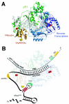
Multiple interactions between HIV-1 RT enzyme, RNA genome, and tRNA3Lys primer. (A) X-ray structure of the p51-p66 heterodimer of the HIV-1 RT enzyme with a double-stranded DNA duplex (26). The enzymatically active p66 domain is shown in blue, and the RNase H domain is shown in yellow. The catalytically active residues in both domains are shown as red dots. The p51 subunit is shown in green. The figure was drawn with Molscript (38) and Raster3D (53) software, using coordinates from the protein data bank (entry 1 HMI [26]). (B) Cartoon of the complex between the HIV-1 RT enzyme, the viral RNA genome, and the self tRNA3Lys primer. The PBS-anti-PBS base-pairing interaction is indicated. We also marked other interaction domains, including the U-rich anticodon (AC) loop of tRNA3Lys, an A-rich segment in the viral RNA genome (green), and the anti-PAS and PAS motifs (yellow). We selected an HIV-1 variant that switched its primer usage from tRNA3Lys to tRNA1,2Lys by simultaneous mutation of the PBS and PAS motifs. This virus acquired a point mutation in the RNase H domain (Gly490Glu; shown in yellow), which implicates this RT domain in primer binding. The sequence of the anticodon stem is the most variable region between tRNA3Lys and tRNA1,2Lys (marked in red; see Fig. 1 for details), and this tRNA domain may interact with the gatekeeper residue 490. The anticodon hairpin is proposed to dock in the cleft between the RNase H and RT domains of the p66 subunit.

Similar articles

Cited by

References

    1. Aiyar, A., D. Cobrinik, Z. Ge, H. J. Kung, and J. Leis. 1992. Interaction between retroviral U5 RNA and the TYC loop of the tRNA(Trp) primer is required for efficient initiation of reverse transcription. J. Virol. 66:2464-2472. - PMC - PubMed
    1. Aiyar, A., Z. Ge, and J. Leis. 1994. A specific orientation of RNA secondary structures is required for initiation of reverse transcription. J. Virol. 68:611-618. - PMC - PubMed
    1. Beerens, N., and B. Berkhout. 2002. The tRNA primer activation signal in the human immunodeficiency virus type 1 genome is important for initiation and processive elongation of reverse transcription. J. Virol. 76:2329-2339. - PMC - PubMed
    1. Beerens, N., and B. Berkhout. 2002. Switching the in vitro tRNA usage of HIV-1 by simultaneous adaptation of the PBS and PAS. RNA 8:357-369. - PMC - PubMed
    1. Beerens, N., F. Groot, and B. Berkhout. 2000. Stabilization of the U5-leader stem in the HIV-1 RNA genome affects initiation and elongation of reverse transcription. Nucleic Acids Res. 28:4130-4137. - PMC - PubMed

Publication types

MeSH terms

LinkOut - more resources