. 2004 Oct;78(19):10724-37.
doi: 10.1128/JVI.78.19.10724-10737.2004.

Structure and mechanistic analysis of the anti-human immunodeficiency virus type 1 antibody 2F5 in complex with its gp41 epitope

Affiliations

Structure and mechanistic analysis of the anti-human immunodeficiency virus type 1 antibody 2F5 in complex with its gp41 epitope

Gilad Ofek et al. J Virol. 2004 Oct.

Abstract

The membrane-proximal region of the ectodomain of the gp41 envelope glycoprotein of human immunodeficiency virus type 1 (HIV-1) is the target of three of the five broadly neutralizing anti-HIV-1 antibodies thus far isolated. We have determined crystal structures of the antigen-binding fragment for one of these antibodies, 2F5, in complex with 7-mer, 11-mer, and 17-mer peptides of the gp41 membrane-proximal region, at 2.0-, 2.1-, and 2.2-A resolutions, respectively. The structures reveal an extended gp41 conformation, which stretches over 30 A in length. Contacts are made with five complementarity-determining regions of the antibody as well as with nonpolymorphic regions. Only one exclusive charged face of the gp41 epitope is bound by 2F5, while the nonbound face, which is hydrophobic, may be hidden due to occlusion by other portions of the ectodomain. The structures reveal that the 2F5 antibody is uniquely built to bind to an epitope that is proximal to a membrane surface and in a manner mostly unaffected by large-scale steric hindrance. Biochemical studies with proteoliposomes confirm the importance of lipid membrane and hydrophobic context in the binding of 2F5 as well as in the binding of 4E10, another broadly neutralizing antibody that recognizes the membrane-proximal region of gp41. Based on these structural and biochemical results, immunization strategies for eliciting 2F5- and 4E10-like broadly neutralizing anti-HIV-1 antibodies are proposed.


Figures

FIG. 1.
FIG. 1.
Overall structure of the 2F5-Fab-gp41654-670 17-mer complex. (A) Cα traces of the 2F5 Fab heavy chain (blue) and light chain (gray) and of the gp41 peptide (red) from residue Glu657 to Trp670; (B) 90° view of the same traces.
FIG. 2.
FIG. 2.
Contact interface between gp41 and 2F5. The orientation shown is similar to that in Fig. 1B, with the CDR H3 protruding off the top of the figure. (A) Electrostatic potential. An atomic bond representation of the peptide (yellow, carbon; red, oxygen; blue, nitrogen) is shown, with gp41 residues labeled. The molecular surface of 2F5 is colored by electrostatic potential: red for electronegative, blue for electropositive, and white for apolar (42). Extensive hydrogen bonds and salt bridges are observed between the peptide and antibody, including contacts with the CDRs of both 2F5 chains, as well as contacts with nonpolymorphic regions of the 2F5 light-chain N terminus. (B) Hydrophobic interactions. A representation of the peptide similar to that for panel A is shown, but in this case it is shown against the Cα worm of the 2F5 heavy (blue) and light (gray) chains, with side chains labeled and shown in green for 2F5 residues that form hydrophobic contacts with gp41. (C) Electron density of the gp41 peptide. Shown (blue) is the electron density (2Fo − Fc) around the gp41 peptide contoured at 1σ. The electron density around residues Glu657 and Asn658 cannot be seen at this contour level, consistent with the tenuous nature of their contacts with 2F5. Beginning with residue Glu659, the density improves and is maintained through Trp670.
FIG. 3.
FIG. 3.
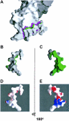
2F5 binding surface on gp41. (A) Molecular surface representations of 2F5 complexed to gp41. The orientation shown is similar to that in Fig. 4. Colored in magenta is the surface on 2F5 that is buried by the interaction with gp41, and colored in green is the surface on gp41 that is buried by the interaction with 2F5. (B and C) Close-ups of the peptide. The gp41 peptide molecular surface from panel A is oriented so that Trp670 is at the bottom of the panel and the N terminus of the peptide is at the top; 180° views are shown. Roughly 40% of the surface of the gp41 peptide is buried by 2F5 (green), while the remaining surface remains hidden (white). (D and E) When the molecular surface of the peptide is colored by electrostatic potential, it becomes apparent that the surface that is bound by 2F5 is charged (E), while the surface that is hidden from 2F5 is hydrophobic (D). The electrostatic potential shown is colored at the same potential contour as in Fig. 2A, with red for electronegative, blue for electropositive, and white for apolar (42).
FIG. 4.
FIG. 4.
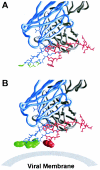
CDR H3 loop of 2F5. 2F5 is shown with the heavy chain in blue and the light chain in gray. Atomic bond representations are shown for the 2F5 CDR H3 loop (blue rods), and the gp41 peptide (red rods). (A) Side chains of hydrophobic residues at the apex of the CDR H3 loop (LeuH100a, PheH100b, ValH100d, and IleH100f) are colored in green. These residues define a hydrophobic surface which if extended as a plane intersects the indole ring of Trp670. (B) The coplanarity of these hydrophobic side chains is evident when the atoms of the side chains are shown in a space-filling representation. The hydrophobic plane defined by these apical CDR H3 residues may be an adaptation that allows 2F5 to bind at or in close proximity to the viral membrane.
FIG. 5.
FIG. 5.
Comparison of the 2F5-bound conformation of gp41 with other gp41 conformations. In blue is the structure of the HR2 helix observed in the structure of the postfusion six-helix bundle (64), in red is the structure of 2F5-bound gp41 presented here, and in magenta is the NMR structure of the membrane-proximal region of gp41 in the context of dodecylphosphocholine micelles (56). The structures are aligned not according to biological context, but relative to the Cα positions of residues Glu657 and Trp670. The helices of both the six-helix bundle and the downstream micellar structure would have to partially unravel for gp41 to adopt the 2F5-bound conformation. The sequences of the three structures are aligned at the top, with structurally ordered residues colored according to their respective structures and the sequences of disordered residues colored gray.
FIG. 6.
FIG. 6.
Transmembrane-proximal region of gp41. This schematic shows the 2F5 epitope (red) with its hidden face occluded by the HIV ectodomain. The conformation of the N-terminal adjoining region is not known. The C-terminal adjoining 4E10 and Z13 epitopes are shown as a helix lying parallel to the viral membrane, with Trp670 and Trp678 (red) embedded in the lipid bilayer. Although the 4E10-Z13 epitope is shown for visual clarity as extending away from the rest of the HIV ectodomain, the angle it makes with the 2F5 epitope is unknown, and it is likely to be partially occluded in the viral spike.
FIG. 7.
FIG. 7.
Biochemical analysis of 2F5 and 4E10 binding: effect of lipid membrane and hydrophobic context. PLs composed of paramagnetic beads conjugated to the 1D4 antibody were used to capture different envelope constructs through C-terminal C9 tags, which are recognized by 1D4. Each of the envelope constructs retained the native JRFL transmembrane domain (TM), so that envelope-TM-captured PLs incubated with lipids simulated the native TM in a lipid bilayer. Alternatively PLs could be washed extensively with detergent to remove bound lipid and expose the naked TM. In this manner, PLs with and without lipid membrane could be prepared. (A) Flow cytometry analysis of b12, 2F5, and 4E10 binding to PLs with and without lipid membrane. Fluorescently labeled antibodies at 2 μg/ml (left panels), 10 μg/ml (middle panels), and 50 μg/ml (right panels) were incubated with JRFL gp145-captured PLs. Histograms (normalized for 100,000 events) of the flow cytometry-sorted PL fluorescence intensity are shown for PLs with membrane (red) and without membrane (blue). (B) Effect of context on the binding of 2F5 and 4E10. Five different PLs were analyzed, each with the 2F5 and 4E10 epitopes placed in a different context. A schematic of each context is shown, with gp120 referring to the cleavage-minus N-terminal gp120 attached to gp41; 2F5, 4E10, and HA referring to the respective epitopes of 2F5, 4E10, and the antihemagglutinin antibody sc-7392; and TM and C9 referring to the JRFL transmembrane region and 1D4-recognized tag, respectively. Each PL was tested for binding to fluorescently labeled antibodies b12, 2F5, 4E10, and HA, either in the presence of membrane or after extensive detergent washing to remove membrane. The relative fluorescence with and without membrane was tested over an antibody concentration range of 2 to 200 μg/ml. The results of two independent experiments are shown (presented as result for experiment 1/result for experiment 2). ++, 10- to 50-fold-greater fluorescence in the presence of membrane; +, 2- to 10-fold-greater fluorescence; −, 0.5- to 2.0-fold-greater fluorescence. Parentheses indicate that the particular antibody epitope was not present on the construct, and asterisks indicate that the overall level of antibody binding was low, both with and without membrane. As can be seen, not only the presence of lipid membrane but also the surrounding sequence influences optimal 2F5 and 4E10 epitope recognition.
FIG. 8.
FIG. 8.
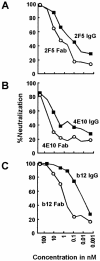
Neutralization of HIV-1 isolate JRFL by IgG versus Fab. In order to assess the existence of steric hindrance of access of IgG molecules to membrane-proximal region epitopes, neutralization capacities of IgGs (squares) versus Fabs (circles) were compared for 2F5 (A), 4E10 (B), and b12 (C) as a control. The concentrations of the IgGs and Fabs ranged from 0.002 to 300 nM. Neutralization data were obtained with CD4 T cells as target cells and flow cytometry of anti-p24 antibody-stained cells to assess the number of infected cells after a single round of virus infection. The percentage of neutralization was calculated as the reduction in the number of infected cells compared to the number of infected cells in wells incubated with mock antibody. Similar results were obtained for HIV-1 strain SF162.
FIG. 9.
FIG. 9.
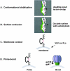
Vaccine immunization strategy. Shown is a four-part strategy to elicit 2F5-like antibodies. (A) Conformational stabilization of the 2F5-bound extended conformation of gp41. The molecular surface of a potential immunogen is shown in a color scheme similar to that in Fig. 3B and C, with the surface bound by 2F5 colored in green and the surface hidden from 2F5 colored in white. Disulfide bonds or lactam bridges that stabilize conformation are shown in magenta. (B) Occlusion of the hidden face of gp41. Carbohydrate (black) is shown occluding the hidden hydrophobic surface of gp41 from humoral immune recognition. Due to the size of the epitope, N-linked glycans may be too large to use here, but smaller O-linked glycans may allow more precise masking. (C) Membrane context. To elicit antibodies that are able to accommodate an epitope that is proximal to membrane, one could immunize with a conformationally stabilized, surface-occluded immunogen in the context of membrane, either on virus-like particles (VLPs) or on PLs. (D) Prime-boost. Various prime-boost strategies could be employed to select only those antibodies that are able to overcome accessibility barriers to the membrane-proximal region. Shown here is one example, with the prime consisting of a conformationally stabilized, surface-occluded immunogen presented in the context of membrane. A boost with the complete Env ectodomain should select antibodies that can bind to the native viral spike.

Similar articles

Cited by

References

    1. Babcock, G. J., T. Mirzabekov, W. Wojtowicz, and J. Sodroski. 2001. Ligand binding characteristics of CXCR4 incorporated into paramagnetic proteoliposomes. J. Biol. Chem. 276:38433-38440. - PubMed
    1. Barbato, G., E. Bianchi, P. Ingallinella, W. H. Hurni, M. D. Miller, G. Ciliberto, R. Cortese, R. Bazzo, J. W. Shiver, and A. Pessi. 2003. Structural analysis of the epitope of the anti-HIV antibody 2F5 sheds light into its mechanism of neutralization and HIV fusion. J. Mol. Biol. 330:1101-1115. - PubMed
    1. Biron, Z., S. Khare, A. O. Samson, Y. Hayek, F. Naider, and J. Anglister. 2002. A monomeric 3(10)-helix is formed in water by a 13-residue peptide representing the neutralizing determinant of HIV-1 on gp41. Biochemistry 41:12687-12696. - PubMed
    1. Brunger, A. T., P. D. Adams, G. M. Clore, W. L. DeLano, P. Gros, R. W. Grosse-Kunstleve, J. S. Jiang, J. Kuszewski, M. Nilges, N. S. Pannu, R. J. Read, L. M. Rice, T. Simonson, and G. L. Warren. 1998. Crystallography & NMR system: a new software suite for macromolecular structure determination. Acta Crystallogr. D 54:905-921. - PubMed
    1. Bryson, S., A. Cunningham, J. Ho, R. C. Hynes, D. E. Isenman, B. H. Barber, R. Kunert, H. Katinger, M. Klein, and E. F. Pai. 2001. Cross-neutralizing human monoclonal anti-HIV-1 antibody 2F5: preparation and crystallographic analysis of the free and epitope-complexed forms of its Fab′ fragment. Protein Peptide Lett. 8:413-418.

Publication types

LinkOut - more resources