. 2004 Oct;78(19):10765-75.
doi: 10.1128/JVI.78.19.10765-10775.2004.

Temporal modulation of an autoprotease is crucial for replication and pathogenicity of an RNA virus

Affiliations

Temporal modulation of an autoprotease is crucial for replication and pathogenicity of an RNA virus

T Lackner et al. J Virol. 2004 Oct.

Abstract

Pestiviruses belong to the family Flaviviridae, and their genome is a single-stranded RNA of positive polarity encoding one large polyprotein which is further processed into mature proteins. Noncytopathogenic (noncp) strains of the pestivirus bovine viral diarrhea virus (BVDV) can establish persistent infection. In persistently infected animals, noncp BVDVs occasionally acquire mutations in viral nonstructural protein 2 (NS2) that give rise to cytopathogenic (cp) BVDV variants, and, eventually, lead to the onset of lethal disease. A molecular marker of cp BVDV infection is a high-level expression of the replicative NS3 protease/helicase that together with NS2 is derived from NS2-3. Here, we present evidence for NS2-3 autoprocessing by a newly identified cysteine protease in NS2 that is distantly related to the NS2-3 autoprotease of hepatitis C and GB viruses. The vital role of this autoprotease in BVDV infection was established, implying an essential function for NS3 in pestiviral RNA replication which cannot be supplied by its NS2-3 precursor. Accordingly, and contrary to a current paradigm, we detected almost complete cleavage of NS2-3 in noncp BVDV at early hours of infection. At 6 to 9 h postinfection, NS2-3 autoprocessing diminished to barely detectable levels for noncp BVDV but decreased only moderately for cp BVDV. Viral RNA synthesis rates strictly correlated with different NS3 levels in noncp and cp BVDV-infected cells, implicating the NS2 autoprotease in RNA replication control. The biotype-specific modulation of NS2-3 autoprocessing indicates a crucial role of the NS2 autoprotease in the pathogenicity of BVDV.


Figures

FIG. 1.
FIG. 1.
The NS2 protease domain is conserved between hepaciviruses and pestiviruses. Fragments of polyproteins (locations are indicated in parentheses) including the C-terminal part of NS2 and five N-terminal amino acid residues of NS3 of a representative set of pestiviruses, hepaciviruses, and GB virus were aligned (see Materials and Methods). The alignment was subsequently manually adjusted to maximize similarity (gaps are indicated by dashes). The Gibbs sampler (29) of the MACAW workbench (48) was used to assess the similarity between pestivirus BVDV CP7 and three hepaciviruses that share <30% identical residues. Blocks A (1.4e−08) and B (5.1e−04) were statistically significant with the NS2-3 protein and the interblock A-NS3-catalytic-Ser searching spaces, respectively. Blocks H, E, and C are named after respective (putative) catalytic residues of NS2 (His, Glu, and Cys) and were recognized in hepaciviruses and, subsequently, in pestiviruses with the catalytic residues of HCV NS2 as anchors. Note that a region separating blocks C and A hosts cellular insertions of variable sizes in some cp BVDV isolates (37) (not shown) supporting this alignment. The position of the NS2-3 cleavage site is indicated below the alignment in yellow. Red characters, (putative) catalytic His, Glu, and Cys residues; blue background, putative Zn2+-coordinating residues of the ZnB site; black background, invariant residues; dark gray background, residues conserved to 100% (see similarity groups) or invariant residues in 80% of the positions; light gray background, residues conserved in not less than 60% of the positions. Amino acid similarity group members: D, N, Q, and E; K, R, and H; F, Y, and W; A and G; S and T; A, C, L, I, V, M, F, and Y. The experimental data (see Results) did not support the putative functions indicated for E1461 (red) and H1483 and C1484 (blue background). Virus names and respective NCBI protein identification numbers are as follows: BVDV-CP7, BVDV strain CP7 (1518836); BDV, border disease virus X818 (20198946); CSFV-E, classical swine fever virus strain Eystrup (12657942); GBV-A, GB virus A (1096574); GBV-B, GB virus B (9628102); GBV-CG, GB virus C-hepatitis G virus (4426796); HCV-J6, HCV isolate HC-J6 (221651); HCV-J8, HCV isolate HC-J8 (221609); HCV-H, HCV isolate H (329738). Numbers in parentheses in the alignment of BVDV-CP7 correspond to amino acid sequences of BVDV strain SD-1 (289507).
FIG. 2.
FIG. 2.
Determinants of NS2-3 processing. (A) Radiosequencing. The diagram depicts the amount of radioactivity released by each cycle of Edman degradation of the partially purified NS3/1596-Met-GST. The amino acid sequence encoded by flagNS2-3/1596-Met-GST in the region of the NS2-3 cleavage site was aligned to the fractions with respect to the methionine residues. The cDNA construct used for protein expression is shown above the diagram; the amino acid sequence downstream of aa 1589 is shown above pflagNS2-3/1596-Met-GST (the number corresponds to amino acid sequences in BVDV strain SD-1); the N-terminal 7 aa of NS3 are followed by a sequence that includes three methionine residues. (B) Scheme of the expression constructs. Bars symbolize proteins; numbers below the bars indicate the amino acid positions in the BVDV polyprotein (the numbers correspond to amino acids in BVDV strain SD-1): 1137, N terminus of NS2; 1590, N terminus of NS3. Truncated proteins are indicated by asterisks. S, signal peptide preceding E2; flag, flag epitope. (C) NS2-3 cleavage studied by transient expression in metabolically labeled BHK-21 cells; the transfected plasmids are indicated above the lanes. For RIP, a GST-specific antibody was applied, and precipitated proteins were analyzed by SDS-PAGE and autoradiography. Arrows indicate the positions of NS2-3/1645GST, NS3/1645GST, NS3/1599GST, and NS3/1596GST.
FIG. 2.
FIG. 2.
Determinants of NS2-3 processing. (A) Radiosequencing. The diagram depicts the amount of radioactivity released by each cycle of Edman degradation of the partially purified NS3/1596-Met-GST. The amino acid sequence encoded by flagNS2-3/1596-Met-GST in the region of the NS2-3 cleavage site was aligned to the fractions with respect to the methionine residues. The cDNA construct used for protein expression is shown above the diagram; the amino acid sequence downstream of aa 1589 is shown above pflagNS2-3/1596-Met-GST (the number corresponds to amino acid sequences in BVDV strain SD-1); the N-terminal 7 aa of NS3 are followed by a sequence that includes three methionine residues. (B) Scheme of the expression constructs. Bars symbolize proteins; numbers below the bars indicate the amino acid positions in the BVDV polyprotein (the numbers correspond to amino acids in BVDV strain SD-1): 1137, N terminus of NS2; 1590, N terminus of NS3. Truncated proteins are indicated by asterisks. S, signal peptide preceding E2; flag, flag epitope. (C) NS2-3 cleavage studied by transient expression in metabolically labeled BHK-21 cells; the transfected plasmids are indicated above the lanes. For RIP, a GST-specific antibody was applied, and precipitated proteins were analyzed by SDS-PAGE and autoradiography. Arrows indicate the positions of NS2-3/1645GST, NS3/1645GST, NS3/1599GST, and NS3/1596GST.
FIG. 3.
FIG. 3.
Effect of single-amino-acid exchanges in NS2 of cp BVDV strain CP7 on NS2-3 cleavage efficiency. Wt i− indicates wt CP7 with the cp-specific insertion deleted. The numbers indicate amino acid residues based on those of strain BVDV SD-1. (A) RIP analysis. Metabolically labeled NS2-3 was isolated from BHK-21 cells by RIP, application of an NS3-specific MAb, and further analysis by SDS-PAGE and phosphorimaging. Cleavage of CP7 wt NS2-3 was set at 100%. The order of the six blocks is indicated by numbers. (B) Summary of the results presented in panel A. Shown is the NS2 amino acid sequence, followed by seven amino acids of NS3; the NS3-derived amino acids are underlined. The indicated NS2-3 cleavage efficiency is based on a single- or triple-quantified experiment. Color code for cleavage (percentage of wt CP7): red, no NS3 visible; orange, <10%; yellow, 10 to 75%; green, >75%. The 9-aa insertion is indicated by italics. Putative functions of residues according to the experimental results are indicated by colors and shading of residue numbers: red characters on black background, catalytic; white characters on blue background, coordination of Zn2+ ion. (C) NS2-3 cleavage efficiency, based on the results of three quantified experiments. Error bars reach from the lowest to the highest values measured. (D) Quantification of NS2-3 cleavage efficiency based one or three (see panel C legend) independent experiments.
FIG. 4.
FIG. 4.
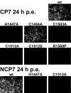
IF analysis of MDBK cells 24 h p.e. with RNA transcribed from full-length cDNA clones of BVDV strains CP7 and NCP7. The wt and mutants indicated above the individual pictures were characterized. A representative part of each dish is shown; primary magnification, ×100.
FIG. 5.
FIG. 5.
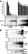
NS2-3 cleavage in cells infected with noncp BVDV NCP7 and cp BVDV CP7. NS3 and NS2-3 are marked with arrows. (A) RIP analysis of MDBK cells infected with strain NCP7 (left) or CP7 (right) after metabolic labeling with [35S]methionine-cysteine for the indicated time periods p.i. The diagram above indicates the NS2-3 cleavage rate as [NS3/(NS3 + NS2-3)], with values in percentages; values were obtained by phosphorimager analysis. (B) Western blot analysis of MDBK cells infected with strain CP7 (top panel) or NCP7 (middle and bottom panels) with an NS3/NS2-3-specific MAb. The lysate separated in each lane represents about 5 × 10⁴ cells (top and middle) or 5 × 10⁵ cells (bottom). Lysates were prepared at the indicated time points p.i.
FIG. 6.
FIG. 6.
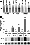
Intracellular accumulation of viral RNA. (A) Analysis of viral RNA synthesis in cells infected with BVDV strain NCP7 or CP7. Metabolically labeled RNA was purified and separated by denaturing agarose gel electrophoresis. Labeling periods in hours are specified below the lanes. The position of a BVDV full-length RNA transcript is indicated by an arrow. (B) (Top) Graph depicting the relative amounts of viral RNA. The amount of BVDV CP7 RNA measured at 48 h p.i. was set at 100%; bovine GAPDH RNA served to standardize the RNA amounts. Values were obtained by phosphorimager analysis. Error bars reach from the lowest to the highest value measured in three independent experiments. (Bottom) Northern blot analysis. Total RNA was prepared from MDBK cells infected with BVDV strains NCP7 or CP7 at the indicated time points p.i. Upon separation by denaturing agarose gel electrophoresis, RNA was blotted onto a membrane and hybridized in parallel against probes specific for BVDV or GAPDH. The entire experiment was done in triplicate, and the data shown is representative of all experiments.
FIG. 7.
FIG. 7.
Growth kinetics for BVDV strains CP7 and NCP7. The graph shows mean values derived from three independent experiments. MDBK cells were infected at an MOI of 10. Culture supernatants were harvested at the indicated times p.i. Virus titers are given as log 50% tissue culture infective doses per milliliter. For cDNA-derived viruses, see previously published results (6).
FIG. 8.
FIG. 8.
NS2-3 cleavage in three genera of the family Flaviviridae. Polyprotein fragments encompassing NS2-3 of the three Flaviviridae genera are depicted schematically. The locations of the helicase and protease domains are indicated; (putative) ZnB motifs (Zn2+) are shaded in gray. The positions of the minimal protease domains capable of cleaving the NS2-3 junction in vitro are shown below the polyproteins by filled ovals (catalytic domains) and rectangles (essential accessory domains). Curved arrows point to the cleavage sites processed by the respective protease.

Similar articles

Cited by

References

    1. Agapov, E. V., I. Frolov, B. D. Lindenbach, B. M. Pragai, S. Schlesinger, and C. M. Rice. 1998. Noncytopathic Sindbis virus RNA vectors for heterologous gene expression. Proc. Natl. Acad. Sci. USA 95:12989-12994. - PMC - PubMed
    1. Agapov, E. V., C. L. Murray, I. Frolov, L. Qu, T. M. Myers, and C. M. Rice. 2004. Uncleaved NS2-3 is required for production of infectious bovine viral diarrhea virus. J. Virol. 78:2414-2425. - PMC - PubMed
    1. Baker, J. C. 1987. Bovine viral diarrhea virus: a review. J. Am. Vet. Med. Assoc. 190:1449-1458. - PubMed
    1. Baroth, M., M. Orlich, H.-J. Thiel, and P. Becher. 2000. Insertion of cellular NEDD8 coding sequences in a pestivirus. Virology 278:456-466. - PubMed
    1. Becher, P., M. Orlich, and H.-J. Thiel. 2000. Mutations in the 5′ nontranslated region of bovine viral diarrhea virus result in altered growth characteristics. J. Virol. 74:7884-7894. - PMC - PubMed

Publication types

MeSH terms

LinkOut - more resources