. 2004 Oct;78(19):10793-802.
doi: 10.1128/JVI.78.19.10793-10802.2004.

Mapping the Golgi targeting and retention signal of Bunyamwera virus glycoproteins

Affiliations

Mapping the Golgi targeting and retention signal of Bunyamwera virus glycoproteins

Xiaohong Shi et al. J Virol. 2004 Oct.

Abstract

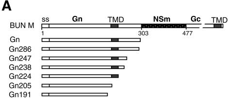
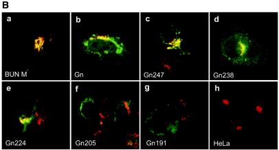
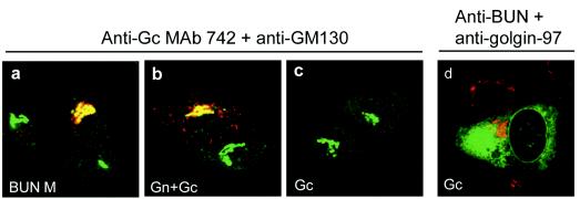
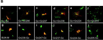
The membrane glycoproteins (Gn and Gc) of Bunyamwera virus (BUN; family Bunyaviridae) accumulate in the Golgi complex, where virion maturation occurs. The Golgi targeting and retention signal has previously been shown to reside within the Gn protein. A series of truncated Gn and glycoprotein precursor cDNAs were constructed by progressively deleting the coding region of the transmembrane domain (TMD) and the cytoplasmic tail. We also constructed chimeric proteins of BUN Gc, enhanced green fluorescent protein (EGFP), and human respiratory syncytial virus (HRSV) fusion (F) protein that contain the Gn TMD with various lengths of its adjacent cytoplasmic tails. The subcellular localization of mutated BUN glycoproteins and chimeric proteins was investigated by double-staining immunofluorescence with antibodies against BUN glycoproteins or the HRSV F protein and with antibodies specific for the Golgi complex. The results revealed that Gn and all truncated Gn proteins that contained the intact TMD (residues 206 to 224) were able to translocate to the Golgi complex and also rescued the Gc protein, which is retained in the endoplasmic reticulum when expressed alone, to this organelle. The rescued Gc proteins acquired endo-beta-N-acetylglucosaminidase H resistance. The Gn TMD could also target chimeric EGFP to the Golgi and retain the F protein, which is characteristically expressed on the surface of HRSV-infected cells, in the Golgi. However, chimeric BUN Gc did not translocate to the Golgi, suggesting that an interaction with Gn is involved in Golgi retention of the Gc protein. Collectively, these data demonstrate that the Golgi targeting and retention signal of BUN glycoproteins resides in the TMD of the Gn protein.


Figures

FIG. 1.
FIG. 1.
Intracellular localization of BUN Gn mutants. (A) Diagram of BUN glycoprotein precursor (BUN M) and Gn mutants. Amino acid positions are indicated beneath the top bar. ss, signal sequence. Gn mutants are designated according to the numbers of residues that they contain. (B) HeLa T4+ cells were infected with vTF7-3, followed by transfection with BUN Gn mutant cDNAs as indicated. The cells were then doubly stained with a mixture of anti-BUN antiserum and a MAb specific for the Golgi marker golgin 97. Merged confocal microscopic images are shown, with the Gn glycoprotein stained green and the Golgi stained red.
FIG. 1.
FIG. 1.
Intracellular localization of BUN Gn mutants. (A) Diagram of BUN glycoprotein precursor (BUN M) and Gn mutants. Amino acid positions are indicated beneath the top bar. ss, signal sequence. Gn mutants are designated according to the numbers of residues that they contain. (B) HeLa T4+ cells were infected with vTF7-3, followed by transfection with BUN Gn mutant cDNAs as indicated. The cells were then doubly stained with a mixture of anti-BUN antiserum and a MAb specific for the Golgi marker golgin 97. Merged confocal microscopic images are shown, with the Gn glycoprotein stained green and the Golgi stained red.
FIG. 2.
FIG. 2.
Intracellular localization of coexpressed and separately expressed BUN Gc proteins. Gc proteins were coexpressed with Gn from either the whole M segment cDNA (a) or separate Gn and Gc cDNAs (b). (c and d) Gc expressed alone from pTM-BUNGc. The cells were doubly stained with the anti-Gc MAb 742 and an anti-GM130 serum (a to c) or with anti-BUN serum and an anti-golgin 97 antibody (d). Merged confocal microscopic images are shown. BUN glycoproteins are stained red and the Golgi is stained green in panels a to c, whereas Gc is stained green and the Golgi is stained red in panel d.
FIG. 3.
FIG. 3.
Rescue of BUN Gc from ER to the Golgi complex by truncated Gn proteins. (A) Diagram of BUN M cDNA constructs containing internal deletions. The amino acid residues deleted from the precursor cDNA are listed on the right. (B) Confocal images of transfected cells. HeLa T4+ cells were transfected with either separate Gc and mutant Gn cDNAs (a to f) or M segment cDNAs with internal deletions (g to l). The cells were doubly stained with a mixture of anti-Gc MAb 742 and anti-GM130 antibodies. Merged confocal microscopic images are shown, with the Gc glycoprotein stained red and the Golgi stained green.
FIG. 3.
FIG. 3.
Rescue of BUN Gc from ER to the Golgi complex by truncated Gn proteins. (A) Diagram of BUN M cDNA constructs containing internal deletions. The amino acid residues deleted from the precursor cDNA are listed on the right. (B) Confocal images of transfected cells. HeLa T4+ cells were transfected with either separate Gc and mutant Gn cDNAs (a to f) or M segment cDNAs with internal deletions (g to l). The cells were doubly stained with a mixture of anti-Gc MAb 742 and anti-GM130 antibodies. Merged confocal microscopic images are shown, with the Gc glycoprotein stained red and the Golgi stained green.
FIG. 4.
FIG. 4.
Endo H resistance of BUN glycoproteins. Vero E6 cells were infected with vTF7-3 and subsequently transfected with the parental BUN M segment cDNA (pTM-BUNM) and mutants carrying internal deletions as indicated. The cells were radiolabeled with [35S]methionine, and radiolabeled proteins were immunoprecipitated with an anti-BUN antiserum. The resulting precipitates were subjected to digestion with endo H (H) or PNGase F (F) as shown and were analyzed by SDS-10% PAGE under reducing conditions. The truncated Gn proteins expressed from M cDNA mutants are marked with asterisks. The gel was exposed to a phosphorimager (Molecular Imager FX; Bio-Rad Laboratories), and the relative intensities of the two forms of Gc were estimated from a computer-enlarged image by the use of Quantity One (v. 4.2.2) software.
FIG. 5.
FIG. 5.
Intracellular localization of chimeric Gc containing the Gn TMD and cytoplasmic tail. (A) Diagram of chimeric Gc constructs containing the Gn TMD and parts of the cytoplasmic tail (Gn-TMC). (B) HeLa T4+ cells were infected with vTF7-3, followed by transfection with full-length Gc (a and e)- or chimeric Gc (b to d and f to h)-expressing plasmids. The cells in panels f to h were cotransfected with a Gn-expressing plasmid. The cells were stained with anti-BUN and anti-golgin 97 antibodies (a to d) or with anti-Gc MAb 742 and anti-GM130 antibodies (e to h) and were examined by confocal microscopy. Merged images are shown in which BUN Gc is stained green and the Golgi is stained red in panels a to d, whereas in panels e to h the Golgi is stained green and Gc is stained red.
FIG. 5.
FIG. 5.
Intracellular localization of chimeric Gc containing the Gn TMD and cytoplasmic tail. (A) Diagram of chimeric Gc constructs containing the Gn TMD and parts of the cytoplasmic tail (Gn-TMC). (B) HeLa T4+ cells were infected with vTF7-3, followed by transfection with full-length Gc (a and e)- or chimeric Gc (b to d and f to h)-expressing plasmids. The cells in panels f to h were cotransfected with a Gn-expressing plasmid. The cells were stained with anti-BUN and anti-golgin 97 antibodies (a to d) or with anti-Gc MAb 742 and anti-GM130 antibodies (e to h) and were examined by confocal microscopy. Merged images are shown in which BUN Gc is stained green and the Golgi is stained red in panels a to d, whereas in panels e to h the Golgi is stained green and Gc is stained red.
FIG. 6.
FIG. 6.
Intracellular localization of chimeric EGFP containing BUN Gn TMD and cytoplasmic tail. (A) Diagram of chimeric EGFP constructs containing the signal sequence (ss) of Hantaan virus Gn, the BUN Gn TMD, and different lengths of the cytoplasmic tail (Gn-TMC). EGFP-GnTM224 has no cytoplasmic tail at its C terminus. (B) Vero E6 cells were transfected with chimeric EGFP cDNAs as indicated and then incubated for 24 h. The cells were stained with an anti-golgin 97 MAb and Cy5-conjugated anti-mouse IgG and then examined by confocal microscopy. The images show GFP fluorescence (green), golgin 97 staining (red), and merged images.
FIG. 6.
FIG. 6.
Intracellular localization of chimeric EGFP containing BUN Gn TMD and cytoplasmic tail. (A) Diagram of chimeric EGFP constructs containing the signal sequence (ss) of Hantaan virus Gn, the BUN Gn TMD, and different lengths of the cytoplasmic tail (Gn-TMC). EGFP-GnTM224 has no cytoplasmic tail at its C terminus. (B) Vero E6 cells were transfected with chimeric EGFP cDNAs as indicated and then incubated for 24 h. The cells were stained with an anti-golgin 97 MAb and Cy5-conjugated anti-mouse IgG and then examined by confocal microscopy. The images show GFP fluorescence (green), golgin 97 staining (red), and merged images.
FIG. 7.
FIG. 7.
Intracellular and cell surface expression of parental HRSV F protein (Fwt) and chimeric F proteins containing the TMD and cytoplasmic tail of BUN Gn protein. (A) Diagram of chimeric HRSV F containing BUN Gn TMD. Gn TMD amino acids are underlined. The remaining original F TMD residues that flank the Gn TMD are shown in gray. (B) HeLa T4+ cells were infected with vTF7-3, followed by transfection with the wild-type F or chimeric F cDNAs. For the examination of cell surface expression, the cells were not permeabilized with Triton X-100 before incubation with antibodies. The cells were then stained with anti-F MAb 19 and anti-GM130 antibody for permeabilized samples or with only MAb 19 for nonpermeabilized samples (cell surface staining). The F protein and chimeric F proteins are stained red and GM130 is stained green.
FIG. 7.
FIG. 7.
Intracellular and cell surface expression of parental HRSV F protein (Fwt) and chimeric F proteins containing the TMD and cytoplasmic tail of BUN Gn protein. (A) Diagram of chimeric HRSV F containing BUN Gn TMD. Gn TMD amino acids are underlined. The remaining original F TMD residues that flank the Gn TMD are shown in gray. (B) HeLa T4+ cells were infected with vTF7-3, followed by transfection with the wild-type F or chimeric F cDNAs. For the examination of cell surface expression, the cells were not permeabilized with Triton X-100 before incubation with antibodies. The cells were then stained with anti-F MAb 19 and anti-GM130 antibody for permeabilized samples or with only MAb 19 for nonpermeabilized samples (cell surface staining). The F protein and chimeric F proteins are stained red and GM130 is stained green.

References

    1. Andersson, A. M., L. Melin, A. Bean, and R. F. Pettersson. 1997. A retention signal necessary and sufficient for Golgi localization maps to the cytoplasmic tail of a Bunyaviridae (Uukuniemi virus) membrane glycoprotein. J. Virol. 71:4717-4727. - PMC - PubMed
    1. Andersson, A. M., and R. F. Pettersson. 1998. Targeting of a short peptide derived from the cytoplasmic tail of the G1 membrane glycoprotein of Uukuniemi virus (Bunyaviridae) to the Golgi complex. J. Virol. 72:9585-9596. - PMC - PubMed
    1. Arkin, I. T. 2002. Structural aspects of oligomerization taking place between the transmembrane alpha-helices of bitopic membrane proteins. Biochim. Biophys. Acta 1565:347-363. - PubMed
    1. Bretscher, M. S., and S. Munro. 1993. Cholesterol and the Golgi apparatus. Science 261:1280-1281. - PubMed
    1. Chalfie, M., Y. Tu, G. Euskirchen, W. W. Ward, and D. C. Prasher. 1994. Green fluorescent protein as a marker for gene expression. Science 263:802-805. - PubMed

Publication types

MeSH terms

LinkOut - more resources