. 2004 Oct;78(19):10803-13.
doi: 10.1128/JVI.78.19.10803-10813.2004.

Construction and characterization of a fluorescently labeled infectious human immunodeficiency virus type 1 derivative

Affiliations

Construction and characterization of a fluorescently labeled infectious human immunodeficiency virus type 1 derivative

Barbara Müller et al. J Virol. 2004 Oct.

Abstract

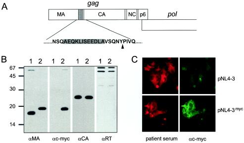
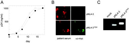
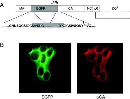
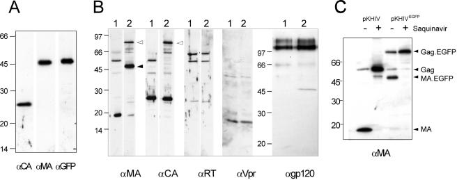
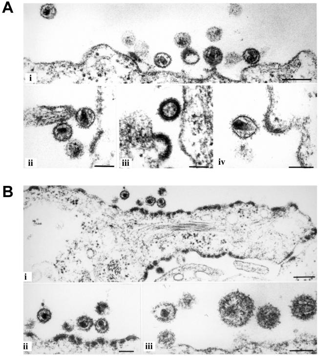
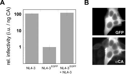
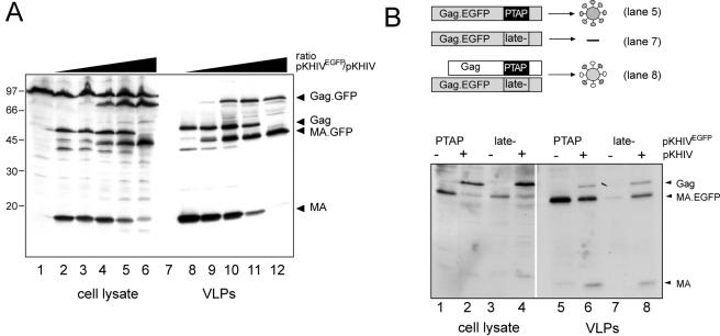
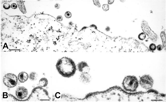
The introduction of a label which can be detected in living cells opens new possibilities for the direct analysis of dynamic processes in virus replication, such as the transport and assembly of structural proteins. Our aim was to generate a tool for the analysis of the trafficking of the main structural protein of human immunodeficiency virus type 1 (HIV-1), Gag, as well as for the analysis of virus-host cell interactions in an authentic setting. We describe here the construction and characterization of infectious HIV derivatives carrying a label within the Gag polyprotein. Based on our initial finding that a short epitope tag could be inserted near the C terminus of the matrix domain of Gag without affecting viral replication, we constructed HIV derivatives carrying the egfp gene at the analogous position, resulting in the expression of a Gag-EGFP fusion protein in the authentic viral context. Particles displaying normal viral protein compositions were released from transfected cells, and Gag-EGFP was efficiently processed by the viral protease, yielding the expected products. Furthermore, particles with mature morphology were observed by thin-section electron microscopy. The modified virus was even found to be infectious, albeit with reduced relative infectivity. By preparing mixed particles containing equimolar amounts of Gag-EGFP and Gag, we were able to obtain highly fluorescently labeled virion preparations which displayed normal morphology and full wild-type infectivity, demonstrating that the process of HIV particle assembly displays a remarkable flexibility. The fluorescent virus derivative is a useful tool for investigating the interaction of HIV with live cells.


Figures

FIG. 1.
FIG. 1.
Insertion of a c-myc epitope tag into the HIV-1 Gag protein. (A) The coding sequence for the c-myc epitope tag sequence, flanked by an alanine residue at either side, was inserted into the gag ORF as indicated, resulting in the introduction of 12 additional amino acids between Gln127 and Val128 of MA. (B) Expression of the myc-tagged MA protein in the viral context. VLPs were prepared from 293T cells transfected with pKHIV (lanes 1) or pKHIVmyc (lanes 2). Viral proteins were separated by SDS-polyacrylamide gel electrophoresis (17.5% polyacrylamide; 200:1), transferred to nitrocellulose, and reacted with the indicated antisera. Molecular mass markers (in kilodaltons) are indicated on the left. (C) Detection of c-myc-tagged MA protein in transfected cells. HeLaP4 cells were transfected with pNL4-3 or pNL4-3myc. Forty-eight hours after transfection, cells were fixed and stained with patient serum or a monoclonal antibody against c-myc, respectively.
FIG. 2.
FIG. 2.
Infectivity of NL4-3myc and stability of the epitope tag. (A) Supernatants from HeLaP4 cells transfected with pNL4-3 (open squares) or pNL4-3myc (filled squares) were used to infect PM-1 cells (10 ng of p24 CA per 106 cells). The release of p24 CA into the supernatant was monitored by ELISA. (B) Supernatants from transfected HeLa cells were used to infect MT-4 cells. Following three passages on MT-4 cells, cells were fixed with patient antiserum and stained with a monoclonal antibody against c-myc. (C) After five rounds of replication in C8166 cells, DNA was prepared from infected cultures, and gag-derived sequences were amplified by PCR with primers flanking the epitope tag insertion site as described in Materials and Methods. Samples were separated by electrophoresis on an agarose gel. Amplification of the wild-type sequence yielded a 210-bp fragment; in the case of the myc-tagged variant, the expected size of the PCR product is 246 bp.
FIG. 3.
FIG. 3.
(A) Construction of an HIV-1 variant carrying the egfp gene. The schematic drawing shows the gag ORF with the egfp gene inserted between the MA and CA coding regions. The expanded regions show the derived amino acid sequence at the domain borders, with HIV sequences displayed in boldface type and EGFP sequences shown in black type on a gray background. An arrowhead indicates the PR cleavage site between MA and CA. (B) Expression of EGFP and Gag in HeLaP4 cells. Cells were transfected with pKHIVEGFP, and immunofluorescence using anti-CA antiserum was carried out 44 h posttransfection. The images show a syncytium displaying EGFP fluorescence and red immunofluorescence.
FIG. 4.
FIG. 4.
Detection of HIV proteins in preparations of wild-type and EGFP-labeled particles. 293T cells were transfected with pKHIV or pKHIVEGFP, and particles were purified from the supernatant 48 h posttransfection by centrifugation through a sucrose cushion. HIV proteins and EGFP were detected in the preparations by enhanced chemiluminescence immunoblotting by using the indicated antisera. Molecular mass standards (in kilodaltons) are indicated at the left of each blot. (A) Immunoblot of a KHIVEGFP preparation, showing that the protein of approximately 45 kDa is recognized by antisera against MA as well as against GFP. (B) Comparison of viral protein patterns of parallel preparations of KHIV (lanes 1) and KHIVEGFP (lanes 2). (C) Inhibition of Gag-GFP processing by the HIV PR inhibitor saquinavir. Particles were prepared from 293T cells grown in the absence or presence of 5 μM saquinavir and analyzed by immunoblotting by using antiserum against MA.
FIG. 5.
FIG. 5.
Morphology of NL4-3EGFP particles. HeLaP4 cells were transfected with the proviral plasmids pNLC4-3 (A) and pNLC4-3EGFP (B). Twenty-four hours posttransfection, cells were harvested, prepared, and analyzed by thin-section electron microscopy as described in Materials and Methods. Bars, 200 nm (panels i) or 100 nm (A, panels ii, iii, and iv, and B, panels ii and iii).
FIG. 6.
FIG. 6.
Infectivity of fluorescently labeled HIV derivatives. (A) 293T cells were transfected with pNLC4-3, pNLC4-3EGFP, or an equimolar mixture of both plasmids. Serial dilutions of supernatants were used to infect TZM cells, and relative infectivities were determined as described in Materials and Methods. Data points are mean values obtained from two independent transfections, with all samples titrated in duplicate. Several independent experiments yielded similar relative differences. (B) TZM cells infected with NL4-3EGFP displayed green fluorescence (top) and expressed HIV-1 Gag as detected by immunofluorescence using polyclonal serum against CA (bottom).
FIG. 7.
FIG. 7.
Generation of mixed particles by cotransfection of the wild type and the EGFP-labeled variant. (A) Variation of the ratio between the wild-type and the fluorescence-labeled variant. 293T cells were transfected with a mixture of pKHIV and pKHIVEGFP, while the total amount of DNA was kept constant. The relative amounts of pKHIVEGFP used were 0% (lanes 2 and 8), 4% (lanes 3 and 9), 20% (lanes 4 and 10), 50% (lanes 5 and 11), and 100% (lanes 6 and 12). At 44 h posttransfection, cells were lysed and particles were purified from the tissue culture supernatant by centrifugation through a sucrose cushion. Gag-derived proteins were detected in the cell lysate and purified particles by enhanced chemiluminescence immunoblotting by using antiserum against MA. (B) Rescue of a release-deficient variant of pKHIVEGFP by cotransfection with wild-type pKHIV. PKHIVEGFP and pKHIVEGFP late− were either transfected alone (lanes 1, 3, 5, and 7) or cotransfected with an equimolar amount of wild-type pKHIV (lanes 2, 4, 6, and 8). At 44 h posttransfection, cells were lysed and particles were prepared from the tissue culture supernatant. Gag-derived proteins were detected in the samples by immunoblotting with polyclonal antiserum against MA.
FIG. 8.
FIG. 8.
Morphology of partly fluorescently labeled particles. HeLaP4 cells were transfected with a mixture of equimolar amounts of pNLC4-3 and pNLC4-3EGFP. At 44 h posttransfection, cells were harvested, prepared, and analyzed by thin-section electron microscopy as described in Materials and Methods. Bars, 200 nm (A) or 100 nm (B and C).

Similar articles

Cited by

References

    1. Adachi, A., H. E. Gendelman, S. Koenig, T. Folks, R. Willey, A. Rabson, and M. A. Martin. 1986. Production of acquired immunodeficiency syndrome-associated retrovirus in human and nonhuman cells transfected with an infectious molecular clone. J. Virol. 59:284-291. - PMC - PubMed
    1. Auerbach, M. R., C. Shu, A. Kaplan, and I. R. Singh. 2003. Functional characterization of a portion of the Moloney murine leukemia virus gag gene by genetic footprinting. Proc. Natl. Acad. Sci. USA 100:11678-11683. - PMC - PubMed
    1. Basyuk, E., T. Galli, M. Mougel, J. M. Blanchard, M. Sitbon, and E. Bertrand. 2003. Retroviral genomic RNAs are transported to the plasma membrane by endosomal vesicles. Dev. Cell 5:161-174. - PubMed
    1. Bearer, E. L., X. O. Breakefield, D. Schuback, T. S. Reese, and J. H. LaVail. 2000. Retrograde axonal transport of herpes simplex virus: evidence for a single mechanism and a role for tegument. Proc. Natl. Acad. Sci. USA 97:8146-8150. - PMC - PubMed
    1. Bohne, J., and H.-G. Kräusslich. 2004. Mutation of the major 5′ splice site renders a CMV-driven HIV-1 proviral clone Tat-dependent: connections between transcription and splicing. FEBS Lett. 563:113-118. - PubMed

Publication types

MeSH terms

LinkOut - more resources