. 2004 Oct;24(19):8342-55.
doi: 10.1128/MCB.24.19.8342-8355.2004.

Swi1 and Swi3 are components of a replication fork protection complex in fission yeast

Affiliations

Swi1 and Swi3 are components of a replication fork protection complex in fission yeast

Eishi Noguchi et al. Mol Cell Biol. 2004 Oct.

Abstract

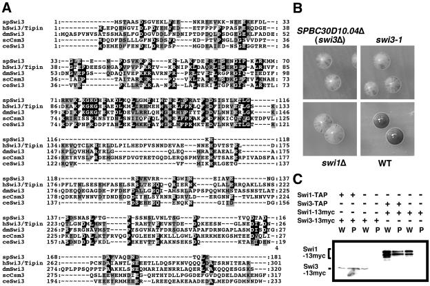
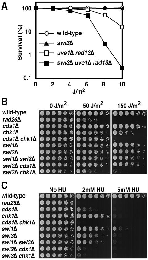
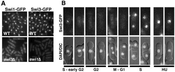
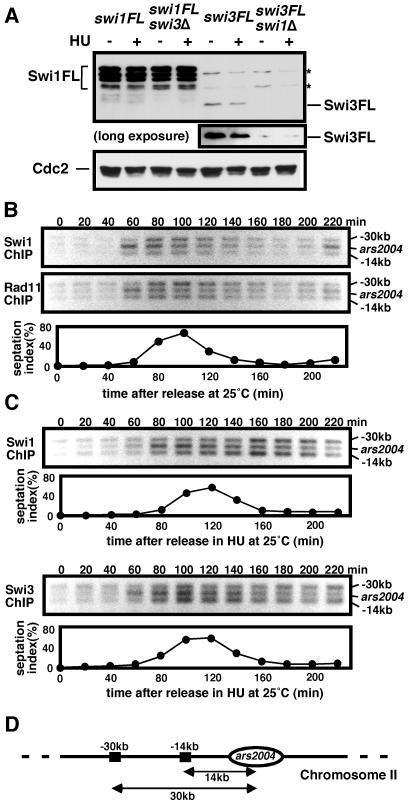
Swi1 is required for programmed pausing of replication forks near the mat1 locus in the fission yeast Schizosaccharomyces pombe. This fork pausing is required to initiate a recombination event that switches mating type. Swi1 is also needed for the replication checkpoint that arrests division in response to fork arrest. How Swi1 accomplishes these tasks is unknown. Here we report that Swi1 copurifies with a 181-amino-acid protein encoded by swi3(+). The Swi1-Swi3 complex is required for survival of fork arrest and for activation of the replication checkpoint kinase Cds1. Association of Swi1 and Swi3 with chromatin during DNA replication correlated with movement of the replication fork. swi1Delta and swi3Delta mutants accumulated Rad22 (Rad52 homolog) DNA repair foci during replication. These foci correlated with the Rad22-dependent appearance of Holliday junction (HJ)-like structures in cells lacking Mus81-Eme1 HJ resolvase. Rhp51 and Rhp54 homologous recombination proteins were not required for viability in swi1Delta or swi3Delta cells, indicating that the HJ-like structures arise from single-strand DNA gaps or rearranged forks instead of broken forks. We propose that Swi1 and Swi3 define a fork protection complex that coordinates leading- and lagging-strand synthesis and stabilizes stalled replication forks.


Figures

FIG. 1.
FIG. 1.
Identification of Swi3 as a Swi1-interacting protein. (A) Swi3 is conserved across evolution. Multiple alignments of Swi3 homologs from S. pombe (spSwi3; GenBank accession no. AY498547), humans (hSwi3/Tipin; GenBank accession no. NP_060328), Drosophila (dmSwi3; GenBank accession no. NP_609895), S. cerevisiae (scCsm3), and C. elegans (ceSwi3; GenBank accession no. NP_490977) are shown. (B) Deletion of SPBC30D10.04 (swi3Δ), swi3-1, and swi1Δ showed inefficient mating-type switching. Homothallic h90 strains with the indicated genotypes were incubated on sporulation medium for 6 to 7 days at 25°C. Plates were exposed to iodine vapors. Colonies that have efficient mating-type switching stain darkly with iodine vapors, whereas inefficient strains show mottled staining. (C) Swi1 and Swi3 form a protein complex in S. pombe. Cells expressing Swi1 or Swi3 as TAP or 13myc fusion proteins were used for coimmunoprecipitation studies. TAP proteins were precipitated and probed with anti-myc antibodies. All proteins were expressed at endogenous levels. W, whole-cell extract; P, precipitated fraction.
FIG. 2.
FIG. 2.
Swi3 is required for survival of fork arrest. (A) Swi3 contributes to survival with UV irradiation in a rad13Δ uve1Δ strain that cannot excise UV damage. Cells of the indicated genotypes were spread on YES agar medium and exposed to the indicated dose of UV. Agar plates were incubated for 3 days at 30°C to measure UV survival. (B) Interaction of swi3Δ and chk1Δ mutations in UV survival assays indicates that Swi1 is required for tolerance of UV damage during DNA replication. Fivefold serial dilutions of cells were plated on YES agar medium and exposed to the indicated dose of UV. Agar plates were incubated for 2 to 5 days at 30°C. (C) Swi3 has a role in tolerating HU-induced fork arrest. swi3Δ cells were highly sensitive to HU. Synergistic interactions of swi3Δ with cds1Δ and chk1Δ mutations indicate that Swi3 has an HU survival function that is at least partially independent of Cds1 and Chk1. Fivefold serial dilutions of cells were incubated on YES agar medium supplemented with the indicated amounts of HU for 2 to 5 days at 30°C.
FIG. 3.
FIG. 3.
Swi3 is required for the replication checkpoint. (A) Indicated strains were incubated in YES liquid medium supplemented with 0 or 12 mM HU for 6 h at 30°C and then stained with DAPI to visualize nuclear DNA as described in Materials and Methods. Wild-type, swi3Δ, swi3Δ swi1Δ, and swi3Δ cds1Δ cells treated with HU underwent checkpoint arrest, as indicated by the appearance of elongated, uninucleate cells without septa. In contrast, swi3Δ chk1Δ cells treated with HU failed to undergo cell cycle arrest and instead displayed aberrant mitosis, as indicated by a cut phenotype. The cut phenotype also appeared in ∼10% of septated swi3Δ chk1Δ cells grown in the absence of HU (indicated by arrow). In some of the HU-treated cells the nuclei were not stained efficiently with DAPI. (B) Cds1 kinase activation is defective in swi1Δ and swi3Δ cells. Cells of the indicated genotypes were incubated in YES liquid medium supplemented with 0 or 12 mM HU for 4 h at 30°C. Kinase activity of immunoprecipitated Cds1 was measured by using myelin basic protein as a substrate. (C) HU sensitivity of swi1Δ or swi3Δ cells was suppressed by a multicopy cds1+ plasmid. The swi1Δ or swi3Δ cells were transformed with the indicated plasmid and plated on YES medium containing 0 or 5 mM HU for 2 to 5 days at 30°C.
FIG. 4.
FIG. 4.
Recruitment of Swi3-GFP to chromatin in S phase. (A) Swi1-GFP and Swi3-GFP are nuclear proteins. Swi1-GFP delocalized from the nucleus in swi3Δ cells. Swi3-GFP was not detectable in the absence of Swi1. Live cells were analyzed for Swi1-GFP or Swi3-GFP fluorescence. (B) In situ chromatin binding assay of Swi3-GFP. Spheroplasts were extracted with Triton X-100 to remove soluble nuclear protein and then fixed for microscopic analysis (41). Representative patterns of fluorescence are shown. Swi3-GFP was detected predominantly in septated cells and unseptated small cells, which are in S phase or possibly early G2 phase. Representative photos of HU-treated cells are shown.
FIG. 5.
FIG. 5.
Swi1 associates with the origin in early S phase. (A) Immunoblot analysis of Swi1-3FLAG (Swi1-FL) and Swi3-3FLAG (Swi3-FL). Cells of the indicated strains were incubated in YES medium supplemented with 0 or 12 mM HU. Protein samples of the indicated cells were analyzed by immunoblotting with anti-FLAG antibody. A longer exposure of the immunoblot showed that a very low amount of Swi3-FL was detected in swi1Δ cells. Asterisks show proteins that were cross-reactive with anti-FLAG antibody. (B) ChIP assays of Swi1-13myc and Rad11-3FLAG were performed at ars2004 and sites located 14 or 30 kb away from ars2004 (42). The cdc25-22 cells were synchronized at the G2-M boundary by incubation at 36°C for 4 h and then released in fresh YES medium at 25°C. An increase in the septation index indicates the onset of S phase. (C) ChIP assays of Swi1-FL and Swi3-FL were performed as described above, except that cells were released into YES medium supplemented with 10 mM HU. (D) Diagram of region used in the ChIP assay. ars2004 and surrounding regions are shown.
FIG. 6.
FIG. 6.
Spontaneous DNA damage occurs during S phase in swi3Δ cells. (A) Multiple Rad22-YFP foci accumulated in swi3Δ cells. Log-phase cells were grown in Edinburgh minimal medium at 25°C. In wild-type cells, 15.8% of the nuclei contained a single Rad22-YFP focus and none showed multiple Rad22-YFP foci. In swi3Δ cells, 65.6% of the nuclei contained at least one Rad22-YFP focus and 48.9% had multiple foci. (B) Quantification of Rad22-YFP foci according to cell cycle stage estimated from cell and nuclear morphology. The percentages of nuclei that have at least one focus or two or more foci are shown. S-phase cells had the most Rad22-YFP foci.
FIG. 7.
FIG. 7.
swi1Δ cells accumulate X-shaped DNA. (A) Model of the fork recapture mechanism. Fork recapture requires strand invasion catalyzed by Rhp51 and Rhp54 followed by HJ resolution by the Mus81-Eme1 complex. (B) X-shaped DNA accumulates in swi1Δ mus81Δ cells. Genomic DNA samples prepared from exponentially growing S. pombe cells with the indicated genotypes were analyzed by 2D gel electrophoresis with the ars3001 HindIII-KpnI fragment as a probe (41). The amount of X-shaped DNA expressed as the percentage of entire replication and recombination intermediates was quantified. The arrow points to X-shaped DNA in swi1Δ mus81Δ cells. (C) The X-shaped DNA branch migrates into linear DNA. Genomic DNA from swi1Δ mus81Δ cells was run in the first-dimension gel and gel slices were incubated in branch migration buffer at 4 or 65°C for 5 h, as described in Materials and Methods, and then DNA was electrophoresed in the 2D gel. The arrow indicates the spot corresponding to the linear DNA products derived from the X-shaped DNA molecules by branch migration. (D) Diagram of the migration pattern of replication and recombination intermediates that can be detected by 2D gel electrophoresis before (4°C) and after (65°C) the branch migration reaction. X-shaped DNA that has the property of HJs can be converted into a linear product that produces a distinct spot which migrates below the X-shaped DNA on the 1N line.
FIG. 8.
FIG. 8.
Two models for Rad22-dependent generation of X-shaped DNA molecules and their resolution by Mus81-Eme1 in swi1Δ and swi3Δ cells. (A) The ssDNA gap left in the lagging strand participates in a recombination event catalyzed by Rad22. Strand invasion by the 3′ end of an Okazaki fragment and following DNA synthesis leads to formation of double HJs that are targeted by Mus81-Eme1. (B) An ssDNA gap may lead to fork regression, creating a chicken foot structure that is similar to an HJ. The exposed nascent strand of DNA may anneal to the template parental strand ahead of the fork, resulting in formation of double HJs.

Similar articles

Cited by

References

    1. Alcasabas, A. A., A. J. Osborn, J. Bachant, F. Hu, P. J. Werler, K. Bousset, K. Furuya, J. F. Diffley, A. M. Carr, and S. J. Elledge. 2001. Mrc1 transduces signals of DNA replication stress to activate Rad53. Nat. Cell Biol. 3:958-965. - PubMed
    1. Alfa, C., P. Fantes, J. Hyams, M. McLeod, and E. Warbrick. 1993. Experiments with fission yeast. Cold Spring Harbor Laboraotry Press, Cold Spring Harbor, N.Y.
    1. al-Khodairy, F., E. Fotou, K. S. Sheldrick, D. J. Griffiths, A. R. Lehmann, and A. M. Carr. 1994. Identification and characterization of new elements involved in checkpoint and feedback controls in fission yeast. Mol. Biol. Cell 5:147-160. - PMC - PubMed
    1. Bähler, J., J. Q. Wu, M. S. Longtine, N. G. Shah, A. McKenzie III, A. B. Steever, A. Wach, P. Philippsen, and J. R. Pringle. 1998. Heterologous modules for efficient and versatile PCR-based gene targeting in Schizosaccharomyces pombe. Yeast 14:943-951. - PubMed
    1. Barnes, J. W., S. A. Tischkau, J. A. Barnes, J. W. Mitchell, P. W. Burgoon, J. R. Hickok, and M. U. Gillette. 2003. Requirement of mammalian Timeless for circadian rhythmicity. Science 302:439-442. - PubMed

Publication types

MeSH terms