. 2004 Oct;24(19):8374-85.
doi: 10.1128/MCB.24.19.8374-8385.2004.

Protein kinases C and D mediate agonist-dependent cardiac hypertrophy through nuclear export of histone deacetylase 5

Affiliations

Protein kinases C and D mediate agonist-dependent cardiac hypertrophy through nuclear export of histone deacetylase 5

Rick B Vega et al. Mol Cell Biol. 2004 Oct.

Abstract

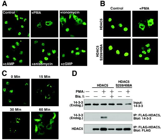
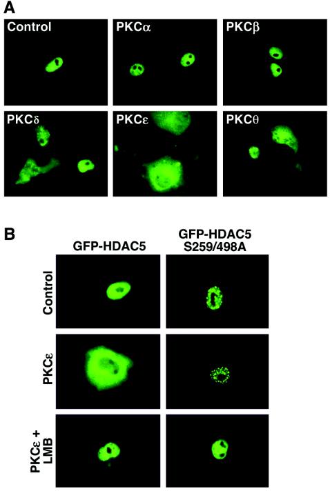
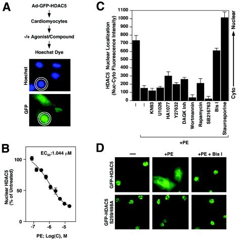
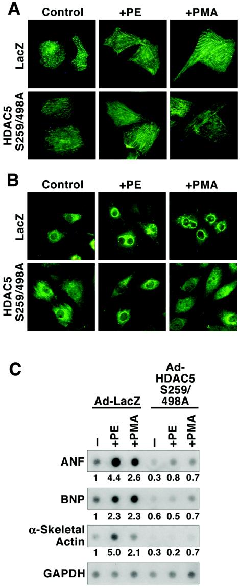
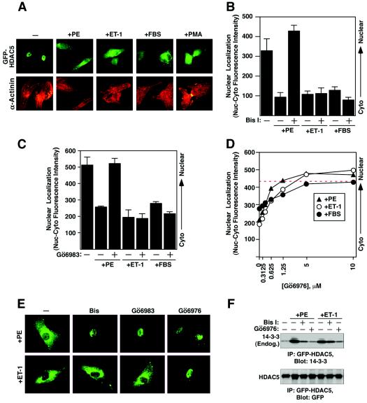
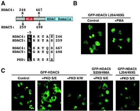
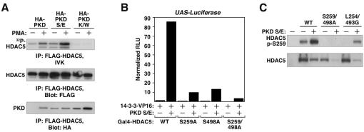
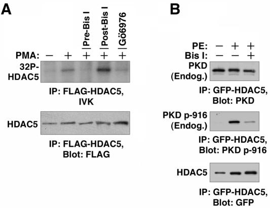
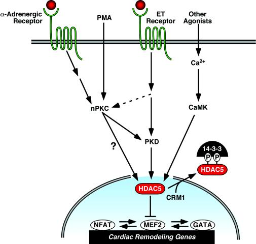
A variety of stress signals stimulate cardiac myocytes to undergo hypertrophy. Persistent cardiac hypertrophy is associated with elevated risk for the development of heart failure. Recently, we showed that class II histone deacetylases (HDACs) suppress cardiac hypertrophy and that stress signals neutralize this repressive function by triggering phosphorylation- and CRM1-dependent nuclear export of these chromatin-modifying enzymes. However, the identities of cardiac HDAC kinases have remained unclear. Here, we demonstrate that signaling by protein kinase C (PKC) is sufficient and, in some cases, necessary to drive nuclear export of class II HDAC5 in cardiomyocytes. Inhibition of PKC prevents nucleocytoplasmic shuttling of HDAC5 in response to a subset of hypertrophic agonists. Moreover, a nonphosphorylatable HDAC5 mutant is refractory to PKC signaling and blocks cardiomyocyte hypertrophy mediated by pharmacological activators of PKC. We also demonstrate that protein kinase D (PKD), a downstream effector of PKC, directly phosphorylates HDAC5 and stimulates its nuclear export. These findings reveal a novel function for the PKC/PKD axis in coupling extracellular cues to chromatin modifications that control cellular growth, and they suggest potential utility for small-molecule inhibitors of this pathway in the treatment of pathological cardiac gene expression.


Figures

FIG. 1.
FIG. 1.
PKC-dependent nuclear export of HDAC5. (A) COS cells were cultured in six-well dishes, transfected with a GFP-HDAC5 expression vector (1 μg), and stimulated with the indicated compounds, as described in Materials and Methods. At 1 h after addition of compounds, GFP-HDAC5 distribution was determined by fluorescence microscopy. PMA stimulation resulted in complete relocalization of GFP-HDAC5 from the nucleus to the cytoplasm, while ionomycin triggered a partial response. (B) COS cells were transfected with expression vectors encoding FLAG-tagged versions of either HDAC5 or an HDAC5 mutant harboring alanines in place of serines 259 and 498 (HDAC5 S259/498A) (1 μg each). The cells were stimulated with PMA for 1 h, and HDAC5 distribution was determined by indirect immunofluorescence with anti-FLAG primary antibody and fluorecein-conjugated secondary antibody. HDAC5 S259/498A is refractory to PMA stimulation. (C) COS cells were transfected with GFP-HDAC5 encoding expression vector (1 μg) and stimulated with PMA for the indicated times. (D) COS cells were transiently transfected with expression vectors encoding FLAG-tagged versions of HDAC5 or HDAC5 S259/498A (1 μg each). The cells were pretreated with the PKC inhibitor Bis I at 10 μM for 30 min and stimulated with PMA for 30 min, as indicated. Association of FLAG-HDAC5 with endogenous 14-3-3 was detected by sequential immunoprecipitation (IP) and immunoblotting (Blot).
FIG. 2.
FIG. 2.
Calcium-independent PKCs trigger the nuclear export of HDAC5. (A) CV-1 cells were transiently transfected with an expression vector encoding GFP-HDAC5 and constructs for PKCα, PKCβ, PKCδ, PKCɛ, or PKCθ (0.5 μg each). The effects of PKC overexpression were assessed by fluorescence microscopy 24 h post-transfection. (B) CV-1 cells were transfected with vectors encoding either GFP-HDAC5 or GFP-HDAC5 S259/498A in the absence or presence of a vector for constitutively active PKCɛ (0.5 μg each). Prior to analysis, some cells received 18.5 nM leptomycin B (LMB) for 1 h.
FIG. 3.
FIG. 3.
PKC inhibition blocks PE-mediated nuclear export of HDAC5 in cardiomyocytes. (A) Schematic representation of a quantitative assay for HDAC5 nuclear export. NVRMs are cultured in 96-well dishes and infected with adenovirus encoding GFP-HDAC5. Cells are serum starved, subjected to agonists and inhibitors, fixed, and stained with Hoechst dye. The relative abundance of GFP-HDAC5 in the nucleus versus the cytoplasm is quantified by using the Cellomics high-content imaging system, which demarcates nuclei based on Hoechst fluorescence and defines a cytoplasmic ring based on these nuclear dimensions. Values represent the mean of nuclear minus cytoplasmic fluorescence intensity. (B) Assay validation. NRVMs were infected with adenovirus encoding GFP-HDAC5 and exposed to PE at concentrations ranging from 0.1 to 20 μM. Cells were prepared for Cellomics analysis following 2 h of stimulation. Mean nuclear minus cytoplasmic fluorescence intensity was determined for at least 50 cells/well in eight wells per condition (400 cells total). The value for untreated cells was set to 100%. PE triggered the dose-dependent nuclear export of HDAC5. EC50, 50% effective concentration. (C) NRVMs were infected with adenovirus encoding GFP-HDAC5 and pretreated with kinase inhibitors (the concentrations of the inhibitors are given in Materials and Methods). The subcellular distribution of HDAC5 was quantified following stimulation with PE (20 μM) for 2 h. Mean nuclear minus cytoplasmic fluorescence intensity was determined for at least 50 cells/well in eight wells per condition (400 cells total). Higher values indicate greater abundance of HDAC5 in the nucleus. Well-to-well standard deviations are shown. Only staurosporine and the PKC inhibitor Bis I effectively blocked HDAC5 nuclear export. (D) Representative images of GFP-HDAC5 and GFP-HDAC5 S259/498A in the absence and presence of Bis I.
FIG. 4.
FIG. 4.
Inhibition of PKC-mediated cardiac hypertrophy by signal-resistant HDAC5. NRVMs were cultured on six-well dishes and infected with adenoviruses (multiplicity of infection, 10) encoding a LacZ control (Ad-LacZ) or FLAG-tagged HDAC5 harboring alanines in place of serines 259 and 498 (Ad-HDAC5 S259/498A), which are required for 14-3-3-mediated nuclear export. Cells were treated with 20 μM PE or 100 nM PMA for 24 h prior to analysis. (A) Cells were fixed, and sarcomeres were visualized by indirect immunofluorescence with primary antibody specific for α-actinin and fluorescein-conjugated secondary antibody. (B) ANF protein was detected by indirect immunofluorescence with anti-ANF primary antibody. (C) Total RNA was harvested from cells and subjected to dot blot analysis with radiolabeled oligonucleotides specific for the indicated transcripts. RNA levels were quantified using a phosphorimager and are depicted as the fold change relative to amounts in unstimulated cells infected with Ad-LacZ. Values were normalized to GAPDH controls.
FIG. 5.
FIG. 5.
Differential repression of agonist-mediated nuclear export of HDAC5 by PKC inhibitors. (A) NRVMs were cultured in six-well dishes and infected with adenovirus encoding GFP-HDAC5. The cells were serum starved for 4 h prior to stimulation with 20 μM PE, 50 nM ET-1, 10% FBS, or 50 nM PMA for 2 h. The cells were fixed and stained with antibodies against sarcomeric α-actinin (red) to confirm that HDAC5 was being visualized in cardiomyocytes. (B) NRVMs were cultured in 96-well dishes and infected with adenovirus encoding GFP-HDAC5. Following serum starvation, infected cells were pretreated with 10 μM Bis I for 30 min and stimulated with the indicated agonists for 2 h. Nuclear export of HDAC5 was quantified using the Cellomics imaging system. Higher values indicate greater abundance of HDAC5 in the nucleus. (C) The experiment was performed as described for panel B, except that the cells received 10 μM Gö6983 before being treated with the agonists. (D) The experiment was performed as described for panel B, except that the cells received increasing doses of Gö6976 before being treated with the agonists. (E) Representative images from each treatment group were captured using a fluorescence microscope equipped with a digital camera. (F) NRVMs were infected with adenovirus encoding GFP-HDAC5 and cultured on 10-cm dishes. At 24 h postinfection, the cells were serum starved for 4 h and pretreated with 10 μM Bis I or 10 μM Gö6976 for 1 h before being stimulated with 20 μM PE or 50 nM ET-1 for 1 h. Whole-cell protein lysates were prepared and subjected to sequential immunoprecipitation (IP) and immunoblotting (Blot) as indicated.
FIG. 6.
FIG. 6.
PKD stimulates the nuclear export of HDAC5. (A) Amino acid sequences surrounding the regulatory phosphorylation sites of class II HDACs. NLS, nuclear localization signal; HDAC domain, deacetylase catalytic domain. The consensus target site for PKD is shown. Leucine at position −5 relative to the phosphorylation site is required for optimal PKD-directed phosphorylation of other proteins. (B) COS cells were transfected with an expression vector encoding GFP fused to HDAC5 harboring glycines in place of leucines 254 and 493 (L254/493G). At 24 h posttransfection, the cells were left untreated (control) or stimulated with PMA for 30 min. (C) COS cells were cotransfected with expression vectors (1 μg each) encoding GFP-HDAC5 or GFP-HDAC5 S259/498A and constitutively active (S/E) or catalytically inactive (K/W) forms of PKD. HDAC5 localization was determined at 24 h posttransfection.
FIG. 7.
FIG. 7.
PKD is an HDAC5 kinase. (A) COS cells were cotransfected with expression vectors (1 μg each) encoding FLAG-HDAC5 and HA-tagged versions of either wild-type, constitutively active (S/E), or catalytically inactive (K/W) PKD. At 24 h posttransfection, the cells were treated with PMA or vehicle control for 30 min. FLAG-HDAC5 was immunoprecipitated (IP) from whole-cell protein lysates and either incorporated into an in vitro kinase assay (IVK) or resolved by SDS-PAGE for Western blot analysis (Blot) to detect associated PKD, as indicated. Phosphorylated HDAC5 was resolved by SDS-PAGE and detected by autoradiography. (B) Mammalian two-hybrid assay. Expression vectors encoding the GAL4 DNA binding domain fused to HDAC5 (Gal4-HDAC5) or the indicated HDAC5 alanine substitution mutants were cotransfected into COS cells with a plasmid encoding 14-3-3 fused to the VP16 transcriptional activation domain (14-3-3-VP16), a Gal4-dependent luciferase reporter (UAS-Luciferase), and a vector encoding constitutively active PKD (S/E). PKD stimulates the association between HDAC5 and 14-3-3, which is dependent on the phospho-acceptors at positions 259 and 498. WT, wild type. (C) COS cells were left untransfected or transfected with expression vectors encoding FLAG-tagged versions of wild-type HDAC5, HDAC5 S259/498A, or HDAC5 L254/493G (1 μg) in the absence or presence of 1 μg of constitutively active PKD S/E. After 24 h of transfection, protein lysates were prepared and subjected to immunoblotting with antibodies directed against either HDAC5 phosphorylated at serine 259 (P-S259) or FLAG, to reveal the total levels of ectopic HDAC5.
FIG. 8.
FIG. 8.
Association of endogenous PKD with HDAC5 in cardiomyocytes. (A) NRVMs were cultured on 10-cm dishes and infected with adenovirus encoding FLAG-HDAC5. At 24 h posttransfection, the cells were stimulated with PMA for 30 min and whole-cell protein lysates were prepared. Some cells were pretreated with 10 μM Bis I (pre-Bis I) for 30 min prior to PMA stimulation. FLAG-HDAC5 was immunoprecipitated (IP) and incorporated into in vitro kinase reaction mixtures supplemented with 10 μM Bis I (post-Bis I) or 10 μM Gö6976 (post-Gö6976), as indicated. Phosphorylation of HDAC5 was blocked when the cells were pretreated with Bis I (pre-Bis I). Gö6976 but not Bis I blocked phosphoryl transfer to HDAC5 when added directly to kinase reaction mixtures. (B) NRVMs were infected with adenovirus encoding GFP-HDAC5 and cultured on 10-cm dishes. At 24 h postinfection, the cells were serum starved for 4 h and pretreated with 10 μM Bis I for 30 min before being stimulated with 20 μM PE for 1 h. HDAC5 was immunoprecipitated from whole-cell lysates, and associated total PKD or PKD autophosphorylated at serine 916 (p-916) were detected by immunoblotting. Blots were reprobed with GFP-specific antibodies to determine the total amounts of immunoprecipitated HDAC5.
FIG. 9.
FIG. 9.
Model of kinase-dependent signaling pathways that regulate the nuclear export of class II HDACs and cardiac hypertrophy. HDAC5 represses pathological cardiac gene expression and remodeling via interactions with MEF2. MEF2 associates with other prohypertrophic transcription factors, including NFAT and GATA4, and thus HDAC5 also indirectly represses genes under the control of these factors. The repressive effects of HDAC5 are neutralized by signals that culminate in phosphorylation of the protein. Phospho-HDAC5 binds 14-3-3 proteins, resulting in nuclear export of HDAC5 through a CRM1-dependent mechanism. Phosphorylation of HDAC5 can be triggered by CaMK and, as shown in the present study, by signaling via calcium-independent PKCs, also referred to as novel (nPKCs). Hypertrophic signaling cascades, including those elicited by the α-adrenergic and ET receptors, stimulate the nuclear export of HDAC5 via activation of nPKCs and their downstream effector PKD. However, ET receptor signaling also appears to activate PKD through a PKC-independent mechanism. PMA directly activates PKC. It remains possible that PKCs bypass PKD and directly phosphorylate HDAC5.

Similar articles

Cited by

References

    1. Allo, S. N., L. L. Carl, and H. E. Morgan. 1992. Acceleration of growth of cultured cardiomyocytes and translocation of protein kinase C. Am. J. Physiol. 263:C319-C325. - PubMed
    1. Antos, C. L., T. A. McKinsey, M. Dreitz, L. M. Hollingsworth, C. L. Zhang, K. Schreiber, H. Rindt, R. J. Gorczynski, and E. N. Olson. 2003. Dose-dependent blockade to cardiomyocyte hypertrophy by histone deacetylase inhibitors. J. Biol. Chem. 278:28930-28937. - PubMed
    1. Bowman, J. C., S. F. Steinberg, T. Jiang, D. L. Geenen, G. I. Fishman, and P. M. Buttrick. 1997. Expression of protein kinase C beta in the heart causes hypertrophy in adult mice and sudden death in neonates. J. Clin. Investig. 100:2189-2195. - PMC - PubMed
    1. Braz, J. C., O. F. Bueno, L. J. De Windt, and J. D. Molkentin. 2002. PKC alpha regulates the hypertrophic growth of cardiomyocytes through extracellular signal-regulated kinase1/2 (ERK1/2). J. Cell Biol. 156:905-919. - PMC - PubMed
    1. Braz, J. C., K. Gregory, A. Pathak, W. Zhao, B. Sahin, R. Klevitsky, T. F. Kimball, J. N. Lorenz, A. C. Nairn, S. B. Liggett, I. Bodi, S. Wang, A. Schwartz, E. G. Lakatta, A. A. DePaoli-Roach, J. Robbins, T. E. Hewett, J. A. Bibb, M. V. Westfall, E. G. Kranias, and J. D. Molkentin. 2004. PKC-alpha regulates cardiac contractility and propensity toward heart failure. Nat. Med. 10:248-254. - PubMed

Publication types

LinkOut - more resources