. 2004 Oct;24(19):8408-17.
doi: 10.1128/MCB.24.19.8408-8417.2004.

Selective decrease in paracellular conductance of tight junctions: role of the first extracellular domain of claudin-5

Affiliations

Selective decrease in paracellular conductance of tight junctions: role of the first extracellular domain of claudin-5

Huajie Wen et al. Mol Cell Biol. 2004 Oct.

Abstract

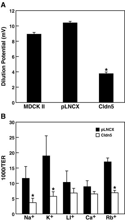
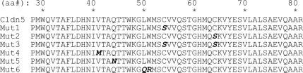
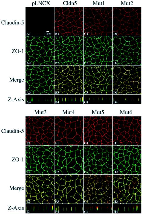
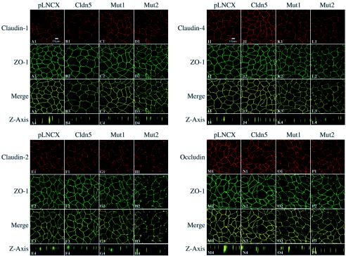
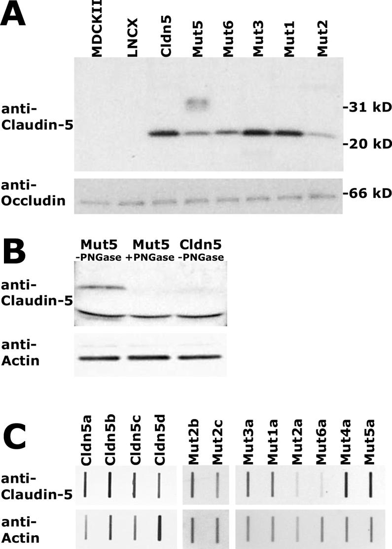
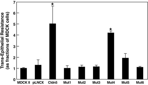
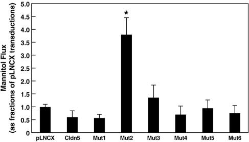
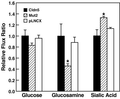
Claudin-5 is a protein component of many endothelial tight junctions, including those at the blood-brain barrier, a barrier that limits molecular exchanges between the central nervous system and the circulatory system. To test the contribution of claudin-5 to this barrier function of tight junctions, we expressed murine claudin-5 in Madin-Darby canine kidney II cells. The result was a fivefold increase in transepithelial resistance in claudin-5 transductants and a reduction in conductance of monovalent cations. However, the paracellular flux of neither neutral nor charged monosaccharides was significantly changed in claudin-5 transductants compared to controls. Therefore, expression of claudin-5 selectively decreased the permeability to ions. Additionally, site-directed mutations of particular amino acid residues in the first extracellular domain of claudin-5 altered the properties of the tight junctions formed in response to claudin-5 expression. In particular, the conserved cysteines were crucial: mutation of either cysteine abolishted the ability of claudin-5 to increase transepithelial resistance, and mutation of Cys(64) strikingly increased the paracellular flux of monosaccharides. These new insights into the functions of claudin-5 at the molecular level in tight junctions may account for some aspects of the blood-brain barrier's selective permeability.


Figures

FIG. 1.
FIG. 1.
Paracellular conductance of ions. (A) The 50% dilution potentials were measured in parental MDCK II cells, control MDCK II cells containing the pLNCX vector, and MDCK II cells expressing claudin-5. Expression of claudin-5 significantly decreased dilution potentials compared to parental cells and to cells with the pLNCX vector (one-way analysis of variance combined with Bonferroni t tests; n = 6, P < 0.001). (B) Paracellular conductance of group Ia alkali cations. Claudin-5 expression dramatically reduced the ionic conductance of sodium and potassium in the paracellular pathway. There was a statistically significant difference in conductance of sodium ions, potassium ions, and rubidium ions between pLNCX transductants and claudin-5 transductants. Data are presented as mean ± standard deviation (one-way analysis of variance combined with Bonferroni t tests; n = 4, P < 0.001).
FIG. 2.
FIG. 2.
Predicted amino acid sequence of the first ECD in wild-type claudin-5 and its mutants. Residues altered by site-directed mutagenesis are indicated in bold italics.
FIG. 3.
FIG. 3.
Confocal microscopic images of claudin-5 wild-type and mutants expressed in MDCK II cells following immunofluorescent labeling for ZO-1 and claudin-5. The TJs were localized by ZO-1 expression visualized with fluorescein isothiocyanate. Claudin-5 expression was visualized with Texas Red. Transductants with a blank pLNCX vector were negative for claudin-5 labeling (A). In wild-type claudin-5 transductants, claudin-5 stained with Texas Red colocalized with ZO-1 (B). The patterns of immunolocalization in the claudin-5 mutants Mut1 to Mut6 are presented in panels C to H. Rows 1 to 3 represent images in the x-y plane taken at the midpoint of ZO-1 localization; row 4 represents computer reconstruction of the x-z plane. Bar, 10 μm.
FIG. 4.
FIG. 4.
Confocal microscopic images of claudin-5 wild-type and mutants expressed in MDCK II cells following immunofluorescent labeling for ZO-1 and other claudins and occludin. The TJs were localized by ZO-1 expression visualized with fluorescein isothiocyanate. Claudin-1 (panels A to D), claudin-2 (panels E to H), claudin-4 (panels I to L), and occludin (panels M to P) were visualized with Texas Red. Images were performed as per the Fig. 3 legend.
FIG. 5.
FIG. 5.
Immunoblot analysis of exogenous claudin-5 protein expression in MDCK II cells. Western blot analysis of wild-type claudin-5, Mut1 to Mut3, and Mut6 revealed a distinct band of 23-kDa, seen after reaction with anti-claudin-5, visualization with ECL, and exposure to X-ray film (A, upper). Mut5, containing a consensus glycosylation site, exhibited an additional diffuse band centered around 35 kDa (A, upper). The blot was stripped and incubated with antioccludin with visualization as above, resulting in a band of 66 kDa (A, lower). Similar Western blot analysis indicating that the slower-migrating species in Mut5-expressing cells is eliminated following digestion with PNGase F (B, upper). The blot was stripped and incubated with antiactin (B, lower). Quantification of claudin-5 expression in individual cell clones was performed by slot blot analysis, with serial dilutions of total cell extracts incubated with anti-claudin-5 or antiactin and visualized by ECL with multiple exposures to film to obtain data in the linear range of densitometry. Examples of the data are shown (C), with the different lowercase letters indicating independent cellular clones expressing a given construct.
FIG. 6.
FIG. 6.
TER of wild-type and mutant claudin-5 transductants. Wild-type claudin-5 transductants exhibited a significant fivefold increase in TER, and Mut4 (with random mutation) transductants had a fourfold increase; the other claudin-5 mutants had no significant alterations. Data are shown as means ± standard deviation (one-way analysis of variance combined with Bonferroni t tests; n = 3, P < 0.001).
FIG. 7.
FIG. 7.
Paracellular flux of mannitol in MDCK II cells expressing claudin-5 or its mutants. Only Mut2 (Cys64) transductants exhibited a significant difference from controls, a 3.7-fold increase in mannitol flux. Data are shown as means ± standard deviation (one-way analysis of variance combined with Bonferroni t tests; n = 3, P < 0.001).
FIG. 8.
FIG. 8.
Relative paracellular flux of uncharged and charged monosaccharides in MDCK II cells expressing the pLNCX vector, wild-type claudin-5, or Mut2. Mut2-expressing cells showed increased paracellular flux of all monosaccharides. Normalized to the flux of mannitol, the Mut2 transductants were significantly more permeable to the negatively charged sialic acid (F = 15.84, P = 0.004) and less permeable to the positively charged glucosamine (F = 12.06, P = 0.008) than were the vector pLNCX or wild-type claudin-5 transductants (one-way analysis of variance combined with post hoc Bonferroni t test; n = 3; P < 0.05 for between-two-group comparisons).

Similar articles

Cited by

References

    1. Anderson, J. M. 2001. Molecular structure of tight junctions and their role in epithelial transport. News Physiol. Sci. 16:126-130. - PubMed
    1. Anderson, J. M., and C. M. Van Itallie. 1995. Tight junctions and the molecular basis for regulation of paracellular permeability. Am. J. Physiol. 269:G467-G475. - PubMed
    1. Antohe, F., L. Dobrila, C. Heltianu, N. Simionescu, and M. Simionescu. 1993. Albumin-binding proteins function in the receptor-mediated binding and transcytosis of albumin across cultured endothelial cells. Eur. J. Cell Biol. 60:268-275. - PubMed
    1. Balda, M. S., C. Flores-Maldonado, M. Cereijido, and K. Matter. 2000. Multiple domains of occludin are involved in the regulation of paracellular permeability. J. Cell Biochem. 78:85-96. - PubMed
    1. Balda, M. S., J. A. Whitney, C. Flores, S. Gonzalez, M. Cereijido, and K. Matter. 1996. Functional dissociation of paracellular permeability and transepithelial electrical resistance and disruption of the apical-basolateral intramembrane diffusion barrier by expression of a mutant tight junction membrane protein. J. Cell Biol. 134:1031-1049. - PMC - PubMed

Publication types

LinkOut - more resources