. 2004 Oct;24(19):8467-76.
doi: 10.1128/MCB.24.19.8467-8476.2004.

Histone deacetylases 5 and 9 govern responsiveness of the heart to a subset of stress signals and play redundant roles in heart development

Affiliations

Histone deacetylases 5 and 9 govern responsiveness of the heart to a subset of stress signals and play redundant roles in heart development

Shurong Chang et al. Mol Cell Biol. 2004 Oct.

Abstract

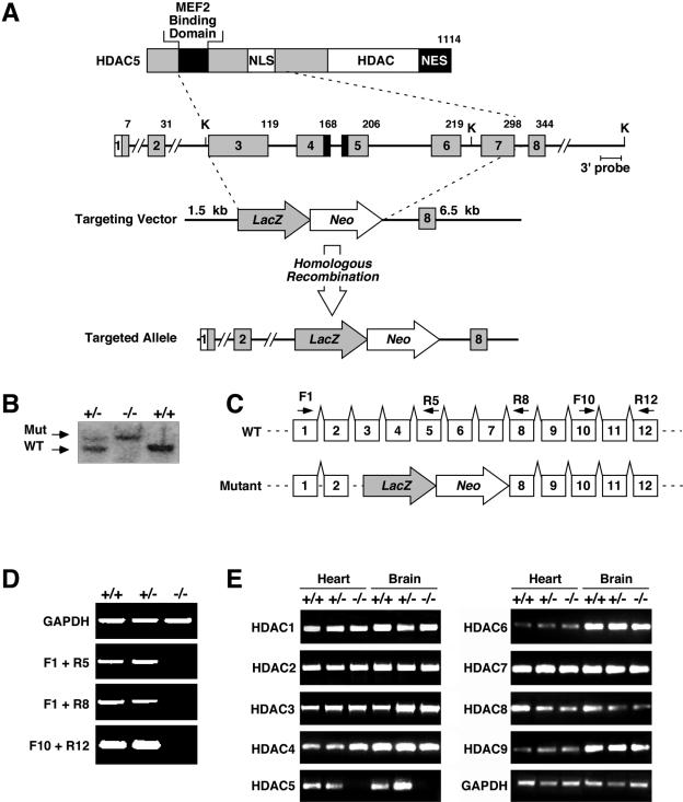
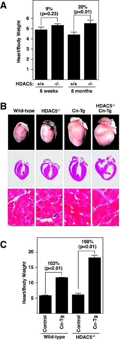
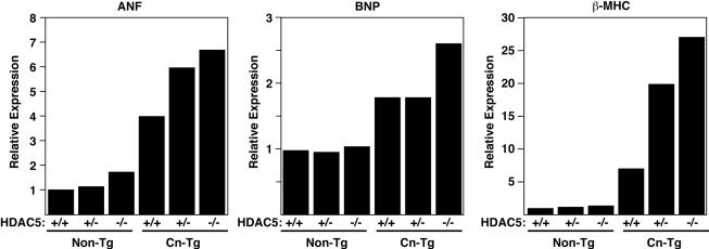
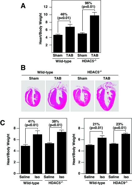
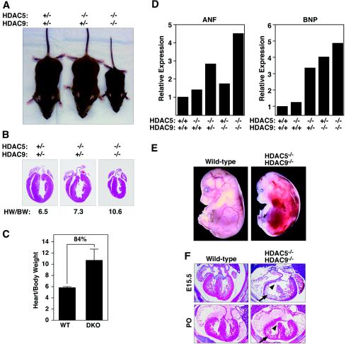
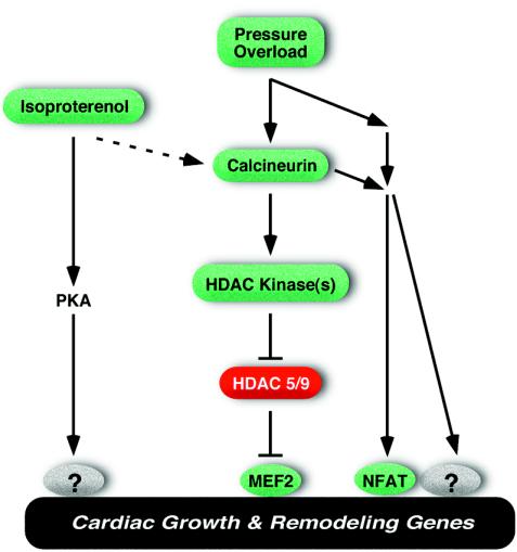
The adult heart responds to stress signals by hypertrophic growth, which is often accompanied by activation of a fetal cardiac gene program and eventual cardiac demise. We showed previously that histone deacetylase 9 (HDAC9) acts as a suppressor of cardiac hypertrophy and that mice lacking HDAC9 are sensitized to cardiac stress signals. Here we report that mice lacking HDAC5 display a similar cardiac phenotype and develop profoundly enlarged hearts in response to pressure overload resulting from aortic constriction or constitutive cardiac activation of calcineurin, a transducer of cardiac stress signals. In contrast, mice lacking either HDAC5 or HDAC9 show a hypertrophic response to chronic beta-adrenergic stimulation identical to that of wild-type littermates, suggesting that these HDACs modulate a specific subset of cardiac stress response pathways. We also show that compound mutant mice lacking both HDAC5 and HDAC9 show a propensity for lethal ventricular septal defects and thin-walled myocardium. These findings reveal central roles for HDACs 5 and 9 in the suppression of a subset of cardiac stress signals as well as redundant functions in the control of cardiac development.


Figures

FIG. 1.
FIG. 1.
Targeting the mouse HDAC5 gene. (A) A diagram of the HDAC5 protein is shown above a portion of the mouse HDAC5 locus encompassing coding exons 1 to 8. Amino acid numbers are shown above the exons. NES, nuclear export sequence; NLS, nuclear localization sequence. In the targeting vector, a nuclear lacZ reporter was inserted in-frame with exon 3. Homologous recombination resulted in deletion of exons 3 to 7, which encompass the MEF2 binding domain and nuclear localization sequence. The structure of the targeted allele is shown. K, KpnI sites. (B) Southern blot analysis of genomic DNA from mice of the indicated genotypes. A 500-bp DNA fragment downstream of 3′ arm sequences was used as to probe KpnI-digested tail DNA. Wild-type (WT) and mutant (Mut) bands of 8.5 and 15.5 kb, respectively, are shown. (C and D) RNA from hearts of mice of the indicated genotypes was analyzed by RT-PCR. The positions of the primers used for RT-PCR are shown above the corresponding exons of the wild-type and mutant HDAC5 alleles (panel C). No functional HDAC5 mRNA was detected in homozygous mutants. Glyceraldehyde-3-phosphate dehydrogenase (GAPDH) transcripts were measured as a control. (E) Transcripts for HDACs 1 to 9 were detected by RT-PCR with RNA isolated from the hearts and brains of mice of the indicated genotypes.
FIG. 2.
FIG. 2.
Expression of lacZ from the targeted HDAC5 allele. Embryos heterozygous for the targeted HDAC5 allele were stained for lacZ expression on the indicated days of embryogenesis. Strong expression of LacZ was seen in the looping heart tube at E9.5 and in the heart and spinal cord at later stages. LacZ expression was also detected in the muscle forming regions of the limbs at E12.5. The lower panels show transverse sections through LacZ-stained embryos visualized in bright field, with LacZ staining indicated in pink. h, heart; nt, neural tube; sm, skeletal muscle.
FIG. 3.
FIG. 3.
Enhanced hypertrophy in HDAC5 mutant mice. (A) Hearts were dissected from mice of the indicated genotypes at 6 weeks and 8 months of age, and heart weight-to-body weight ratios were determined. Values represent the mean ± standard deviation. The heart sizes of wild-type (n = 4) and HDAC5 mutant (n = 4) mice were similar at 6 weeks, but by 8 months, the hearts of the mutant mice (n = 5) were enlarged compared to those of the wild-type mice (n = 3). (B) HDAC5 mutant mice were bred with mice harboring theαMHC-calcineurin transgene (Cn-Tg). Hearts from 1-month-old mice of the indicated genotypes were isolated (top images), sectioned, and stained with hematoxylin and eosin (bottom images). (C) Heart weight/body weight ratios of mice of wild-type control (n = 6), αMHC-calcineurin transgene (n = 10), HDAC5−/− (n = 5), and HDAC5−/−/ αMHC-calcineurin transgene (n = 4) mice are shown.
FIG. 4.
FIG. 4.
Fetal gene expression in HDAC5 knockout mice. RNA was isolated from the hearts of mice with the indicated genotypes, and expression of fetal cardiac genes was measured by dot blot analysis. Values are expressed as the level of expression of each transcript relative to that in hearts from wild-type mice.
FIG. 5.
FIG. 5.
Cardiac responses to thoracic aortic banding and chronic isoproterenol administration. (A and B) Wild-type (non-Tg) and HDAC5 null mice at 6 weeks of age were subjected to thoracic aortic banding (TAB) for 21 days, at which time hearts were dissected and heart weight-to-body weight ratios were determined (A). Values represent the mean ± standard deviation. We tested sham-treated wild-type mice (n = 3), TAB-treated wild-type mice (n = 3), HDAC5−/− sham-treated mice (n = 4), and TAB-treated HDAC5−/− mice (n = 4). (B) Hearts were sectioned and stained with hematoxylin and eosin. (C) Wild-type or HDAC5 and HDAC9 mutant mice at 8 weeks of age were infused with isoproterenol (Iso) or saline alone for 7 days, at which time the hearts were dissected and heart weight-to-body weight ratios were determined. The hypertrophic responses of the HDAC5 and HDAC9 mutant mice were not statistically different from those of wild-type mice. The left panel shows saline-treated wild-type mice (n = 3), isoproterenol-treated wild-type mice (n = 4), sham-treated HDAC5−/− mice (n = 4), and isoproterenol-treated HDAC5−/− mice (n = 6). The right panel shows saline-treated wild-type mice (n = 4), wild-type isoproterenol-treated mice (n = 4), sham-treated HDAC9−/− mice (n = 4), and isoproterenol-treated HDAC9−/− mice (n = 4).
FIG. 6.
FIG. 6.
Growth defects and cardiac abnormalities in HDAC5/HDAC9 double mutant mice. (A) One-month-old mice of the indicated genotypes are shown. The HDAC5/HDAC9 double mutant animals are severely growth retarded. (B) Hearts were dissected from the wild-type and HDAC5/HDAC9 double mutant mice shown in panel A, and heart weight-to-body weight ratios were determined. The double mutant mice had smaller hearts than the wild-type mice, but they were enlarged compared to body weight. Heart weight/body weight (HW/BW) ratios are shown. (C) Hearts were dissected from wild-type (n = 6) and HDAC5/HDAC9 double knockout (DKO) (n = 5) mice at 6 months of age, and heart/body weight ratios were determined. Values represent the mean ± standard deviation. (D) RNA was isolated from the hearts of adult mice with the indicated genotypes, and expression of fetal cardiac genes was measured by dot blot analysis. Values are expressed as the level of expression of each transcript relative to that in hearts from wild-type mice. (E) Wild-type and HDAC5/HDAC9 double mutant embryos at E15.5. The double mutant shows multifocal hemorrhages. (F) Hematoxylin and eosin staining of cardiac sections from wild-type and HDAC5/HDAC9 double mutant mice at E15.5 (top panels) and at birth (P0, bottom panels). Note the ventricular septal defect (arrowhead) and thin-walled myocardium (arrow) in the double mutant.
FIG. 7.
FIG. 7.
Schematic of the roles of HDACs 5 and 9 as antagonists of hypertrophic signaling. PKA, protein kinase A.

Similar articles

Cited by

References

    1. Antos, C. L., T. A. McKinsey, M. Dreitz, L. M. Hollingsworth, C. L. Zhang, K. Schreiber, H. Rindt, R. J. Gorczynski, and E. N. Olson. 2003. Dose-dependent blockade to cardiomyocyte hypertrophy by histone deacetylase inhibitors. J. Biol. Chem. 278:28930-28937. - PubMed
    1. Chien, K. R. 1999. Stress pathways and heart failure. Cell 98:555-558. - PubMed
    1. Davis, F. J., M. Gupta, B. Camoretti-Mercado, R. J. Schwartz, and M. P. Gupta. 2003. Calcium/calmodulin-dependent protein kinase activates serum response factor transcription activity by its dissociation from histone deacetylase, HDAC4. Implications in cardiac muscle gene regulation during hypertrophy. J. Biol. Chem. 278:20047-20058. - PubMed
    1. De Windt, L. J., H. W. Lim, O. F. Bueno, Q. Liang, U. Delling, J. C. Braz, B. J. Glascock, T. F. Kimball, F. del Monte, R. J. Hajjar, and J. D. Molkentin. 2001. Targeted inhibition of calcineurin attenuates cardiac hypertrophy in vivo. Proc. Natl. Acad. Sci. USA 98:3322-3327. - PMC - PubMed
    1. Fentzke, R. C., C. E. Korcarz, R. M. Lang, H. Lin, and J. M. Leiden. 1998. Dilated cardiomyopathy in transgenic mice expressing a dominant-negative CREB transcription factor in the heart. J. Clin. Investig. 101:2415-2426. - PMC - PubMed

Publication types