. 2004 Oct;24(19):8477-86.
doi: 10.1128/MCB.24.19.8477-8486.2004.

The Keap1-BTB protein is an adaptor that bridges Nrf2 to a Cul3-based E3 ligase: oxidative stress sensing by a Cul3-Keap1 ligase

Affiliations

The Keap1-BTB protein is an adaptor that bridges Nrf2 to a Cul3-based E3 ligase: oxidative stress sensing by a Cul3-Keap1 ligase

Sara B Cullinan et al. Mol Cell Biol. 2004 Oct.

Abstract

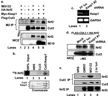
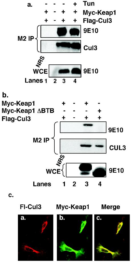
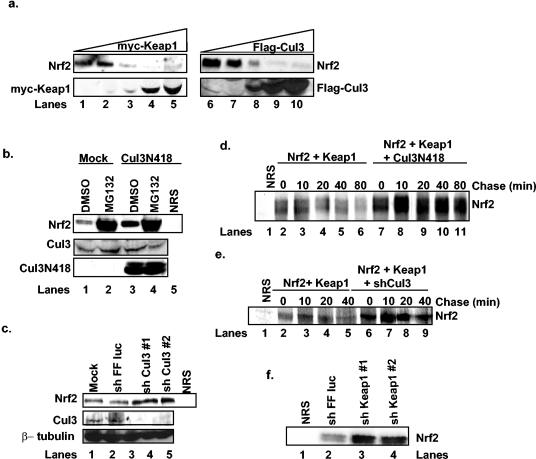
The Nrf2 transcription factor promotes survival following cellular insults that trigger oxidative damage. Nrf2 activity is opposed by the BTB/POZ domain protein Keap1. Keap1 is proposed to regulate Nrf2 activity strictly through its capacity to inhibit Nrf2 nuclear import. Recent work suggests that inhibition of Nrf2 may also depend upon ubiquitin-mediated proteolysis. To address the contribution of Keap1-dependent sequestration versus Nrf2 proteolysis, we identified the E3 ligase that regulates Nrf2 ubiquitination. We demonstrate that Keap1 is not solely a cytosolic anchor; rather, Keap1 is an adaptor that bridges Nrf2 to Cul3. We demonstrate that Cul3-Keap1 complexes regulate Nrf2 polyubiquitination both in vitro and in vivo. Inhibition of either Keap1 or Cul3 increases Nrf2 nuclear accumulation, leading to promiscuous activation of Nrf2-dependent gene expression. Our data demonstrate that Keap1 restrains Nrf2 activity via its capacity to target Nrf2 to a cytoplasmic Cul3-based E3 ligase and suggest a model in which Keap1 coordinately regulates both Nrf2 accumulation and access to target genes.


Figures

FIG. 1.
FIG. 1.
Nrf2 associates with a Cul3 complex in a Keap1-dependent manner. (a) 293T cells were transfected with plasmids encoding HA-Nrf2, Flag-Cul3, and Myc-Keap1 and treated as indicated. Flag-Cul3 was precipitated from whole-cell extracts (WCE) with the M2 monoclonal antibody. Cul3-associated Nrf2 was detected via immunoblot analysis. The presence of ectopic proteins was confirmed via immunoblot analysis. Lane 1 shows a control precipitation with a nonspecific antiserum (NRS). (b) Nrf2, in vitro transcribed and translated in the presence of [35S]methionine, was mixed with purified Flag-Cul3 (lane 3) or His-Keap1-Flag-Cul3 complexes (lane 4) from insect Sf9 cells. Lane 1 shows 100% input, and lane 2 shows a control precipitation with a nonspecific antiserum. Nrf2 was visualized by autoradiography. The bottom panel shows Coomassie staining. The asterisk indicates a nonspecific coprecipitating protein. (c) Following transfection of 293T cells with the indicated short hairpin RNA vectors, Keap1 mRNA levels were assessed by reverse transcription-PCR. (d) 293T cells were transfected with Flag-Cul3 and HA-Nrf2 in combination with short hairpin RNA vectors against firefly luciferase (lane 3) or Keap1 (lane 4). Following MG132 treatment, Flag-Cul3 was precipitated from cell lysates with the M2 antibody; associated Nrf2 was assessed via immunoblot analysis. Lane 1 shows a control precipitation with a nonspecific antiserum. (e) 293T cells were treated with 10 μM MG132 for 4 h (lane 3), 5 μg of tunicamycin per ml for 2 h (lane 4), or vehicle alone (lane 2). Cul3 was precipitated from whole-cell lysates with a Cul3-specific antibody, and Cul3-associated Nrf2 was assessed via immunoblot analysis. Total Nrf2 and Cul3 levels were determined via immunoblot analysis. Lane 1 shows a control precipitation with a nonspecific antiserum.
FIG. 2.
FIG. 2.
Cul3 interacts with Keap1 through its BTB domain. (a) 293T cells transfected with plasmids encoding Myc-Keap1 and Flag-Cul3 were either left untreated (lanes 1 to 3) or treated with 5 μg/ of tunicamycin per ml for 1 h (lane 4). Flag-Cul3 was precipitated from whole-cell extracts with the M2 monoclonal antibody; Flag-Cul3 (middle panel) and Cul3-associated Keap1 were detected by immunoblot analysis with the 9E10 antibody (top panel). The presence of ectopically expressed Myc-Keap1 was confirmed by immunoblot analysis with the 9E10 antibody (bottom panel). Lane 1 is a control precipitation with an irrelevant antibody. (b) 293T cells were transfected with plasmids encoding Myc-Keap1, Myc-Keap1ΔBTB, or Flag-Cul3. Flag-Cul3 was precipitated from whole-cell lysates with the M2 monoclonal antibody, and Cul3-associated Keap1 was detected by immunoblot with the 9E10 antibody. Expression of ectopically expressed proteins was confirmed via immunoblot analysis. Lane 1 is a control precipitation with a nonspecific antiserum. (c) NIH 3T3 cells proliferating on glass coverslips were transfected with plasmids expressing Myc-Keap1 and Flag-Cul3. Cells were fixed and examined by indirect immunofluorescence for the presence of the Myc and Flag epitopes.
FIG. 3.
FIG. 3.
Inactivation of Cul3 stabilizes Nrf2. (a) 293T cells were transfected with increasing concentrations of plasmids encoding Myc-Keap1 (lanes 1 to 5) or Flag-Cul3 (lanes 6 to 10). The expression of Nrf2, Myc-Keap1, and Flag-Cul3 was assessed via immunoblot. (b) 293T cells were mock transfected (lanes 1 and 2) or transfected with a plasmid encoding the N-terminal 418 residues of Cul3 (lanes 3 to 5) and treated with dimethyl sulfoxide (DMSO) or 10 μM MG132 for 4 h. Total Nrf2 levels were detected via immunoprecipitation followed by immunoblot analysis, and total Cul3 levels were determined by immunoblot analysis. Lane 5 shows a control precipitation with a nonspecific antiserum. (c) 293T cells were mock transfected (lane 1) or transfected with plasmids expressing short hairpin RNAs against firefly luciferase (lane 2) or Cul3 (lanes 3 and 4). Total Nrf2 levels were detected by immunoprecipitation followed by immunoblot analysis. Total Cul3 and β-tubulin levels were determined via immunoblot analysis. Lane 5 shows a control precipitation with a nonspecific antiserum. (d) 293T cells transfected with plasmids encoding HA-Nrf2 and Myc-Keap1 in the absence (lanes 2 to 6) or presence of a plasmid encoding the N-terminal 418 residues of Cul3 (lanes 7 to 11) were pulsed with [35S]methionine followed by the addition of complete methionine-containing medium for the indicated intervals. Nrf2 was immunoprecipitated from whole-cell lysates, and proteins were resolved via SDS-PAGE and visualized by autoradiography. Lane 1 shows a control precipitation with a nonspecific antiserum. (e) 293T cells transfected with plasmids encoding HA-Nrf2 and Myc-Keap1 in the absence (lanes 2 to 5) or presence of a plasmid encoding short hairpin RNA against Cul3 (lanes 6 to 9) were pulsed with [35S]methionine followed by the addition of complete, methionine-containing medium for the indicated intervals. Nrf2 was precipitated from whole-cell lysates, and proteins were resolved via SDS-PAGE and visualized via phosphorimaging. Lane 1 shows a control precipitation with a nonspecific antiserum. (f) 293T cells were transfected with plasmids encoding short hairpin RNAs against firefly luciferase (lane 2) or Keap1 (lanes 3 and 4). Total Nrf2 levels were determined via immunoprecipitation followed by immunoblot analysis.
FIG. 4.
FIG. 4.
Cul3 contributes to the regulation of Nrf2-dependent gene expression. (a) 293T cells were transfected with the 4×ARE firefly luciferase reporter and a plasmid encoding Renilla luciferase in the absence or presence of plasmids expressing short hairpin RNA against Keap1 or Cul3. Cells were collected, and luciferase activity was measured with a luminometer. Error bars represent the standard deviation for three independent experiments. (b) Same as panel a except that cells were either mock treated or treated with 100 μM tBHQ for 16 h. (c) Mock-transfected wild-type MEFs and PERK−/− MEFs transfected as indicated were treated with 2.5 μg of tunicamycin (Tun) per ml for 0, 2, or 4 h. Cells were stained with propidium iodide, and the percentage of propidium iodide-positive cells (y axis) was determined. Error bars represent the standard deviation for three independent experiments.
FIG. 5.
FIG. 5.
Endoplasmic reticulum or oxidative stress stabilizes Nrf2. (a) 293T cells were mock treated (lane 1), treated with 100 μM tBHQ for 8 h (lane 2), or treated with 5 μg of tunicamycin (Tun) (lane 3) per ml for 1 h. Total Nrf2 levels were detected via immunoprecipitation followed by immunoblot analysis. Lane 4 shows a control precipitation with a nonspecific antiserum. (b) NIH 3T3 cells were treated for 30 min with dimethyl sulfoxide (DMSO) (lanes 2 to 5) or with 5 μg of tunicamycin per ml (lanes 6 to 9) before the addition of 10 μM cycloheximide (CHX) for the indicated intervals. Nrf2 was detected by immunoprecipitation followed by immunoblot analysis. Lane 1 shows a control precipitation with a nonspecific antiserum.
FIG. 6.
FIG. 6.
Cul3 promotes Nrf2 polyubiquitination. (a) Lysates were collected from 293T cells that were mock treated (lane 2) or treated with 10 μM MG132 (lane 3) for 2 h. Nrf2 was detected via immunoprecipitation followed by immunoblot analysis. Lane 1 shows a control precipitation. (b) 293T cells were transfected with plasmids encoding 6xHis-ubiquitin, Nrf2, Myc-Keap1, Flag-Cul3, and Cul3N418 in the indicated combinations. Cells were lysed under denaturing conditions, and ubiquitin-containing complexes were affinity purified and resolved via SDS-PAGE. The presence of Nrf2, Cul3, and Keap1 was determined via immunoblot analysis.
FIG. 7.
FIG. 7.
In vitro reconstitution of Nrf2 ubiquitination. (a) In vitro-transcribed and -translated Nrf2 was mixed with ATP, ubiquitin, Ubc5, and in vitro-transcribed and -translated Keap1 in in vitro ubiquitination assays for 1 h at 30oC. In lanes 7 to 10, the reactions were carried out for 15, 30, 60, and 90 min, respectively. (b) In vitro-transcribed and -translated Keap1 (lane 1), Keap1ΔBTB (lane 4), Keap1ΔKelch (lane 3), or Calicin (lane 2) was mixed with in vitro-transcribed and -translated Nrf2, ATP, ubiquitin, and Ubc5 in in vitro ubiquitination assays, as in a, for 1 h at 30°C. (c) In vitro-transcribed and -translated Keap1 (lanes 1 to 3), Keap1ΔBTB (lanes 4 to 6), Keap1ΔKelch (lanes 7 to 9), and Calicin (lanes 10 to 12) were mixed with GST-Nrf2 (lanes 3, 6, 9, and 12) or the negative control, GST (lanes 2, 5, 8, and 11) and washed, and proteins were resolved via SDS-PAGE. Lanes 1, 4, 7, and 10 show 10% input. Proteins were visualized via autoradiography.

Similar articles

Cited by

References

    1. Alam, J., D. Stewart, C. Touchard, S. Boinapally, A. M. Choi, and J. L. Cook. 1999. Nrf2, a cap'n'collar transcription factor, regulates induction of the heme oxygenase-1 gene. J. Biol. Chem. 274:26071-26078. - PubMed
    1. Buetler, T. M., E. P. Gallagher, C. Wang, D. L. Stahl, J. D. Hayes, and D. L. Eaton. 1995. Induction of phase I and phase II drug-metabolizing enzyme mRNA, protein, and activity by BHA, ethoxyquin, and oltipraz. Toxicol. Appl. Pharmacol. 135:45-57. - PubMed
    1. Chan, J. Y., and M. Kwong. 2000. Impaired expression of glutathione synthetic enzyme genes in mice with targeted deletion of the Nrf2 basic-leucine zipper protein. Biochim. Biophys. Acta 1517:19-26. - PubMed
    1. Chan, K., and Y. W. Kan. 1999. Nrf2 is essential for protection against acute pulmonary injury in mice. Proc. Natl. Acad. Sci. USA 96:12731-12736. - PMC - PubMed
    1. Cullinan, S. B., and J. A. Diehl. 2004. PERK-dependent activation of Nrf2 contributes to redox homeostasis and cell survival following ER stress. J. Biol. Chem. 279:20076-20087. - PubMed

Publication types

MeSH terms