. 2004 Oct;24(19):8600-10.
doi: 10.1128/MCB.24.19.8600-8610.2004.

Role of pre-rRNA base pairing and 80S complex formation in subnucleolar localization of the U3 snoRNP

Affiliations

Role of pre-rRNA base pairing and 80S complex formation in subnucleolar localization of the U3 snoRNP

Sander Granneman et al. Mol Cell Biol. 2004 Oct.

Abstract

In the nucleolus the U3 snoRNA is recruited to the 80S pre-rRNA processing complex in the dense fibrillar component (DFC). The U3 snoRNA is found throughout the nucleolus and has been proposed to move with the preribosomes to the granular component (GC). In contrast, the localization of other RNAs, such as the U8 snoRNA, is restricted to the DFC. Here we show that the incorporation of the U3 snoRNA into the 80S processing complex is not dependent on pre-rRNA base pairing sequences but requires the B/C motif, a U3-specific protein-binding element. We also show that the binding of Mpp10 to the 80S U3 complex is dependent on sequences within the U3 snoRNA that base pair with the pre-rRNA adjacent to the initial cleavage site. Furthermore, mutations that inhibit 80S complex formation and/or the association of Mpp10 result in retention of the U3 snoRNA in the DFC. From this we propose that the GC localization of the U3 snoRNA is a direct result of its active involvement in the initial steps of ribosome biogenesis.


Figures

FIG. 1.
FIG. 1.
The box C′/D motif is essential for stable U3 snoRNA production. (A) Proposed secondary structure of the human U3 snoRNA. The secondary structure of the box C′/D and box B/C motifs were drawn as described previously (14). The dotted lines in the C′/D and B/C motifs indicate non-Watson-Crick base pairs. The conserved nucleotides (white on black background) in the box C′/D and box B/C motifs and the GAC, A and A′ boxes, as well as the 5′ and 3′ hinge sequences, are indicated. The proposed secondary structure of the StreptoTag sequence and its location in the U3 msl2 construct are shown. (B) Schematic representation of the mutations introduced into either the 5′ or 3′ domain of the U3 msl2 construct. The sequence and structure of each mutation are indicated in a separate box. The conserved sequence elements are marked as described for panel A. The nucleotide numbering corresponds to the full-length U3 snoRNA. (C) HEp-2 cells were transiently transfected with either wild-type (lane 1) or mutant U3 msl2 constructs (lanes 3 to 10). The cells were then cultured for 16 h. Total RNA was extracted from the cells, separated by denaturing polyacrylamide gel electrophoresis, and analyzed by Northern hybridization by using a U3-specific probe. The U3 msl2 construct used is indicated above each lane. The positions of the endogenous U3 snoRNA and transfected U3 msl2 construct are indicated on the left of the panel. (D) Quantitation of msl2 U3 snoRNA expression levels. For each transfection, the relative amount of plasmid recovered from HeLa cells was determined by Southern blotting by using the StreptoTag probe. In each case, the levels of msl2 U3 snoRNA were normalized relative to the amount of DNA transfected into the cells. The transfected construct is indicated on the horizontal axis and the amount of transcript, relative to the wild type, is indicated on the vertical axis. wt, wild-type U3 msl2; 3′H, U3 msl2 mut 3′ hinge; 5′H, U3 msl2 mut 5′ hinge.
FIG. 2.
FIG. 2.
Association of core box C/D and U3-specific proteins with the mutant U3 msl2 RNAs. (A and B) HEp-2 cells were transiently transfected with either wild-type or mutant U3 msl2 constructs. The snoRNPs were then immunoprecipitated from total cell extracts by using either anti-hU3-55K, antifibrillarin, anti-hNOP56, anti-hNOP58, or anti-hMpp10 antibodies. The copurifying RNAs were analyzed by Northern blot hybridization. The identity of the RNA shown in each panel is indicated on the left. Note that shorter exposures were used for the endogenous U3 snoRNA. The U3 msl2 construct used is indicated above each lane. The antibodies used are indicated on the right. (C) Quantitative analysis of the effects of the mutations on U3 msl2 on protein association. Signal intensities obtained by phosphorimaging were used to calculate the ratio between the relative amount of U3 msl2 and endogenous U3 snoRNA coimmunoprecipitated by each antibody (vertical axis). The transfected construct is indicated on the horizontal axis. wt, wild-type U3 msl2; 3′H, U3 msl2 mut 3′ hinge; 5′H, U3 msl2 mut 5′ hinge; beads, protein A Sepharose beads alone.
FIG. 3.
FIG. 3.
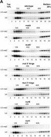
Effects of the U3 snoRNA mutations on the association with 80S particles. HEp-2 cells were transiently transfected with either wild-type or mutant U3 msl2 constructs. Cell extracts were prepared and separated by glycerol gradient centrifugation. (A) RNA was isolated from each fraction and analyzed by Northern blot hybridization by using either a probe specific for the StreptoTag or a probe specific for the U3 snoRNA. The RNA detected is indicated on the left of each panel. The transfected U3 msl2 construct is indicated above each panel. The peak positions of the 40S and 60S ribosomal subunits as well as of the 12S U1 snRNP, present in the gradient fractions, are indicated at the top of each panel. (B) Quantitative analysis of the effects of the mutations on 80S complex formation. Signal intensities obtained by phosphorimaging were used to calculate the ratio between the relative amount of U3 msl2 and endogenous U3 snoRNA associated with 80S complexes (vertical axis). The transfected construct is indicated on the horizontal axis. The average values derived from two independent experiments are depicted in the graph. Error bars indicate the variation between the two experiments.
FIG. 3.
FIG. 3.
Effects of the U3 snoRNA mutations on the association with 80S particles. HEp-2 cells were transiently transfected with either wild-type or mutant U3 msl2 constructs. Cell extracts were prepared and separated by glycerol gradient centrifugation. (A) RNA was isolated from each fraction and analyzed by Northern blot hybridization by using either a probe specific for the StreptoTag or a probe specific for the U3 snoRNA. The RNA detected is indicated on the left of each panel. The transfected U3 msl2 construct is indicated above each panel. The peak positions of the 40S and 60S ribosomal subunits as well as of the 12S U1 snRNP, present in the gradient fractions, are indicated at the top of each panel. (B) Quantitative analysis of the effects of the mutations on 80S complex formation. Signal intensities obtained by phosphorimaging were used to calculate the ratio between the relative amount of U3 msl2 and endogenous U3 snoRNA associated with 80S complexes (vertical axis). The transfected construct is indicated on the horizontal axis. The average values derived from two independent experiments are depicted in the graph. Error bars indicate the variation between the two experiments.
FIG. 4.
FIG. 4.
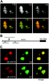
Subnucleolar localization of the U3 and U8 snoRNAs, snoRNP proteins, and pre-rRNAs. (A) Confocal microscopy was performed by using a stable HeLa cell line expressing ECFP-fibrillarin hybridized with oligonucleotides complementary to U3 and U8 snoRNAs. The localization of fibrillarin and the individual snoRNAs is shown in the upper panels. Pairwise confocal overlays of the three components are shown in the lower panels. The identities of the probes are indicated in each panel. The color of the label represents the pseudocolor of the corresponding image in each overlay. (B) Schematic representation of the 5′ ETS sequence showing the localization of the DS 5′ ETS and US 5′ ETS probes relative to the primary processing site. The mature 18S rRNA is shown in black. (C) Confocal microscopy analysis of HeLa cells hybridized with an oligonucleotide complementary to U3 and an oligonucleotide complementary to either the US 5′ ETS or DS 5′ ETS of the primary cleavage site in the 5′ ETS. Shown are the hybridization patterns of the 5′ ETS probes alone (left), U3 snoRNA localization (middle), and a confocal overlay of both (right).
FIG. 5.
FIG. 5.
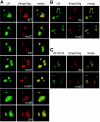
The box B/C motif and the 3′ hinge sequence are required for the correct subnuclear localization of the U3 snoRNA. (A) Confocal analysis of HeLa cells transfected with either wild-type or mutant U3 msl2 constructs and hybridized with oligonucleotides specific for the StreptoTag sequence and the U3 snoRNA. Shown are the localization of the total U3 snoRNA (left), the StreptoTag staining (middle), and a confocal overlay of both (right). The identity of the U3 msl2 construct is indicated in the middle panels. (B) Fluorescence microscopy of HeLa cells transfected with either mut 3′ hinge or mutC msl2 constructs. Cells were hybridized with oligonucleotides specific for the StreptoTag sequence and the U8 snoRNA. Shown are the localization of the U8 snoRNA (left), the StreptoTag U3 msl2 RNA (middle), and a confocal overlay of both (right). The identity of the U3 msl2 construct is indicated in the middle panel. (C) Fluorescence microscopy of HeLa cells transfected with either mut 3′ hinge or mutC msl2 constructs. Cells were hybridized with oligonucleotides specific for the StreptoTag sequence and the US 5′ETS. Shown are the localization of the US 5′ETS (left), the StreptoTag U3 msl2 RNA (middle), and a confocal overlay of both (right). The identity of the U3 msl2 construct is indicated in the middle panel. WT, wild-type U3 msl2; 3′H, U3 msl2 mut 3′ hinge; 5′H, U3 msl2 mut 5′ hinge.

Similar articles

Cited by

References

    1. Bachellerie, J. P., J. Cavaille, and A. Huttenhofer. 2002. The expanding snoRNA world. Biochimie 84:775-790. - PubMed
    1. Bachler, M., R. Schroeder, and U. von Ahsen. 1999. StreptoTag: a novel method for the isolation of RNA-binding proteins. RNA 5:1509-1516. - PMC - PubMed
    1. Beltrame, M., and D. Tollervey. 1995. Base pairing between U3 and the pre-ribosomal RNA is required for 18S rRNA synthesis. EMBO J. 14:4350-4356. - PMC - PubMed
    1. Beven, A. F., R. Lee, M. Razaz, D. J. Leader, J. W. Brown, and P. J. Shaw. 1996. The organization of ribosomal RNA processing correlates with the distribution of nucleolar snRNAs. J. Cell Sci. 109:1241-1251. - PubMed
    1. Billy, E., T. Wegierski, F. Nasr, and W. Filipowicz. 2000. Rcl1p, the yeast protein similar to the RNA 3′-phosphate cyclase, associates with U3 snoRNP and is required for 18S rRNA biogenesis. EMBO J. 19:2115-2126. - PMC - PubMed

Publication types

Substances

LinkOut - more resources