. 2004 Oct;24(19):8681-90.
doi: 10.1128/MCB.24.19.8681-8690.2004.

Forkhead box transcription factor FOXO3a regulates estrogen receptor alpha expression and is repressed by the Her-2/neu/phosphatidylinositol 3-kinase/Akt signaling pathway

Affiliations

Forkhead box transcription factor FOXO3a regulates estrogen receptor alpha expression and is repressed by the Her-2/neu/phosphatidylinositol 3-kinase/Akt signaling pathway

Shangqin Guo et al. Mol Cell Biol. 2004 Oct.

Abstract

The expression status of the estrogen receptor alpha (ERalpha) and that of the epidermal growth factor receptor Her-2/neu frequently correlate inversely in breast cancers. While ERalpha-dependent cancers respond to antiestrogen therapy, Her-2/neu-overexpressing cancers typically display resistance to antiestrogens and poor prognosis. In this report we have explored the mechanism linking the loss of expression of ERalpha in breast cancer cells with overexpression of Her-2/neu, which signals constitutively via a phosphatidylinositol 3-kinase (PI3K)/Akt kinase pathway. We identify for the first time the Forkhead box protein FOXO3a (formerly termed FKHRL-1), which is inactivated by Akt, as a key regulator of ERalpha gene transcription. In breast cancer cell lines, expression of ERalpha was correlated with active FOXO3a levels. Ectopic FOXO3a expression induced ERalpha protein levels and promoter activity, while a dominant negative FOXO3a decreased ERalpha levels. By using transient transfection, mobility shift assays, and site-directed mutagenesis, two major functional Forkhead binding sites were identified in the human ERalpha promoter B. A chromatin immunoprecipitation assay confirmed FOXO3a binding at these two sites. Ectopic FOXO3a induced estrogen response element-driven reporter activity and expression of ERalpha target genes. The constitutively activated myristylated Akt reduced ERalpha expression, whereas agents that negatively affect the PI3K/Akt pathway, i.e., wortmannin, celecoxib, and the green tea polyphenol epigallocatechin-3 gallate, induced ERalpha. Thus, FOXO3a represents an important intracellular mediator of ERalpha expression, suggesting possible therapeutic intervention strategies for Her-2/neu-overexpressing refractory breast tumors.


Figures

FIG. 1.
FIG. 1.
FOXO3a regulates ERα expression. (A) Samples (40 μg) of WCE or nuclear extracts (NE) from the indicated cell lines were analyzed by immunoblotting for FOXO3a (FOX), ERα, or β-actin (actin) as control for equal loading. (B) NF639 cells were transfected with 8 μg of pECE EV, WT FOXO3a, or A3 FOXO3a. Forty-eight hours after transfection, WCE were analyzed by immunoblotting for ERα, FOXO3a, and β-actin. (C) MCF-7 cells were infected with an increasing multiplicity of infection (MOI) of dnFOXO3a-expressing adenovirus. After 40 h, WCE were prepared and samples (40 μg) were analyzed by immunoblotting for FOXO3a and ERα. Equal loading was verified using β-actin immunoblotting and Ponceau S (Ponc.) staining.
FIG. 2.
FIG. 2.
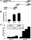
FOXO3a induces ERα promoter activity. (A) Schematic representation of the ER3500-210Luc and ERα promoter A and B reporter constructs. (B) NF639 cells were cotransfected, in triplicate, with 0.5 μg of ER3500-210Luc in the presence of 2 μg of pECE parental vector, WT, or A3 FOXO3a expression vector. Equal amounts of protein were assessed for luciferase activity. Data are presented as fold induction (± SD) relative to that for the parental pECE vector, which was set as 1, and are representative of three independent experiments. (C) NF639 cells were transfected, in triplicate, with 0.5 μg of ERα promoter A or B luciferase reporter vectors and 0, 0.5, 1.0, or 1.5 μg of A3 FOXO3a expression vector and enough pECE to make up a total of 2 μg of DNA. After 48 h, cells were harvested and luciferase activities were measured. Data are presented as fold induction (± SD) relative to that with the pECE EV, which was set as 1, and are representative of three independent experiments. (Inset) WCE used for the luciferase assays were subjected to immunoblot analysis for FOXO3a protein.
FIG. 3.
FIG. 3.
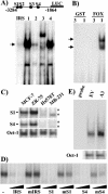
FOXO3a binding sites are present upstream of ERα promoter B. (A) Schematic representation of the four putative FOXO3a binding sites in ERα promoter B. Each individual binding site, as well as the IRS, was end labeled and used in an EMSA with 15 μg of nuclear extract from ZR-75 cells. The arrow indicates the position of the major band observed. (B) Oligonucleotides containing sites S1 (1) and S3 (3) were incubated with GST or GST-FOXO3a (FOX) protein and subjected to EMSA. (C) Samples (15 μg) of nuclear extracts from MCF-7 and ZR-75 ERα-positive cells and Hs578T and MDA-MB-231 (MB-231) ERα-negative breast cancer cell lines were subjected to EMSA using as probe the oligonucleotides containing either the S1 and S4 binding sites or the Oct-1 sequence, as loading control. −, position of the protein complex unique to ERα-positive breast cancer cells; *, positions of protein complexes present in ERα-negative cell extracts. (D) Samples (15 μg) of nuclear extracts from ZR-75 cells were incubated with labeled IRS probe in the absence or presence of a 10- or 50-fold molar excess of WT or mutant (m) forms of the IRS, S1, or S4 oligonucleotides. (E) NF639 cells were transfected with pECE EV or A3 FOXO3a. After 40 h, cells were harvested and extracts were analyzed by EMSA. (Lower panel) Oct-1 was used as control probe and confirmed the presence of similar amounts of protein in each extract.
FIG. 4.
FIG. 4.
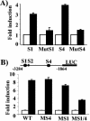
The S1 and S4 sites contain functional Forkhead elements. (A) NF639 cells were cotransfected, in triplicate, with 0.5 μg of S1-CAT, MutS1-CAT, S4-CAT, or MutS4-CAT construct DNA with 2 μg of either pECE or A3 FOXO3a and processed as described above. Values for A3 FOXO3a are presented as the fold induction (± SD) relative to that of the pECE-cotransfected sample, which was set as 1. Data presented are representative of three independent experiments. (B) (Upper panel) Scheme of the proB-Luc construct. S1 and S4 Forkhead binding sites are indicated by the large black circles, and S2 is indicated by a smaller circle. (Bottom panel) WT, MutS4 (MS4), MutS1 (MS1), or MutS1/4 (MS1/4) proB-Luc constructs were cotransfected (1 μg), in triplicate, into NF639 cells with 1 μg of A3 FOXO3a or pECE vector. After 48 h, cells were harvested and processed as for Fig. 2. Data are presented as fold induction (± SD) relative to samples cotransfected with parental pECE vector, which was set as 1, and are representative of three independent experiments.
FIG. 5.
FIG. 5.
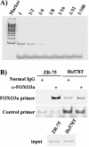
FOXO3a binds to the endogenous S1/S4 sites in the ERα promoter. (A) PCR conditions used to amplify the S1/S4 fragment. (B) ChIP was performed with either ERα-positive ZR-75 cells or ERα-negative Hs578T cells using FOXO3a antibody or normal rabbit IgG as control, as described in Materials and Methods. The FOXO3a primers are located in between the S1 and S4 sites, which are 550 bp from each other. The control primers, located at an irrelevant region, were used to show that a similar amount of DNA was present in each paired sample. (Lower panel) Input DNA before immunoprecipitation.
FIG. 6.
FIG. 6.
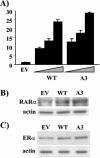
FOXO3a induces ERα activity. (A) NF639 cells were plated in 12-well plates and transfected with pECE and 0.1, 0.2, or 0.4 μg of WT or A3 FOXO3a, together with 0.2 μg of TK-ERE luciferase vector. After 30 h, cells were harvested and analyzed for luciferase activity. Data are presented as fold induction (± SD) relative to samples cotransfected with parental pECE vector, which was set as 1, and are representative of three independent experiments. (B and C) NF639 cells were transfected with the indicated expression vectors, and after 30 h whole-cell or nuclear extracts were prepared. Samples of WCE (50 μg) were analyzed by immunoblotting for RARα (B), and nuclear extracts (15 μg) were analyzed for ERα (C). Analysis of β-actin levels confirmed equal loading.
FIG. 7.
FIG. 7.
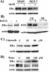
ERα expression is subjected to regulation by Akt kinase activity. (A) NF639 and MCF-7 cells were transfected with a vector expressing AktM, the constitutively active version of Akt. WCE were prepared after 48 h and subjected to immunoblot analysis for ERα and β-actin, as above. (B) NF639 cells were treated with 60 μg of EGCG/ml or the carrier solution, dimethyl sulfoxide, for 24 h, and WCE were analyzed by immunoblotting for ERα and β-actin (left panel) or cytoplasmic (CE) and nuclear (NE) proteins for FOXO3a and β-actin (right panel). (C) NF639 cells were treated with 25 or 50 μM celecoxib for 7 h, and WCE were analyzed by immunoblotting for ERα and β-actin. (D) NF639 cells were treated with 100 or 130 nM wortmannin for 7 h, and WCE were analyzed by immunoblotting for ERα, FOXO3a, and β-actin.

Similar articles

Cited by

References

    1. Biswas, D. K., A. P. Cruz, E. Gansberger, and A. B. Pardee. 2000. Epidermal growth factor-induced nuclear factor kappa B activation: a major pathway of cell-cycle progression in estrogen-receptor negative breast cancer cells. Proc. Natl. Acad. Sci. USA 97:8542-8547. - PMC - PubMed
    1. Bois, P. R., and G. C. Grosveld. 2003. FKHR (FOXO1a) is required for myotube fusion of primary mouse myoblasts. EMBO J. 22:1147-1157. - PMC - PubMed
    1. Brunet, A., A. Bonni, M. J. Zigmond, M. Z. Lin, P. Juo, L. S. Hu, M. J. Anderson, K. C. Arden, J. Blenis, and M. E. Greenberg. 1999. Akt promotes cell survival by phosphorylating and inhibiting a Forkhead transcription factor. Cell 96:857-868. - PubMed
    1. Brunet, A., G. Pages, and J. Pouyssegur. 1994. Constitutively active mutants of MAP kinase kinase (MEK1) induce growth factor-relaxation and oncogenicity when expressed in fibroblasts. Oncogene 9:3379-3387. - PubMed
    1. Burgess, A. W., H. S. Cho, C. Eigenbrot, K. M. Ferguson, T. P. Garrett, D. J. Leahy, M. A. Lemmon, M. X. Sliwkowski, C. W. Ward, and S. Yokoyama. 2003. An open-and-shut case? Recent insights into the activation of EGF/ErbB receptors. Mol. Cell 12:541-552. - PubMed

Publication types

MeSH terms