. 2004 Oct;24(19):8753-64.
doi: 10.1128/MCB.24.19.8753-8764.2004.

Activity of the Yap1 transcription factor in Saccharomyces cerevisiae is modulated by methylglyoxal, a metabolite derived from glycolysis

Affiliations

Activity of the Yap1 transcription factor in Saccharomyces cerevisiae is modulated by methylglyoxal, a metabolite derived from glycolysis

Kazuhiro Maeta et al. Mol Cell Biol. 2004 Oct.

Abstract

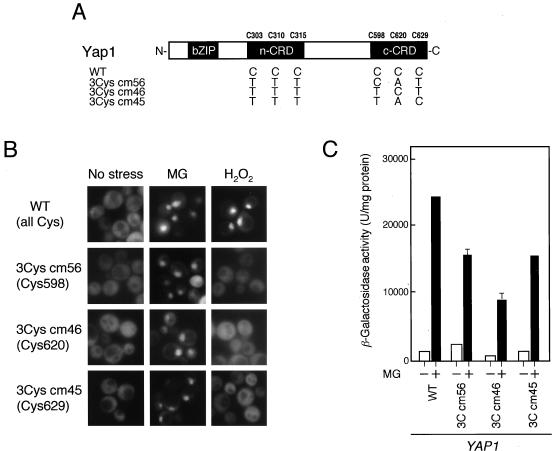
Methylglyoxal (MG) is synthesized during glycolysis, although it inhibits cell growth in all types of organisms. Hence, it has long been asked why such a toxic metabolite is synthesized in vivo. Glyoxalase I is a major enzyme detoxifying MG. Here we show that the Yap1 transcription factor, which is critical for the oxidative-stress response in Saccharomyces cerevisiae, is constitutively concentrated in the nucleus and activates the expression of its target genes in a glyoxalase I-deficient mutant. Yap1 contains six cysteine residues in two cysteine-rich domains (CRDs), i.e., three cysteine residues clustering near the N terminus (n-CRD) and the remaining three cysteine residues near the C terminus (c-CRD). We reveal that any of the three cysteine residues in the c-CRD is sufficient for MG to allow Yap1 to translocate into the nucleus and to activate the expression of its target gene. A Yap1 mutant possessing only one cysteine residue in the c-CRD but no cysteine in the n-CRD and deletion of the basic leucine zipper domain can concentrate in the nucleus with MG treatment. However, substitution of all the cysteine residues in Yap1 abolishes the ability of this transcription factor to concentrate in the nucleus following MG treatment. The redox status of Yap1 is substantially unchanged, and protein(s) interaction with Yap1 through disulfide bond is hardly detected in cells treated with MG. Collectively, neither intermolecular nor intramolecular disulfide bond formation seems to be involved in Yap1 activation by MG. Moreover, we show that nucleocytoplasmic localization of Yap1 closely correlates with growth phase and intracellular MG level. We propose a novel regulatory pathway underlying Yap1 activation by a natural metabolite in the cell.


Figures

FIG. 1.
FIG. 1.
Effect of MG on activation of Yap1. (A) Cells of yap1Δ and yap1Δ glo1Δ carrying the GFP-Yap1 plasmid were cultured in SD medium to an A610 of 0.5, and localization of GFP-Yap1 was observed. (B) (Left panel) Cells of the wild type (WT) carrying the AP-1-CYC1-lacZ reporter plasmid were cultured in SD medium to an A610 of 0.5, and 8 mM MG was added. After a 3-h incubation, β-galactosidase activity was measured. (Right panel) For anaerobic culture, after cells of the wild type and the glo1Δ mutant with reporter gene were inoculated into SD medium, nitrogen gas was flushed through the flask, which was then sealed, and the cells were cultured without shaking at 28°C. (C) GFP-tagged Yap1 was expressed in each mutant. All mutants have a yap1Δ background. (D) GFP-Yap1 was expressed in the yap1Δ and yap1Δ gpx3Δ mutants. Cells were cultured in SD medium to an A610 of 0.5, and 8 mM MG or 0.4 mM H2O2 was added. Localization of GFP-Yap1 was monitored for 60 min (for MG) or 15 min (for H2O2) after the addition of each chemical. (E) Cells were cultured in YPD medium to an A610 of 0.5, diluted with 0.85% NaCl, and spotted onto YPD agar plates with or without 15 mM MG. The cells were cultured at 28°C for 2 days.
FIG. 2.
FIG. 2.
The function of the Yap1 nuclear export machinery and the Yap1 protein level are normal in the glo1Δ mutant. (A) Temperature-sensitive xpo1-1 cells carrying pKW430 (NLSSV40-NESPKI-2×GFP) were cultured at 28°C (permissive temperature) to an A610 of 0.5 and shifted to 37°C (nonpermissive temperature). After 15 min, the localization of NLSSV40-NESPKI-2×GFP was monitored. Cells of the wild type (WT) and the glo1Δ mutant carrying pKW430 were cultured in SD medium to an A610 of 0.5, and localization of NLSSV40-NESPKI-2×GFP was observed. To determine the effect of exogenously added MG on the function of Crm1, wild-type cells carrying pKW430 were treated with 8 mM MG. No change in the localization of NLSSV40-NESPKI-2×GFP was seen at least up to 120 min after the addition of MG. The pictures shown in the figure were taken 60 min after the addition of MG. (B) To compare the steady-state level of Yap1 protein, cells of the yap1Δ (lane 1) and yap1Δ glo1Δ (lane 2) mutants carrying pRS315-Yap1-9Myc were cultured in SD medium to an A610 of 1.0, and cell extracts were prepared. To determine the effect of MG on the Yap1 protein level, yap1Δ cells carrying pRS315-Yap1-9Myc were cultured in SD medium to an A610 of 0.5. After the addition of 8 mM MG, the cells were incubated at 28°C and collected periodically. Lane 3, without MG for 1 h; lane 4, with 8 mM MG for 1 h. We confirmed that the Yap1 protein level did not change at least up to 3 h after MG treatment.
FIG. 3.
FIG. 3.
Yap1 activation by MG is reversible. GFP-Yap1 was expressed in the yap1Δ mutant and cultured in SD medium to an A610 of 0.5. At this point, the localization of GFP-Yap1 was monitored (time, 0 min), 8 mM MG was added to the culture medium, and the cells were incubated to monitor the localization of GFP-Yap1 periodically (time, 5 to 60 min). After 60 min, cells were collected by centrifugation, washed with 0.85% NaCl, and resuspended in fresh SD medium, and localization of GFP-Yap1 was monitored periodically (time, 1 to 5 min). The cellular MG content was measured when the A610 had reached 0.5, and 8 mM MG was added. The MG level was monitored after the addition of MG (30 and 60 min) and also after the removal of MG (5 min).
FIG. 4.
FIG. 4.
Importance of Cys residues in Yap1 for activation by MG. (A) Schematic of the Yap1 mutant protein. (B) GFP-tagged Yap1 of the wild type (WT) and each mutant was expressed in the yap1Δ and yap1Δ glo1Δ mutants. Cells were cultured in SD medium to an A610 of 0.5, and localization of GFP-Yap1 in yap1Δ glo1Δ cells was monitored. For yap1Δ cells, 8 mM MG or 0.4 mM H2O2 was added to the culture medium, the cells were incubated for a further 60 min (MG) or 15 min (H2O2), and localization of GFP-Yap1 was monitored. (C) Each GFP-tagged protein was also expressed in the yap1Δ mutant carrying the FLR1-lacZ reporter gene. After treatment with 8 mM MG for 3 h, the β-galactosidase activity was measured.
FIG. 5.
FIG. 5.
Importance of c-CRD for the nuclear localization of Yap1 following MG treatment. (A) Schematic of Yap1 and the Gal4-GFP-c-CRD reporter protein. (B) The Gal4-GFP-c-CRD reporter gene was expressed in wild-type (WT) and glo1Δ cells. Cells were cultured in SD medium to an A610 of 0.5, and localization of the reporter protein was monitored. Then 8 mM MG was added, and the cells were incubated for 60 min, at which time the localization of the reporter protein was again determined.
FIG. 6.
FIG. 6.
Any one of three Cys residues in the c-CRD is sufficient for Yap1 activation by MG. (A) Schematic of the Yap1 mutant protein. (B) GFP-tagged Yap1 of the wild type (WT) and each mutant was expressed in yap1Δ cells. When the A610 of the culture reached 0.5, 8 mM MG or 0.4 mM H2O2 was added to the culture medium, the cells were incubated for a further 60 min (MG) or 15 min (H2O2), and localization of GFP-Yap1 was then monitored. (C) Each GFP-tagged protein was also expressed in the yap1Δ mutant carrying the FLR1-lacZ reporter gene. After treatment with 8 mM MG for 3 h, the β-galactosidase activity was measured.
FIG. 7.
FIG. 7.
No disulfide formation is involved in Yap1 activation by MG. (A) Schematic of the Yap1 mutant protein. (B) GFP-tagged Yap1 of the wild type (WT) and each mutant was expressed in yap1Δ cells. When the A610 of the culture reached 0.5, 8 mM MG or 0.4 mM H2O2 was added to the culture medium, cells were incubated for a further 60 min (MG) or 15 min (H2O2), and localization of GFP-Yap1 was then monitored.
FIG. 8.
FIG. 8.
Analysis of biochemical changes in Yap1. yap1Δ cells carrying HA-tagged Yap1 were cultured in SD medium to an A610 of 1.0 and then treated with 8 mM MG for 60 min or 0.4 mM H2O2 for 15 min. Cellular proteins were extracted as described in the text and subjected to nonreducing SDS-PAGE followed by Western blotting with anti-HA monoclonal antibody to detect HA-Yap1. The immunoreactive protein was detected using the ECL kit (Amersham Biosciences). The low-mobility band that appeared in H2O2-treated samples was assumed to be a Yap1-Gpx3 complex.
FIG. 9.
FIG. 9.
Correlation between cell growth, cellular MG level, and Yap1 localization. Cells were cultured in SD minimal medium, and cell growth (A610), glucose concentration in the medium, β-galactosidase activity driven by GLO1-lacZ, intracellular MG content, and subcellular localization of GFP-Yap1 were monitored periodically. The glucose concentration was determined using a kit (Glucose B-test Wako; Wako Pure Chemical Industries, Tokyo, Japan). The number of cells counted in each observation for localization of GFP-Yap1 was 500 to 1,000.

Similar articles

Cited by

References

    1. Abdulnur, S. F. 1976. The interactions of glyoxals with proteins and DNA in relation to cancer. Int. J. Quant. Chem. Quant. Biol. Symp. 3:59-64.
    1. Azevedo, D., F. Tacnet, A. Delaunay, C. Rodrigues-Pousada, and M. B. Toledano. 2003. Two redox centers within Yap1 for H2O2 and thiol-reactive chemicals signaling. Free Radic. Biol. Med. 35:889-900. - PubMed
    1. Bito, A., M. Haider, I. Hadler, and M. Breitenbach. 1997. Identification and phenotypic analysis of two glyoxalase II encoding genes from Saccharomyces cerevisiae, GLO2 and GLO4, and intracellular localization of the corresponding proteins. J. Biol. Chem. 272:21509-21519. - PubMed
    1. Bradford, M. M. 1976. A rapid and sensitive method for the quantitation of microgram quantities of protein utilizing the principle of protein-dye binding. Anal. Biochem. 72:248-254. - PubMed
    1. Carmel-Harel, O., R. Stearman, A. P. Gasch, D. Botstein, P. O. Brown, and G. Storz. 2001. Role of thioredoxin reductase in the Yap1-dependent response to oxidative stress in Saccharomyces cerevisiae. Mol. Microbiol. 39:595-605. - PubMed

Publication types

MeSH terms

LinkOut - more resources