. 2004 Sep 15;18(18):2225-30.
doi: 10.1101/gad.317604. Epub 2004 Sep 1.

Essential role of BCL9-2 in the switch between beta-catenin's adhesive and transcriptional functions

Affiliations

Essential role of BCL9-2 in the switch between beta-catenin's adhesive and transcriptional functions

Felix H Brembeck et al. Genes Dev. .

Abstract

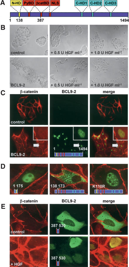
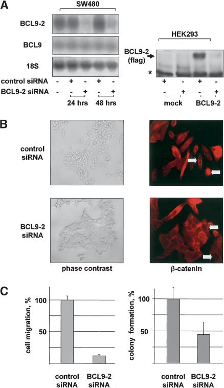
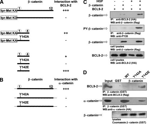
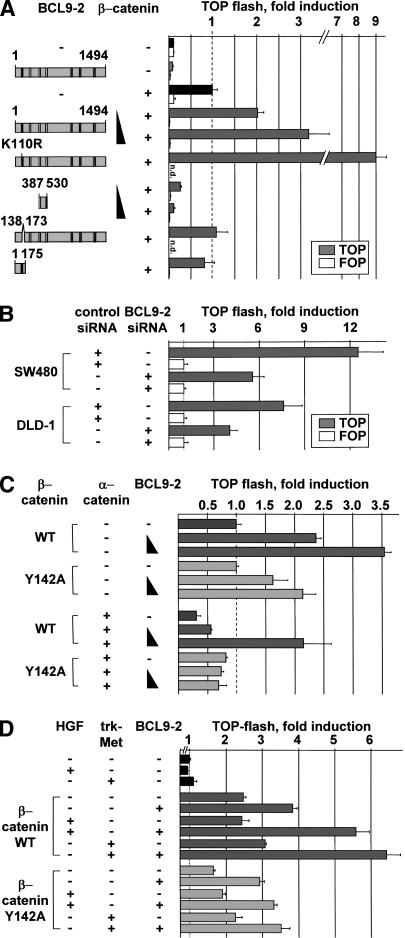
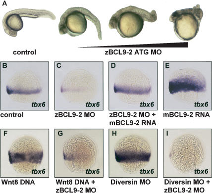
beta-Catenin controls both cadherin-mediated cell adhesion and activation of Wnt target genes. We demonstrate here that the beta-catenin-binding protein BCL9-2, a homolog of the human proto-oncogene product BCL9, induces epithelial-mesenchymal transitions of nontransformed cells and increases beta-catenin-dependent transcription. RNA interference of BCL9-2 in carcinoma cells induces an epithelial phenotype and translocates beta-catenin from the nucleus to the cell membrane. The switch between beta-catenin's adhesive and transcriptional functions is modulated by phosphorylation of Tyr 142 of beta-catenin, which favors BCL9-2 binding and precludes interaction with alpha-catenin. During zebrafish embryogenesis, BCL9-2 acts in the Wnt8-signaling pathway and regulates mesoderm patterning.


Figures

Figure 1.
Figure 1.
BCL9-2 induces epithelial-mesenchymal transition and nuclear translocation of β-catenin in MDCK cells. (A) Domain structure of the vertebrate BCL9-2 protein. The seven conserved domains are highlighted; positions of amino acids are indicated. (B) Morphology of BCL9-2-transfected MDCK cells and controls. HGF treatment was for 18 h. (C) Immunofluorescence microscopy of MDCK cells for endogenous β-catenin (red), transfected BCL9-2 (green), and merged fluorescence (yellow). Arrows mark β-catenin collocation with BCL9-2 (see also inset). (D) Cellular localization of deletions and point mutants of BCL9-2. (E) Localization of the isolated β-catenin-binding fragment of BCL9-2 (amino acids 387-530) in MDCK cells. Treatment with 2 U mL-1 HGF was for 6 h. The used BCL9-2 constructs are schematically indicated in the figures.
Figure 2.
Figure 2.
RNA interference of BCL9-2 in transformed cells reverts cells to the epithelial phenotype, translocates β-catenin from the nucleus to the plasma membrane, and reverts transformed properties. (A, left) Northern blot of SW480 cells that were transfected with BCL9-2 siRNAs or controls for 24 and 48 h. The used probes are indicated on the left. (Right) Western blot of nuclear extracts of HEK293 cells that were stably transfected with BCL9-2 or control vector, and treated for 96 h with BCL9-2 siRNAs. (B) Morphology and immunofluorescence of endogenous β-catenin of SW480 cells that were transfected with BCL9-2 siRNAs or controls for 72 h. (C, left) Cell migration of SW480 cells that were treated with BCL9-2 siRNAs or controls for 72 h. The number of migrating cells (from three independent transfections) is expressed as percent of control cells. (Right) Colony formation of DLD-1 cells that were treated with BCL9-2 siRNAs or controls for 72 h. Colonies in soft agar (from three independent transfections) were counted after 10 d and expressed as percent of controls.
Figure 3.
Figure 3.
Phosphorylation of Tyr 142 of β-catenin mediates complex formation with BCL9-2 and reduces α-catenin binding. (A) Interaction of tpr-Met-β-catenin fusion proteins, of β-catenin repeats 1-4, and Y142 mutations thereof with BCL9-2 (amino acids 387-530), as determined by yeast two-hybrid assays. Arm repeats of β-catenin are depicted by numbers. (KA) kinase-active; (KD) kinase-defective fusion proteins. (B) Interaction of β-catenin and Y142 mutations thereof with α-catenin. Note that constructs without tpr-met were in the prey, BCL9-2 in the bait vectors. Interactions (+ or -) were quantified by growth of yeast on selective medium and by β-galactosidase activity in solution. (C) Interaction of BCL9-2 and tyrosine-phosphorylated β-catenin in COS-7 cells in response to HGF treatment (5 U mL-1 for 18 h), as determined by coimmunoprecipitation. BCL9-2 (amino acids 387-530) was cotransfected with full-length β-catenin. (D) Interaction of BCL9-2 and α-catenin with wild-type and Y142 mutant β-catenin, as determined by GST-β-catenin pull-down experiments. BCL9-2 (amino acids 387-530) and full size α-catenin were prepared from transfected COS-7 cells.
Figure 4.
Figure 4.
BCL9-2 is an essential nuclear coactivator of β-catenin signaling. (A) Transcriptional activation of β-catenin and BCL9-2 in HEK293 cells. Deletions and mutants of BCL9-2 are shown schematically on the left. A total of 0.5 μg of S33A β-catenin and 1.0-3.0 μg of BCL9-2 were cotransfected with the TOP (gray bars) or the control FOP reporter (open bars). (B) Transcriptional activation of β-catenin in SW480 and DLD-1 colon carcinoma cells that were treated with BCL9-2 siRNAs or controls for 48 and 72 h. Transfection with the reporters was for another 24 h. (C) Transcriptional activation in HEK293 cells of wild-type or Y142A mutant β-catenin, and effects of cotransfected BCL9-2 and α-catenin; 0.5 μg β-catenin, 1.0 μg α-catenin, and 0.25-1.0 μg BCL9-2 were transfected. (D) Transcriptional activation of β-catenin and the Y142A mutant in response to HGF treatment (50 U mL-1 for 18 h) or by cotransfected trk-Met (0.15 μg); 0.25 μg β-catenin and 3.0 μg BCL9-2 were transfected. HEK293 cells were cultured at reduced serum concentrations (50% DMEM and 50% OptimemI, Invitrogen).
Figure 5.
Figure 5.
BCL9-2 in zebrafish embryos acts downstream in the Wnt8/β-catenin pathway to pattern the ventro-lateral mesoderm. (A) Malformations of trunk and tail in embryos treated with BCL9-2 ATG MOs at 26 h post-fertilization. Dependence on the amount of injected MOs. (B-E) Effects of BCL9-2 MOs on the expression of the ventro-lateral mesoderm marker tbx6 at 70% epiboly. BCL9-2 MOs (0.03 pmole; C), BCL9-2 MOs in combination with mouse BCL9-2 RNA (1.5 ng; D), and mBCL9-2 RNA alone (E). Control is in B. (F-I) Epistasis of the action of Wnt8, Diversin, and BCL9-2, as examined by expression of tbx6. Wnt8 DNA (0.06 ng; F), Diversin MOs (0.6 pmole; H), and in combination with BCL9-2 MOs (G,I). Lateral view, anterior up, dorsal to the right.

References

    1. Aberle H., Schwartz, H., Hoschuetzky, H., and Kemler, R. 1996. Single amino acid substitutions in proteins of the armadillo gene family abolish their binding to α-catenin. J. Biol. Chem. 271: 1520-1526. - PubMed
    1. Behrens J., Vakaet, L., Friis, R., Winterhager, E., Van Roy, F., Mareel, M.M., and Birchmeier, W. 1993. Loss of epithelial differentiation and gain of invasiveness correlates with tyrosine phosphorylation of the E-cadherin/β-catenin complex in cells transformed with a temperature-sensitive v-SRC gene. J. Cell Biol. 120: 757-766. - PMC - PubMed
    1. Behrens J., von Kries, J.P., Kuhl, M., Bruhn, L., Wedlich, D., Grosschedl, R., and Birchmeier, W. 1996. Functional interaction of β-catenin with the transcription factor LEF-1. Nature 382: 638-642. - PubMed
    1. Behrens J., Jerchow, B.A., Wurtele, M., Grimm, J., Asbrand, C., Wirtz, R., Kuhl, M., Wedlich, D., and Birchmeier, W. 1998. Functional interaction of an axin homolog, conductin, with β-catenin, APC, and GSK3β. Science 280: 596-599. - PubMed
    1. Bienz M. and Clevers, H. 2000. Linking colorectal cancer to Wnt signaling. Cell 103: 311-320. - PubMed

Publication types

MeSH terms