. 2004 Sep 15;18(18):2302-13.
doi: 10.1101/gad.1230804.

Connecting two-component regulatory systems by a protein that protects a response regulator from dephosphorylation by its cognate sensor

Affiliations

Connecting two-component regulatory systems by a protein that protects a response regulator from dephosphorylation by its cognate sensor

Akinori Kato et al. Genes Dev. .

Abstract

A fundamental question in signal transduction is how an organism integrates multiple signals into a cellular response. Here we report the mechanism by which the Salmonella PmrA/PmrB two-component system responds to the signal controlling the PhoP/PhoQ two-component system. We establish that the PhoP-activated PmrD protein binds to the phosphorylated form of the response regulator PmrA, preventing both its intrinsic dephosphorylation and that promoted by its cognate sensor kinase PmrB. This results in PmrA-mediated transcription because phosphorylated PmrA exhibits higher affinity for its target promoters than unphosphorylated PmrA. A PmrD-independent form of the PmrA protein was resistant to PmrB-catalyzed dephosphorylation and promoted transcription of PmrA-activated genes in the absence of inducing signals. This is the first example of a protein that enables a two-component system to respond to the signal governing a different two-component system by protecting the phosphorylated form of a response regulator.


Figures

Figure 1.
Figure 1.
Model illustrating the regulatory interactions between the PhoP/PhoQ and PmrA/PmrB two-component systems and the PmrD protein. The PmrA protein promotes transcription of the pbgP gene during growth in low Mg2+ via the PhoP/PhoQ system, which turns on expression of the PhoP-activated PmrD protein, which, in turn, activates the PmrA/PmrB system at a posttranslational level. The PmrB protein responds to Fe3+ by activating the PmrA protein independently of the PhoP/PhoQ system and PmrD protein, which promotes transcription of the pbgP gene and represses transcription of the pmrD gene.
Figure 2.
Figure 2.
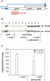
The PmrD protein activates the PmrA/PmrB system at a posttranslational level. (A) Genetic organization of the pmrCAB operon. The blue arrow indicates transcription from a promoter that is positively autoregulated by the PmrA/PmrB system; the black arrow denotes a constitutive promoter that produces basal levels of PmrA and PmrB proteins even under noninducing conditions. The red box indicates the location of the MudJ transposon insertion in strains EG9460 and EG11785. (B) Western blot analysis of extracts prepared from wild type (14028s), pmrD (EG11491), pmrC::MudJ (EG9460), pmrC::MudJ pmrD (EG11785), pmrC::MudJ (EG9460) harboring pUH-pmrAB, and pmrC::MudJ pmrD (EG11785) harboring pUH-pmrAB strains grown in N-minimal medium at pH 7.7 with 10 mM Mg2+ (H) or 10 μM Mg2+ (L) detected with anti-PmrBc (upper panel) or anti-PmrA (lower panel) antibodies. (C) β-galactosidase activity (Miller units) from a pmrC-lac transcriptional fusion expressed by bacteria grown in N-minimal medium at pH 7.7 with 10 mM Mg2+ (solid bar) or 10 μM Mg2+ (open bar) was determined in pmrC::MudJ (EG9460), pmrC::MudJ pmrD (EG11785), pmrC::MudJ (EG9460) harboring pUH-pmrAB, and pmrC::MudJ pmrD (EG11785) harboring pUH-pmrAB strains. The data correspond to mean values of three independent experiments performed in duplicate. Error bars correspond to the standard deviation (and are only shown if greater than the resolution of the figure).
Figure 3.
Figure 3.
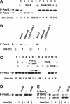
The PmrD protein specifically promotes the phosphorylated state of the PmrA protein by inhibiting dephosphorylation of phospho-PmrA catalyzed by the PmrB protein. (A) Phosphotransfer assay from phospho-PmrBc to the PmrA and PmrAD51A proteins was performed with 5 μM of regulator (PmrA and PmrAD51A) and 2.5 μM of PmrBc proteins. (B) Kinase/phosphatase assay of the PmrBc and PmrA proteins was performed in the presence (PmrD) or absence (-) of the PmrD protein. The concentrations of the PmrA, PmrBc, and PmrD proteins were 5, 2.5, and 5 μM, respectively. The small cationic proteins RNase A and cytochrome C were used as negative controls at a final concentration of 5 μM. (C) Phosphatase assay of phospho-PmrA protein was performed as follows: 32P-labeled phosphorylated GST-PmrBcT156R beads were used as phosphodonor for the PmrA protein and removed before the phosphatase assay. The concentrations of the PmrA, histidine kinase (i.e., PmrBc, PmrBcT156R), and PmrD proteins were 2.5, 5, and 2.5 μM, respectively. The top band corresponds to phospho-PmrBcT156R or phospho-PmrBc due to reverse-phosphorylation from phospho-PmrA. (D) Autokinase assay of the PmrBc protein was performed in the presence (PmrD) or absence (-) of the PmrD protein. The concentrations of the PmrBc and PmrD proteins were 2.5 and 5 μM, respectively. (E) Kinase/phosphatase assay of the PmrBcT156R and PmrA proteins was performed in the presence (PmrD) or absence (-) of the PmrD protein. The concentrations of the PmrA, PmrBcT156R, and PmrD proteins were 5, 2.5, and 5 μM, respectively. Samples were analyzed by 10% SDS-PAGE.
Figure 4.
Figure 4.
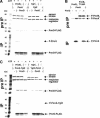
The PmrD protein interacts with the N-terminal domain of phospho-PmrA in a phosphorylation-dependent manner. (A) Immunoprecipitation of PmrD-Flag protein complexed with phospho-PmrA. Regulator (PmrA and YgiX), sensor (PmrBc and YgiYc), and PmrD-Flag proteins were incubated with ATP for 16 h before immunoprecipitation (pre-IP). The PmrD-Flag protein was precipitated from the phosphorylation reaction mixture using anti-Flag beads (IP). (B) Immunoprecipitation of the PmrD-Flag protein complexed with phospho-PmrA after phosphorylation with [γ-32P] ATP. PmrA, sensor (PmrBc and PmrBcT156R), and PmrD-Flag proteins were incubated with [γ-32P] ATP for 4 h before immunoprecipitation (pre-IP). PmrD-Flag protein was precipitated from the phosphorylation reaction mixture using anti-Flag beads (IP). Coimmunoprecipitated PmrA protein retains phosphoryl group. (C) Immunoprecipitation of the PmrD-Flag protein complexed with phospho-PmrA-YgiX. Chimeric regulator (PmrA-YgiX and YgiX-PmrA), sensor (PmrBc and YgiYc), and PmrD-Flag proteins were incubated with ATP for 16 h before immunoprecipitation (pre-IP). The PmrD-Flag protein was precipitated from the phosphorylation reaction mixture using anti-Flag beads (IP).
Figure 5.
Figure 5.
The PmrD protein specifically inhibits dephosphorylation of phospho-PmrA by targeting its N-terminal region. (A) Spontaneous dephosphorylation of phospho-PmrA protein in the presence (PmrD) or absence (-) of the PmrD protein: 32P-labeled phosphorylated GST-PmrBcT156R beads were used as phosphodonor for the PmrA protein and removed before the assay. The concentrations of the PmrA and PmrD proteins were 2.5 μM. (B) Spontaneous dephosphorylation of phospho-YgiX protein was performed in the presence (PmrD) or absence (-) of the PmrD protein. 32P-labeled phosphorylated GST-YgiYc beads were incubated with the YgiX protein for 1 h to produce phospho-YgiX protein and removed before the assay. The concentrations of the YgiX and PmrD proteins were 2.5 μM. (C) Spontaneous dephosphorylation of phospho-PmrA protein was performed in the presence (PmrD) or absence (-) of PmrD protein. All proteins used in this assay were not in contact with PmrB or derivatives, because they were independently overexpressed in E. coli strain EG13796, which lacks the entire pmrAB homolog basRS, and purified as described in Materials and Methods. 32P-labeled phosphorylated GST-YgiYc beads were used as phosphodonor for the PmrA protein and removed before the assay. The concentration of the PmrA and PmrD proteins was 2.5 μM. (D) Quantitative analysis of the phosphatase assay of phospho-PmrA protein. 32P-labeled phosphorylated GST-YgiYc beads were used as phosphodonor for the PmrA protein and removed before the assay. The concentrations of the PmrA, PmrBc, and PmrD proteins were 2.5, 5, and 2.5 μM, respectively. The small cationic proteins RNase A and cytochrome C were used as negative controls at a final concentration of 2.5 μM. (E) Quantitative analysis of the phosphatase assay of phospho-YgiX protein. 32P-labeled phosphorylated GST-PmrBcT156R beads were incubated with the YgiX protein for 45 min to produce phospho-YgiX protein and removed before the assay. The concentrations of YgiX, YgiYc, and PmrD proteins were 2.5, 5, and 2.5 μM, respectively. (F) Quantitative analysis of the phosphatase assay of phospho-PmrA-YgiX protein. 32P-labeled phosphorylated GST-YgiYc beads were incubated with the PmrA-YgiX protein for 2 h to produce phospho-PmrA-YgiX protein and removed before the assay. The concentrations of the PmrA-YgiX, PmrBc, and PmrD proteins were 2.5, 5, and 2.5 μM, respectively. (G) Quantitative analysis of the phosphatase assay of phospho-YgiX-PmrA protein. 32P-labeled phosphorylated GST-PmrBcT156R beads were incubated with the YgiX-PmrA protein for 2 h to produce phospho-YgiX-PmrA protein and removed before the assay. The concentrations of the YgiX-PmrA, YgiYc, and PmrD proteins were 2.5, 5 and 2.5 μM, respectively. Samples were analyzed by 10% SDS-PAGE and quantified as described in Materials and Methods.
Figure 6.
Figure 6.
Transcription of PmrA-activated genes is rendered pmrD-independent upon inactivation of the phosphatase activity of the PmrB protein or in a PmrA mutant protein resistant to PmrBc's phosphatase activity. (A) β-galactosidase activity (Miller units) from a pmrC-lac transcriptional fusion expressed by bacteria grown in N-minimal medium at pH 7.7 with 10 mM Mg2+ (solid bars), 10 μM Mg2+ (hatched bars), or 10 μM Mg2+ and 100 μM Fe2+ (open bars) was determined in a pmrB strain (EG14087) harboring pCCR9 (vector), pCCR9-pmrB (ppmrB), or pCCR9-pmrBT156R2 (ppmrBT156R). (B) β-galactosidase activity (Miller units) from a pmrC-lac transcriptional fusion expressed by bacteria grown in N-minimal medium at pH 7.7 with 10 mM Mg2+ (solid bars), 10 μM Mg2+ (hatched bars), or 10 μM Mg2+ and 100 μM Fe2+ (opened bars) was determined in the pmrB pmrD strain EG14090 harboring pCCR9 (vector), pCCR9-pmrB (ppmrB), or pCCR9-pmrBT156R2 (ppmrBT156R). (C) β-galactosidase activity (Miller units) from a pbgP-lac transcriptional fusion expressed by bacteria grown in N-minimal medium at pH 7.7 with 10 mM Mg2+ (solid bars) or 10 μM Mg2+ (opened bars) was determined in wild-type (EG9888), pmrB (EG13735), pmrA505 (EG9868), or pmrA505 and pmrB (EG13641) strains. The data correspond to mean values of three independent experiments performed in duplicate. Error bars show standar deviation (and are only shown if greater than the resolution of the figure). (D) Quantitative analysis in the phosphatase assay of phospho-PmrA and phospho-PmrA505 proteins was performed as follows: 32P-labeled phosphorylated GST-PmrBcT156R beads were used as phosphodonor for the PmrA and PmrA505 proteins and removed before the assay. The concentrations of the PmrA, PmrA505, PmrBc, and PmrD proteins were 2.5, 2.5, 5, and 2.5 μM, respectively. Samples were analyzed by 10% SDS-PAGE.

Similar articles

Cited by

References

    1. Atkinson M.R., Kamberov, E.S., Weiss, R.L., and Ninfa, A.J. 1994. Reversible uridylylation of the Escherichia coli PII signal transduction protein regulates its ability to stimulate the dephosphorylation of the transcription factor nitrogen regulator I (NRI or NtrC). J. Biol. Chem. 269: 28288-28293. - PubMed
    1. Bijlsma J.J. and Groisman, E.A. 2003. Making informed decisions: Regulatory interactions between two-component systems. Trends Microbiol. 11: 359-366. - PubMed
    1. Burkholder W.F., Kurtser, I., and Grossman, A.D. 2001. Replication initiation proteins regulate a developmental checkpoint in Bacillus subtilis. Cell 104: 269-279. - PubMed
    1. Cherepanov P.P. and Wackernagel, W. 1995. Gene disruption in Escherichia coli: TcR and KmR cassettes with the option of Flp-catalyzed excision of the antibiotic-resistance determinant. Gene 158: 9-14. - PubMed
    1. Datsenko K.A. and Wanner, B.L. 2000. One-step inactivation of chromosomal genes in Escherichia coli K-12 using PCR products. Proc. Natl. Acad. Sci. 97: 6640-6645. - PMC - PubMed

Publication types