. 2004 Dec;168(4):1951-62.
doi: 10.1534/genetics.104.031781. Epub 2004 Sep 15.

The Caenorhabditis elegans Ror RTK CAM-1 inhibits EGL-20/Wnt signaling in cell migration

Affiliations

The Caenorhabditis elegans Ror RTK CAM-1 inhibits EGL-20/Wnt signaling in cell migration

Wayne C Forrester et al. Genetics. 2004 Dec.

Abstract

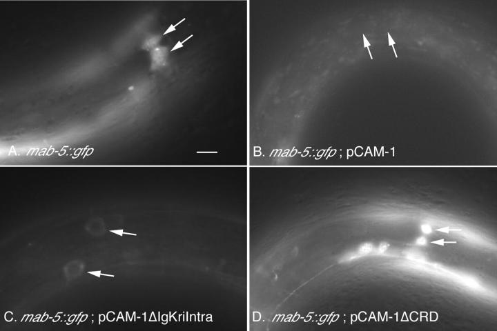
During Caenorhabditis elegans development, the HSN neurons and the right Q neuroblast and its descendants undergo long-range anteriorly directed migrations. Both of these migrations require EGL-20, a C. elegans Wnt homolog. Through a canonical Wnt signaling pathway, EGL-20/Wnt transcriptionally activates the Hox gene mab-5 in the left Q neuroblast and its descendants, causing the cells to migrate posteriorly. In this report, we show that CAM-1, a Ror receptor tyrosine kinase (RTK) family member, inhibits EGL-20 signaling. Excess EGL-20, like loss of cam-1, caused the HSNs to migrate too far anteriorly. Excess CAM-1, like loss of egl-20, shifted the final positions of the HSNs posteriorly and caused the left Q neuroblast descendants to migrate anteriorly. The reversal in the migration of the left Q neuroblast and its descendants resulted from a failure to express mab-5, an egl-20 mutant phenotype. Our data suggest that CAM-1 negatively regulates EGL-20.


Figures

F<sc>igure</sc> 1.—
Figure 1.—
C. elegans cell migrations. Anterior is to the left and dorsal is up. (A) Neuronal embryonic cell migrations. Schematic lateral view of a newly hatched first larval stage (L1) hermaphrodite is shown. Both the final positions of the ALM, BDU, CAN, and HSN cell bodies (ovals and circles) and their migration routes (arrows) are indicated. Dashed ovals show the positions of some of the landmark hypodermal nuclei (V cells) used in assessing cell position. (B) Q neuroblast migrations. Schematic lateral view of an older L1 animal is shown after the QL descendants have completed their migrations. Indicated are the final positions of the QR descendants, SDQ, AVM, and AQR (solid circles) and their migration routes (solid arrows) and of the QL descendants, SDQ, PVM, and PQR (shaded circles) and their migration routes (shaded arrows). Cell divisions and cell deaths in the Q lineages are not shown. Dashed ovals and circles show location of landmark hypodermal nuclei, Vn.a and Vn.p, used in assessing cell position.
F<sc>igure</sc> 2.—
Figure 2.—
HSN cell migration in wild-type, cam-1, and egl-20 hermaphrodites. Anterior is to the left and dorsal is up. Shown at the top is a schematic of the middle section of a hermaphrodite with the HSN cell (solid circle) and its migration route (arrow). Bars represent percentage of HSN cells located at that position along the anterior-posterior axis of L1 larvae as assessed by Nomarski optics (see materials and methods). Long tick marks on the x-axis denote location of V cell nuclei and short tick marks denote the location of P cell nuclei. Each V cell and Q (QL if on left side of animal or QR if on right) cell is named on the diagram at the top. Tick mark on the y-axis indicates 100%. n, number of HSN cells tallied.
F<sc>igure</sc> 3.—
Figure 3.—
Excess egl-20 drives the HSNs to more anterior positions. Anterior is to the left and dorsal is up. Data are presented as described in the Figure 2 legend.
F<sc>igure</sc> 4.—
Figure 4.—
HSN cell migration in cam-1 and Wnt signaling mutants. Anterior is to the left and dorsal is up. Data are presented as described in the Figure 2 legend.
F<sc>igure</sc> 5.—
Figure 5.—
mab-5::gfp expression in the QL descendants is reduced in animals that overexpress the CAM-1 CRD. Anterior is to the left and dorsal is up. L1 cam-1(gm122); mab-5::gfp animals of indicated genotypes were examined for levels of GFP expression in QL descendants. Arrows show positions of two QL descendants. (A) No CAM-1 transgene. MAB-5::GFP is expressed at high levels in QL descendants. (B) Expression of a full-length cam-1 transgene. MAB-5::GFP is undetectable in QL descendants. (C) CRD expression (pCAM-1ΔIgKriIntra). MAB-5::GFP is expressed at low levels. (D) Expression of a cam-1 transgene that lacks the CRD (pCAM-1ΔCRD). MAB-5::GFP is expressed at high levels. Bar, 10 μm.
F<sc>igure</sc> 6.—
Figure 6.—
QL cell migration in wild-type, cam-1, and egl-20 animals. At the top is a schematic lateral view of the middle section of a late L1 animal. Anterior is to the left and dorsal is up. Lightly shaded ovals show position of landmark Vn.a and Vn.p nuclei (each Vn.p nucleus is labeled). The final positions of the cell bodies of the QL descendants, SDQ and PVM (shaded circles) and their migration routes (shaded arrows) are indicated. Cell divisions and deaths are not shown. Bars represent percentage of QL descendants located at that position along the anterior-posterior axis of L1 larvae. The long tick marks on the x-axis indicate the location of Vn.p nuclei and the short tick marks indicate the location of Vn.a nuclei. The tick mark on the y-axis denotes 100%. Data for SDQ and PVM were combined. PQR was not included because it migrates to a location near other neurons in the head, making its position difficult to score.
F<sc>igure</sc> 7.—
Figure 7.—
QL cell migration in cam-1 and Wnt signaling mutants. Data are presented as described in the Figure 6 legend.

References

    1. Afzal, A. R., A. Rajab, C. D. Fenske, M. Oldridge, N. Elanko et al., 2000. Recessive Robinow syndrome, allelic to dominant brachydactyly type B, is caused by mutation of ROR2. Nat. Genet. 25: 419–422. - PubMed
    1. Ailion, M., and J. H. Thomas, 2003. Isolation and characterization of high-temperature-induced dauer formation mutants in Caenorhabditis elegans. Genetics 165: 127–144. - PMC - PubMed
    1. Al-Shawi, R., S. V. Ashton, C. Underwood and J. P. Simons, 2001. Expression of the Ror1 and Ror2 receptor tyrosine kinase genes during mouse development. Dev. Genes Evol. 211: 161–171. - PubMed
    1. Baum, P. D., C. Guenther, C. A. Frank, B. V. Pham and G. Garriga, 1999. The Caenorhabditis elegans gene ham-2 links Hox patterning to migration of the HSN motor neuron. Genes Dev. 13: 472–483. - PMC - PubMed
    1. Bhanot, P., M. Brink, C. H. Samos, J. C. Hsieh, Y. Wang et al., 1996. A new member of the frizzled family from Drosophila functions as a Wingless receptor. Nature 382: 225–230. - PubMed

Publication types

MeSH terms

Substances