. 2004 Sep 15;24(37):8141-52.
doi: 10.1523/JNEUROSCI.1607-04.2004.

Glucagon-like peptide 1 excites hypocretin/orexin neurons by direct and indirect mechanisms: implications for viscera-mediated arousal

Affiliations

Glucagon-like peptide 1 excites hypocretin/orexin neurons by direct and indirect mechanisms: implications for viscera-mediated arousal

Claudio Acuna-Goycolea et al. J Neurosci. .

Abstract

Glucagon-like peptide 1 (GLP-1) is produced by neurons in the caudal brainstem that receive sensory information from the gut and project to several hypothalamic regions involved in arousal, interoceptive stress, and energy homeostasis. GLP-1 axons and receptors have been detected in the lateral hypothalamus, where hypocretin neurons are found. The electrophysiological actions of GLP-1 in the CNS have not been studied. Here, we explored the GLP-1 effects on GFP (green fluorescent protein)-expressing hypocretin neurons in mouse hypothalamic slices. GLP-1 receptor agonists depolarized hypocretin neurons and increased their spike frequency; the antagonist exendin (9-39) blocked this depolarization. Direct GLP-1 agonist actions on membrane potential were abolished by choline substitution for extracellular Na+, and dependent on intracellular GDP, suggesting that they were mediated by sodium-dependent conductances in a G-protein-dependent manner. In voltage clamp, the GLP-1 agonist Exn4 (exendin-4) induced an inward current that reversed near -28 mV and persisted in nominally Ca2+-free extracellular solution, consistent with a nonselective cationic conductance. GLP-1 decreased afterhyperpolarization currents. GLP-1 agonists enhanced the frequency of miniature and spontaneous EPSCs with no effect on their amplitude, suggesting presynaptic modulation of glutamate axons innervating hypocretin neurons. Paraventricular hypothalamic neurons were also directly excited by GLP-1 agonists. In contrast, GLP-1 agonists had no detectable effect on neurons that synthesize melanin-concentrating hormone (MCH). Together, our results show that GLP-1 agonists modulate the activity of hypocretin, but not MCH, neurons in the lateral hypothalamus, suggesting a role for GLP-1 in the excitation of the hypothalamic arousal system possibly initiated by activation by viscera sensory input.


Figures

Figure 1.
Figure 1.
GLP-1 excites hypocretin neurons. A, Representative traces showing the excitatory effects of GLP-1 on the spike frequency. B, Time course of GLP-1 actions on firing rate in a typical hypocretin neuron. C, Bar graph shows the effect of different concentrations of GLP-1 on the firing rate of hypocretin neurons.
Figure 2.
Figure 2.
The protease-resistant long-acting GLP-1 receptor agonist Exn4 depolarizes and enhances spike frequency of hypocretin neurons. A, Exn4 depolarized the membrane potential and increased the spike frequency of hypocretin cells. B, Time course of the Exn4 actions on the frequency of action potentials in a typical cell. C, The Exn4 effects on spike frequency were dose dependent.
Figure 3.
Figure 3.
Direct actions of GLP-1 agonist on hypocretin neurons. A, Trace 1, With 0.5 μm TTX in the bath, Exn4 depolarized the hypocretin cell membrane potential. Trace 2, In the presence of the GLP-1 receptor antagonist Exn9-39 in the external solution, Exn4 failed to depolarize the membrane potential. Trace 3, The depolarizing actions of Exn4 were blocked by intracellular GDPβS. Trace 4, When extracellular NaCl was replaced by an equimolar concentration of choline chloride, the depolarizing effects of Exn4 were depressed. B, Bar graph summarizing the Exn4 action on the membrane potential in normal, Exn9-39-containing ACSF, low intracellular GTP, and Na+-free extracellular conditions. C, Exn4 (1 μm) induced an inward current at voltages between -100 and -60 mV (top graph). The Exn4-induced current showed a reversal potential consistent with a nonspecific cationic conductance (trace from one cell shown in bottom graph). D-F, Exn4 did not evoke a detectable change on potassium currents activated by a voltage step from -80 to 0 mV for 200 msec. n.s., Not significant. G, Exn4 depressed afterhyperpolarization currents in hypocretin neurons. In the presence of Exn4, the time integral of IAHP was reduced by 17.5% (bar graph). H-L, Exn4 depressed calcium currents in hypocretin neurons. H, Exn4 depressed the current responses evoked by a voltage step from -80 to 0 mV for 200 msec. The time course and mean effect of Exn4 on calcium currents are shown in I and J, respectively. *p < 0.05. K, Current response in the presence and the absence of Exn4 in a typical hypocretin neuron after a voltage ramp from -40 to +50 mV. L, When the relative inhibitory effect of Exn4 on calcium currents was compared at -20 and +20 mV, a significant difference was detected suggesting that the GLP-1 agonist actions were voltage dependent. Ctrl, Control; Wash, washout. *Statistically significant, p < 0.05.
Figure 4.
Figure 4.
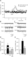
GLP-1 modulates excitatory synaptic inputs to hypocretin neurons. A, Time course of Exn4 effects on the frequency of hypocretin postsynaptic currents in normal (unfilled circles) and Exn9-39-containing (filled circles) ACSF. In this experiments, the cells were held at -60 mV. B, Raw traces show the excitatory effect of Exn4 on the PSC frequency. C, With the GLP-1 receptor antagonist in the bath, Exn4 failed to increase the PSC frequency. D, This bar graph shows the mean effect of Exn4, Exn9-39, and Exn4 plus Exn9-39 on the PSC frequency. E, In the presence of 30 μm BIC in the bath and using KMeSO4 pipettes, EPSCs were recorded. Exn4 increases the EPSC frequency, as shown in this bar graph. *Statistically significant. Ctrl, Control; Wash, washout.
Figure 5.
Figure 5.
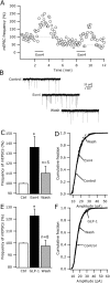
Presynaptic modulation of glutamate release by GLP-1 receptor agonists. A, Exn4 enhanced the frequency of mEPSCs. In this particular case, the GLP-1 agonist was applied twice to the recorded cell, evoking repeatable increases in the frequency of mEPSCs. B, Raw traces showing the excitatory effect of Exn4 on the frequency of mEPSCs. C, This bar graph shows the mean effect of Exn4 on the mEPSC frequency. D, The application of Exn4 to the bath failed to evoke a change in the cumulative distribution of the mEPSC amplitude in five cells tested. E, GLP-1 also enhanced the mEPSC frequency in hypocretin neurons, but did not modify the cumulative distribution of them EPSCs amplitude (F). *Statistically significant. Ctrl, control; Wash, washout.
Figure 6.
Figure 6.
GLP-1 agonist enhances inhibitory, GABA-mediated, synaptic transmission in hypocretin neurons. A, Exn4 enhanced the frequency of IPSCs. In these experiments, KCl was used in the recording pipette and IPSCs were detected as fast inward currents. B, Time course of the Exn4 actions on IPSC frequency. C, Exn4 increased the frequency of IPSCs in five cells tested. D, E, Exn4 actions on the frequency and the amplitude of mIPSCs in hypocretin cells. The GLP-1 agonist increased the frequency of mIPSCs by 42.5 ± 14.3% (D). The cumulative distributions of the mIPSCs were also significantly altered in all of the cells tested (E). *Statistically significant. Ctrl, control; Wash, washout.
Figure 7.
Figure 7.
Effects of GLP-1 agonist on MCH and paraventricular hypothalamic neurons. A, Trace 1, Exn4 (1 μm) did not evoke any detectable change in the firing rate or the membrane potential in an MCH neuron. Trace 2, 50 μm glutamate (Glut) induced a robust depolarization followed by a burst of action potentials in an MCH neuron. The action potentials were truncated in this figure. Trace 3 shows the lack of Exn4 effects on the membrane potential in the presence of the TTX (0.5 μm). B, Bar graph showing the effects of Exn4 and glutamate on the membrane potential of MCH neurons in the presence of 0.5 μm TTX in the bath. C, Exn4 showed no effect on the synaptic inputs to MCH neurons, because it did not increase the frequency of PSCs in these cells. D, E, In the presence of TTX (0.5 μm) in the bath, Exn4 did not evoke any significant change in the frequency (D) or the amplitude (E) of mEPSCs recorded in MCH neurons. F, G, Diverse Exn4 responses were detected in PVN cells. An excitatory (increase in spike frequency) response (F1) was detected in 57% of the recorded neurons, whereas an inhibitory (depressing in the firing rate) response (F2) was observed in 14% of the recorded cells. We did not detect any change in the spike frequency or membrane potential in the remaining 28% of the recorded cells (F3). G, In the presence of 0.5 μm TTX in the bath, only depolarizing responses were detected in 45% of the cells (top trace). The remaining cells (bottom trace) did not show a significant change in their membrane potential with Exn4 application. Ctrl, control; n.s., not significant; Wash, washout.

References

    1. Acuna-Goycolea C, Li Y, van den Pol AN (2004) Group III metabotropic glutamate receptors maintain tonic inhibition of excitatory synaptic input to hypocretin/orexin neurons. J Neurosci 24: 3013-3022. - PMC - PubMed
    1. Bekkers JM, Stevens CF (1995) Quantal analysis of EPSCs recorded from small numbers of synapses in hippocampal cultures. J Neurophysiol 73: 1145-1156. - PubMed
    1. Brandon NJ, Jovanovic JN, Smart TG, Moss SJ (2002) Receptor for activated C kinase-1 facilitates protein kinase C-dependent phosphorylation and functional modulation of GABAA receptors with the activation of G-protein-coupled receptors. J Neurosci 22: 6353-6361. - PMC - PubMed
    1. Broberger C, de Lecea L, Sutcliffe JG, Hökfelt T (1998) Hypocretin/orexin and melanin-concentrating hormone-expressing cells form distinct populations in the rodent lateral hypothalamus: relationship to the neuropeptide Y and Agouti gene-related protein systems. J Comp Neurol 402: 460-474. - PubMed
    1. Borowsky B, Durkin MM, Ogozalek K, Marzabadi MR, DeLeon J, Lagu B, Heurich R, Lichtblau H, Shaposhnik Z, Daniewska I, Blackburn TP, Branchek TA, Gerald C, Vaysse PJ, Forray C (2002) Antidepressant, anxiolytic and anorectic effects of a melanin-concentrating hormone-1 receptor antagonist. Nat Med 8: 825-830. - PubMed

Publication types

MeSH terms

Substances

LinkOut - more resources