. 2004 Dec;15(12):5231-41.
doi: 10.1091/mbc.e04-06-0522. Epub 2004 Sep 15.

Apatite-mediated actin dynamics in resorbing osteoclasts

Affiliations

Apatite-mediated actin dynamics in resorbing osteoclasts

Frédéric Saltel et al. Mol Biol Cell. 2004 Dec.

Abstract

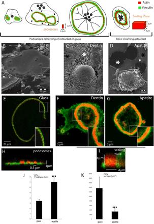
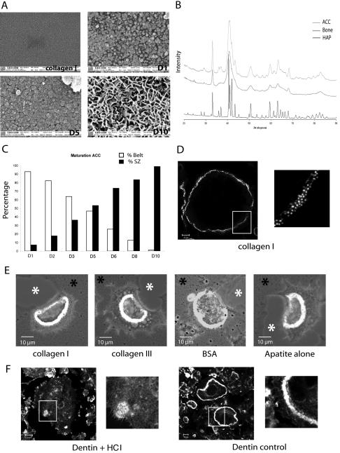
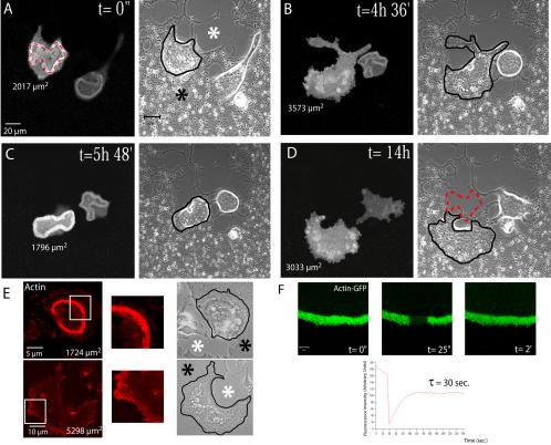
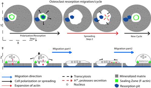
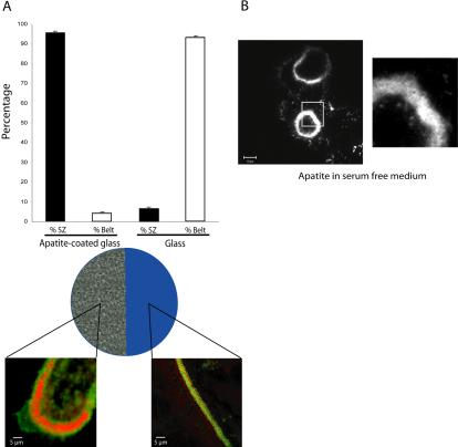
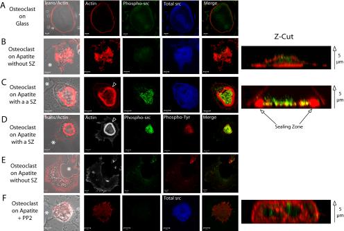
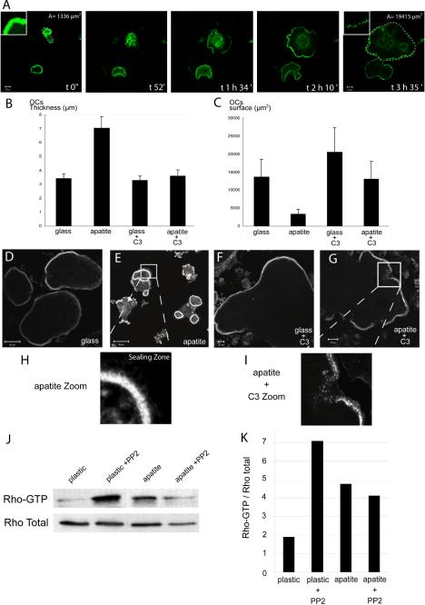
The actin cytoskeleton is essential for osteoclasts main function, bone resorption. Two different organizations of actin have been described in osteoclasts, the podosomes belt corresponding to numerous F-actin columns arranged at the cell periphery, and the sealing zone defined as a unique large band of actin. To compare the role of these two different actin organizations, we imaged osteoclasts on various substrata: glass, dentin, and apatite. Using primary osteoclasts expressing GFP-actin, we found that podosome belts and sealing zones, both very dynamic actin structures, were present in mature osteoclasts; podosome belts were observed only in spread osteoclasts adhering onto glass, whereas sealing zone were seen in apico-basal polarized osteoclasts adherent on mineralized matrix. Dynamic observations of several resorption cycles of osteoclasts seeded on apatite revealed that 1) podosomes do not fuse together to form the sealing zone; 2) osteoclasts alternate successive stationary polarized resorption phases with a sealing zone and migration, nonresorption phases without any specific actin structure; and 3) apatite itself promotes sealing zone formation though c-src and Rho signaling. Finally, our work suggests that apatite-mediated sealing zone formation is dependent on both c-src and Rho whereas apico-basal polarization requires only Rho.


Figures

Figure 1.
Figure 1.
Formation of podosome belt in spread osteoclasts or sealing zone in apico-basal polarized osteoclasts is dependent on extracellular matrix. (A) Scheme of the different actin structures observed in osteoclasts. Osteoclasts seeded on glass form podosomes, small cylinders of actin surrounded by vinculin. Podosomes organize into three different structures along differentiation namely clusters, rings, and belts into mature osteoclasts (Destaing et al., 2003). On bone when resorbing, they form a sealing zone, a large circular band of actin surrounded by vinculin. Differentiated osteoclasts were plated on either glass (B, E, and H), dentin (C–F), or apatite (D, G, and I); scanning electron microscopy images of osteoclasts adherent on their respective substratum are shown (B–D). Mature osteoclasts adherent on glass are large flat cells with a swollen area at site of podosome belt (B, inset). In contrast, on dentin or apatite substrate-resorbing osteoclasts are contracted. Black asterisks indicate nonresorbed matrix and white asterisks resorption pits (C and D). (E) A single osteoclast on a glass coverslip is shown with a podosome belt; the inset is a zoom of the area outlined to show podosomes (bar, 20 μm). (F and G) Osteoclast on dentin and apatite exhibiting a sealing zone; inset is a zoom of the area outlined showing the absence of podosomes and the presence of a large sealing zone (bar, 5 μm). (H and I) Z-cuts of podosome belt and sealing zone. Actin is shown in red and vinculin in green. (J and K) Graphical representation of surface area and thickness of osteoclasts adherent on either glass coverslips or apatite-coated glass coverslips, measured using Zeiss LSM 510 software. Stars indicate statistically significant results using a Student' t test (p < 0.0001; n = 30).
Figure 3.
Figure 3.
Apatite mineral triggers sealing zone formation. (A) Scanning electron microscopy showing the nature of the coating formed along the mineralization process of ACC slides. Apatite particles appeared on day 1, grew, and covered the entire surface by day 5. On day 10, three-dimensional bone-like apatite structures were formed. (B) X-ray diffraction patterns for the substrate ACC in the range of 20–90° 2 theta is presented. As can be seen, ACC substrate showed the characteristic peaks of apatite (002, 310) with some unresolved large broad peaks typical of a poorly crystalline apatitic calcium phosphate. By comparison of the ACC diagram with those of bone samples and hydroxyapatite, we could see that, if the same apatitic phase is common to all the samples, bone as ACC showed a poorly crystalline diagram, compared with hydroxyapatite, which logically presents a highly crystalline phase. (C) Mature osteoclasts were seeded on coverslips at different time points during the mineralization process and stained for actin 24 h later. The percentage of osteoclasts exhibiting podosome belts vs. sealing zones along the mineralization process is shown (n = 200 at each day). (D and E) Whereas osteoclasts seeded on collagen I exhibited podosome belts, they formed sealing zone as long as organic coating (collagen I, collagen III or BSA) was mineralized. (F) In contrast to dentin, osteoclasts adherent on HCl demineralized dentin slices did not form clearly recognizable actin structures.
Figure 2.
Figure 2.
Resorbing osteoclasts on apatite alternate resorption with apico-basal polarization and sealing zone together with spread phases to migrate. (A–D) Osteoclasts derived from the spleen of actin-GFP mice were plated on mineralized substratum (ACC). Images were extracted from Supplementary Video 1 and time of imaging is indicated. Mineralized matrix is indicated by the black asterisk and resorbed area by the white asterisk. Cells were successively imaged using a fluorescein-type filter set and by phase contrast. Contours of osteoclasts are underlined in black. Delineation of the sealing zone by the red dotted line (A) indicates that it matches perfectly with resorbed areas (see the same red line on the final light transmission, image D). Osteoclast surface area at each time point is indicated below the cell. A large surface area, >3000 μm2, corresponded to spreading of nonresorbing osteoclasts without sealing zone. In contrast, cell surfaces between 1700 and 2000 μm2 corresponded to actively resorbing osteoclasts exhibiting a sealing zone (bar, 20 μm). (E) Actin of osteoclasts seeded on apatite was stained with phalloidin-RITC to confirming that on apatite, osteoclasts exhibit either sealing zone when polarized or no recognizable actin structures when spread during migration. (F) FRAP analysis of the sealing zone. Images are extracted from one of several experiments. Experimental data fit an exponential law with characteristic time of 30 s.
Figure 7.
Figure 7.
Representation of osteoclast and its actin dynamic during resorption and migration. Schematic representation of osteoclast and its actin cytoskeleton during resorption on apatite mineralized substrate. They alternate between polarized morphology with sealing zone during resorption and spread morphology without any specific actin structures during migration.
Figure 4.
Figure 4.
Sealing zone formation is not triggered by microenvironmental modifications. (A) To further confirm the role of extracellular matrix on osteoclast actin organization, we used apatite-coated slides. Half the slide was first immersed in HCl (6 N) to remove apatite crystals. Mature osteoclasts were then seeded on these slides for 24 h. Osteoclasts exhibit sealing zones when adherent on the mineral part and podosome belts on glass. The graph represents the percentage of osteoclasts exhibiting podosome belts or sealing zones according to the substratum (n = 150). (B) Mature adherent osteoclasts still exhibit a sealing zone on apatite in serum-free medium ruling out the putative role of medium components.
Figure 5.
Figure 5.
c-src is required for apatite-dependent sealing zone formation but not for polarization. Mature osteoclasts seeded for 24 h on glass or on ACC and associated with resorption pits (asterisk) were observed by confocal microscopy after actin staining by phalloidin or immunolabelling with antic-src or antiphosphotyrosine antibodies. Z-cut sections are provided to confirm polarization (actin in red) and phosphorylated c-src localization (green). Arrow: from bottom to top of the cells. (A) On glass, osteoclasts exhibited typical peripheral podosome belts with diffuse total and activated c-src (bar, 20 μm). (B) On apatite, in the absence of sealing zone, actin and c-src were distributed throughout the cells, whereas (C) in the presence of a sealing zone (arrowhead), activated c-src labeling was more intense and localized within the area delineated by the sealing zone. Merged images of actin and phosphorylated c-src are presented (A and F; bar, 10 μm). (D) Phosphorylated c-src is the main phosphotyrosine target in bone resorbing osteoclasts as shown by their precise colocalization within the sealing zone area, whereas (E) they are diffusely distributed in its absence (bar, 10 μm). (F) After 1 h of PP2 treatment of osteoclasts adherent on apatite, osteoclasts still associated with resorption pits had lost their sealing zone, and activated c-src and phosphotyrosine labeling was diffuse (bar, 10 μm). Nevertheless, cells were still polarized as shown by the Z-cut section. Merged images of phosphorylated c-src and phosphotyrosine proteins are presented (D and E).
Figure 6.
Figure 6.
Rho is required for sealing zone formation and polarization of osteoclast on apatite. Mature osteoclasts adherent on glass or apatite-coated slides were either untreated or treated with exoenzyme C3 (100 nM) for 5 h before being observed by confocal microscopy after actin staining. (A) Images of resorbing osteoclasts on apatite taken at different time point show the transition from polarized (1336 μm2 surface area) to largely spread cells (1945 μm2 at time 3 h 35 min) as well as the transition from sealing zone to podosome belt (insets). (B and C) Graphical representation of the thickness and surface area of osteoclasts, seeded either on glass or apatite and treated, or not, with C3. Mineral substratum increased osteoclast average thickness (B) and decreased surface area (C and E) compared with glass, (C and D) indicating that they were polarized (n = 30), as measured using Zeiss LSM 510 software. Exoenzyme C3–mediated Rho inhibition completely abrogated apatite-dependent sealing zone formation. In the presence of exoenzyme C3, osteoclasts were flat and spread (B, C, F, and G). Osteoclasts seeded on apatite, in the presence of C3, exhibited podosome belts (G and I) instead of the sealing zones seen in untreated cells (E and H). (J) Differentiated osteoclasts were seeded on plastic or apatite for 12 h, and Rho activity was assessed by pull-down binding assays. After 12 h on apatite, the level of Rho-GTP was clearly increased compared with plastic. In presence of PP2 after 12 h on plastic, the level of Rho-GTP increased, but was maintained on the apatite substratum. This experiment has been repeated three times with similar results, and one of them is shown here. (K) Rho GTP vs. total Rho ratio was evaluated by Western blotting and quantitated using Image Quant software.

References

    1. Bourette, R.P., Mouchiroud, G., Ouazana, R., Morle, F., Godet, J., and Blanchet, J.P. (1993). Expression of human colony-stimulating factor-1 (CSF-1) receptor in murine pluripotent hematopoietic NFS-60 cells induces long-term proliferation in response to CSF-1 without loss of erythroid differentiation potential. Blood 81, 2511–2520. - PubMed
    1. Chambers, T.J., Thomson, B.M., and Fuller, K. (1984). Effect of substrate composition on bone resorption by rabbit osteoclasts. J. Cell Sci. 70, 61–71. - PubMed
    1. Chellaiah, M.A., Biswas, R.S., Rittling, S.R., Denhardt, D.T., and Hruska, K.A. (2003a). Rho-dependent Rho Kinase activation increases CD44 surface expression and bone resorption in osteoclasts. J. Biol. Chem. 278, 29086–29097. - PubMed
    1. Chellaiah, M.A., Kizer, N., Biswas, R., Alvarez, U., Strauss-Schoenberger, J., Rifas, L., Rittling, S.R., Denhardt, D.T., and Hruska, K.A. (2003b). Osteopontin deficiency produces osteoclast dysfunction due to reduced CD44 surface expression. Mol. Biol. Cell 14, 173–189. - PMC - PubMed
    1. Chellaiah, M.A., Soga, N., Swanson, S., McAllister, S., Alvarez, U., Wang, D., Dowdy, S.F., and Hruska, K.A. (2000). Rho-A is critical for osteoclast podosome organization, motility, and bone resorption. J. Biol. Chem. 275, 11993–12002. - PubMed

Publication types

MeSH terms

LinkOut - more resources