. 2004 Sep;114(6):795-804.
doi: 10.1172/JCI20921.

The renal papilla is a niche for adult kidney stem cells

Affiliations

The renal papilla is a niche for adult kidney stem cells

Juan A Oliver et al. J Clin Invest. 2004 Sep.

Abstract

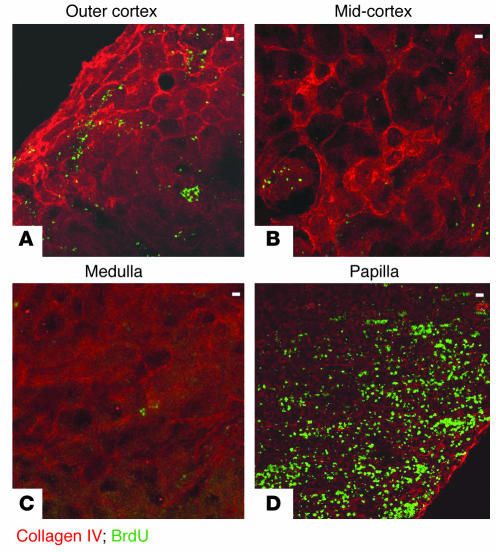
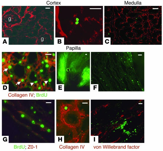
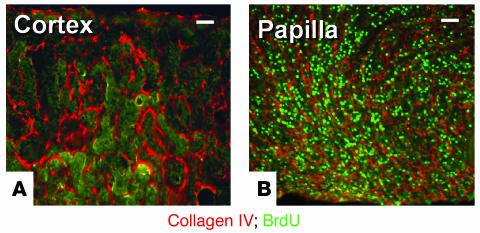
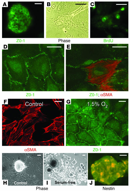
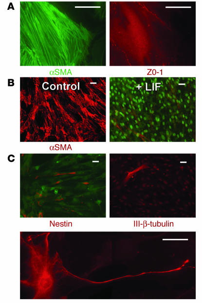
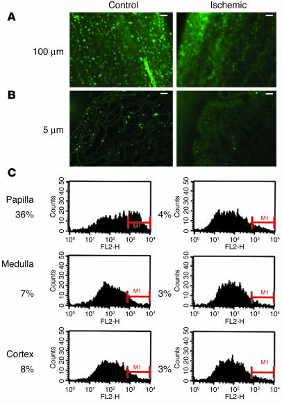
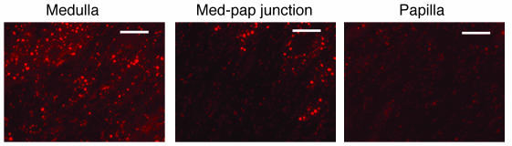
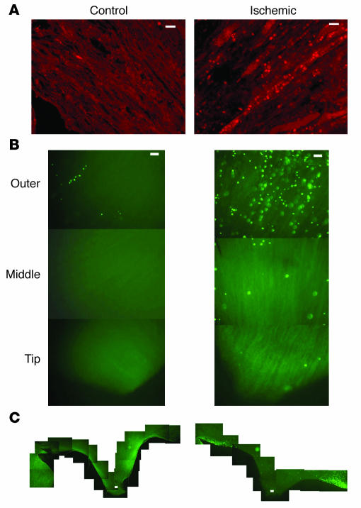
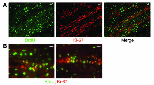
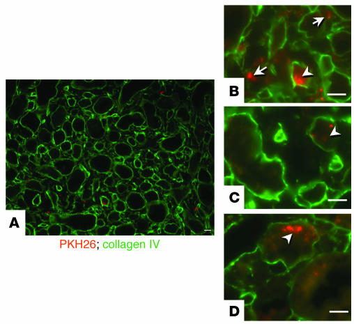
Many adult organs contain stem cells, which are pluripotent and are involved in organ maintenance and repair after injury. In situ, these cells often have a low cycling rate and locate in specialized regions (niches). To detect such cells in the kidney, we administered a pulse of the nucleotide bromodeoxyuridine (BrdU) to rat and mouse pups and, after a long (more than 2-month) chase, examined whether the kidney contained a population of low-cycling cells. We found that in the adult kidney, BrdU-retaining cells were very sparse except in the renal papilla, where they were numerous. During the repair phase of transient renal ischemia, these cells entered the cell cycle and the BrdU signal quickly disappeared from the papilla, despite the absence of apoptosis in this part of the kidney. In vitro isolation of renal papillary cells showed them to have a plastic phenotype that could be modulated by oxygen tension and that when injected into the renal cortex, they incorporated into the renal parenchyma. In addition, like other stem cells, papillary cells spontaneously formed spheres. Single-cell clones of these cells coexpressed mesenchymal and epithelial proteins and gave rise to myofibroblasts, cells expressing neuronal markers, and cells of uncharacterized phenotype. These data indicate that the renal papilla is a niche for adult kidney stem cells.


Figures

Figure 1
Figure 1
BrdU-retaining cells in the adult kidney. Composite views 100 μm in thickness of a kidney from a 2-month-old rat, obtained by confocal microscopy with 2-μm optical sections. (A) Outer cortex, showing few scattered BrdU-retaining cells with an occasional cell aggregate. (B) Mid-cortex, showing a glomerulus with BrdU-positive cells. (C) Medulla with very rare BrdU-positive cells. (D) Papilla with numerous BrdU-retaining cells. Colored labels indicate staining with FITC fluorescin (green) and rhodamine (red) as described in Results. Scale bars: 50 μm.
Figure 2
Figure 2
BrdU-retaining cells in the adult kidney. (A_F) Composite views obtained by confocal microscopy with 1-μm optical sections, except where indicated otherwise (E and F). (A) Composite view 20 μm in thickness of a kidney cortex from a 3-month-old rat showing no BrdU-positive cells. g, glomerulus. (B) Composite view 10 μm in thickness of a glomerulus from a 2-month-old rat with a single BrdU-retaining cell in the glomerular tuft. (C) Composite view 20 μm in thickness of a kidney medulla from a 2-month-old rat with a single BrdU-retaining cell. (D) Composite view 20 μm in thickness of a kidney papilla from a 2-month-old rat showing numerous BrdU-positive cells. Arrows, interstitial cells; arrowheads, tubular cells. (E) Section 100 μm in thickness of a kidney papilla from a 3-month-old rat showing more fluorescent signal (i.e., BrdU) in the outer parts of the papilla. (F) Section 5 μm in thickness of the kidney papilla from a 5-month-old rat. (G) Composite view 20 μm in thickness of kidney papillary tubules from a 4-month-old rat showing that some of the BrdU-retaining cells are tubular epithelial cells expressing ZO-1 in their tight junctions. (H) Composite view 10 μm in thickness of a kidney medulla from a 3-month-old rat showing a single interstitial BrdU-retaining cell surrounded by collagen IV. (I) Composite view 20 μm in thickness of a kidney papilla from a 4-month-old rat showing BrdU-retaining cells in close association with endothelial cells. Scale bars: 50 μm.
Figure 3
Figure 3
BrdU-retaining cells in adult mouse kidney. (A and B) Composite views 60 μm in thickness of kidney sections from a 2-month-old mouse obtained by confocal microscopy with 2-μm optical sections. (A) Outer cortex with no BrdU-positive cells. (B) Outer papilla with abundant BrdU-retaining cells. Scale bars: 50 μm.
Figure 4
Figure 4
Renal papillary cells grown in cell culture. (A) Isolated renal papillary cells grown in standard cell culture conditions formed cell aggregates within about 24 hours of their isolation and expressed ZO-1 in their tight junctions. (B and C) Phase-contrast (Phase; B) and fluorescence (C) microphotographs of a group of renal papillary cells growing in cell culture conditions 4 days after cell isolation; about 40% of the papillary cells isolated from BrdU-loaded animals were BrdU-retaining cells. (D and E) During the first several days of culture, most cells had an epithelial phenotype expressing ZO-1 in their tight junctions (D), but some cells, in addition to ZO-1, also expressed mesenchymal proteins such as α-smooth muscle actin (αSMA) (E). (F) After more than a week in culture under control conditions with 5% CO2 and 95% room air, most cells were spindle-shaped and stained strongly for α-smooth muscle actin. (G) However, when cells were grown under hypoxic conditions (5% CO2, 1.5% O2, and 93.5% N2), most cells retained an epithelial phenotype, with prominent ZO-1 expression. (H) When grown in standard control cell culture media on plastic, most cells adhered to the culture dish but frequently formed cell aggregates that resembled neurospheres. (I) The tendency for the cells to aggregate was markedly enhanced by growth of the cells in the absence of sera. The picture shows a 3-week-old culture with many cellular aggregates. (J) Many of the cells inside the aggregates were positive for nestin. Scale bars: 50 μm.
Figure 5
Figure 5
Characterization of renal papillary cell clones. (A) As the example shows, many cells coexpressed mesenchymal proteins such as α-smooth muscle actin (left) as well as epithelial proteins such as ZO-1 (right). (B) In standard control conditions in the absence of sera, all cells were spindle-shaped and stained strongly for α-smooth muscle actin (Control; left) but when LIF was added to the media, two phenotypes were apparent (+ LIF; right): the majority of cells grew in a monolayer and were negative for α-smooth muscle actin (nuclei stained with 10 nM Sytox green), but a few cells grew on top of the monolayer of cells, were spindle-shaped, and expressed α-smooth muscle actin. (C) In standard control culture conditions, a small number of cells expressed the intermediate filament nestin. Occasional cells also expressed the neuronal marker class III β-tubulin (III-β-tubulin) and some cells even acquired the morphological characteristics of neurons. Scale bars: A, 20 μm; B and C, 50 μm.
Figure 6
Figure 6
Effect of transient renal ischemia on papillary BrdU-retaining cells. The presence of BrdU-retaining cells was examined in papillae of control kidneys and of kidneys subjected to transient ischemia. (A) In 100-μm papillary sections 3 weeks after ischemia, compared with control, there was a marked decrease in BrdU-retaining cells in the papilla of the ischemic kidney. (B) Sections 5 μm in thickness showed that while the papillary tip of the control kidney had numerous BrdU-retaining cells, that of the ischemic kidney had very few. (C) For quantification of the effect of renal ischemia on the number of BrdU-retaining cells, 6 rats 5 months of age were subjected to transient ischemia of the left kidney; 3 weeks later their kidneys were harvested and cells from different parts of both kidneys were stained for BrdU and analyzed by flow cytometry. In all graphs, the y axis shows the number of cells, while the x axis (FL2-H) shows the fluorescent intensity. MI is the area of positive cells. In the papillae of the nonischemic kidneys, the number of BrdU-retaining cells averaged 36% of the total cells, while in the papillae of the kidneys subjected to transient ischemia, this number was only 4%, as shown to the left of each histogram. Compared with the control kidney, the ischemic kidney also had a slight decrease in the number of BrdU-positive cells in the medulla and cortex, suggesting that the papillary BrdU-retaining cells had not simply migrated to other parts of the kidney. Scale bars: 50 μm.
Figure 7
Figure 7
TUNEL staining of apoptotic cells after transient renal ischemia. Representative example of 5-μm sections of different areas of the same kidney 24 hours after ischemia. All three photographs were obtained under identical conditions. The papilla is the only part of the kidney with no significant apoptotic signal. Med-pap, medullary-papillary. Scale bars: 50 μm.
Figure 8
Figure 8
Cellular proliferation in the renal papilla after transient renal ischemia. (A_C) For detection of whether cellular proliferation occurs in the renal papilla after transient renal ischemia, 36 hours after the episode, kidneys were harvested and papillae were examined. (A) Control and ischemic papillae of the same animal stained with an antibody against the proliferation cell marker Ki-67 (rhodamine). Only the papillae of the post-ischemic kidney showed significant proliferation. (B and C) Control and ischemic papillae of two different experiments in which rats were given a dose of BrdU 1 hour before euthanasia and sections of papilla were examined for BrdU incorporation as marker of DNA synthesis. Sections of renal papillae are 100 μm in thickness. (B) Papillary sections from the outer or juxtamedullary area (top), middle area (middle), and tip (bottom) from control and ischemic kidneys. As shown, DNA synthesis was only detected in the papilla of the post-ischemic kidney, particularly in the outer papilla, the area next to the medulla. (C) In the kidney subjected to transient ischemia, DNA synthesis in papillary cells was most prominent in the region adjacent to the urinary space of the outer papilla. Scale bars: 50 μm.
Figure 9
Figure 9
Cellular proliferation of papillary BrdU-retaining cells after transient renal ischemia. (A and B) For determination of whether the papillary cell proliferation observed after ischemia involved the BrdU-retaining cells, transient ischemia was induced in adult rats loaded with BrdU when they were pups. Then, 36 hours after the ischemia, the kidneys were harvested and 5-μm sections of their papillae were examined for BrdU (FITC fluorescin) and the Ki-67 antigen (rhodamine). In the far right panel of A, and the higher power views of B, merged images show that many BrdU-retaining cells began cycling after ischemia. Scale bars: 50 μm.
Figure 10
Figure 10
Intrarenal injection of renal papillary cells. (A_D) Papillary cells were labeled with PKH26 (rhodamine) and were injected into the subcapsular space. Seven days later, sections 3 μm in thickness showed that many cells had scattered into the renal medulla (A). Some cells were located in the interstitial space (arrows in B), but many incorporated inside tubules (arrowheads, B_D), which are highlighted by staining of the collagen IV (FITC fluorescin) in their basement membranes. Scale bars: 50 μm.

Similar articles

Cited by

References

    1. Witzgall R, Brown D, Schwarz C, Bonventre JV. Localization of proliferating cell nuclear antigen, vimentin, c-Fos, and clusterin in the postischemic kidney. Evidence for a heterogenous genetic response among nephron segments, and a large pool of mitotically active and dedifferentiated cells. J. Clin. Invest. 1994;93:2175–2188. - PMC - PubMed
    1. Finn WF, Chevalier RL. Recovery from postischemic acute renal failure in the rat. Kidney Int. 1979;16:113–123. - PubMed
    1. Oliver J, MacDowell M, Tracy A. The pathogenesis of acute renal failure associated with trauma and toxic injury, renal ischemia, nephrotoxic damage and the ischemuric episode. J. Clin. Invest. 1951;30:1307–1440. - PMC - PubMed
    1. Houghton DC, Hartnett M, Campbell-Boswell M, Porter G, Bennett W. A light and electron microscopic analysis of gentamicin nephrotoxicity in rats. Am. J. Pathol. 1976;82:589–612. - PMC - PubMed
    1. Gupta S, Verfaillie C, Chmielewski D, Kim Y, Rosenberg ME. A role for extrarenal cells in the regeneration following acute renal failure. Kidney Int. 2002;62:1285–1290. - PubMed

Publication types

Substances