. 2004 Oct;186(19):6465-76.
doi: 10.1128/JB.186.19.6465-6476.2004.

Insecticidal pilin subunit from the insect pathogen Xenorhabdus nematophila

Affiliations

Insecticidal pilin subunit from the insect pathogen Xenorhabdus nematophila

Puneet Khandelwal et al. J Bacteriol. 2004 Oct.

Abstract

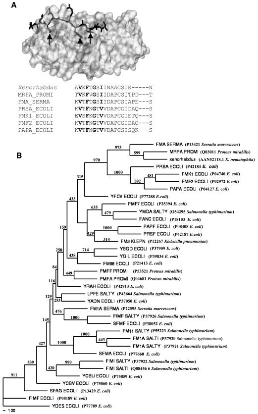
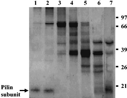
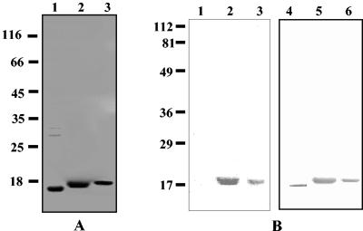
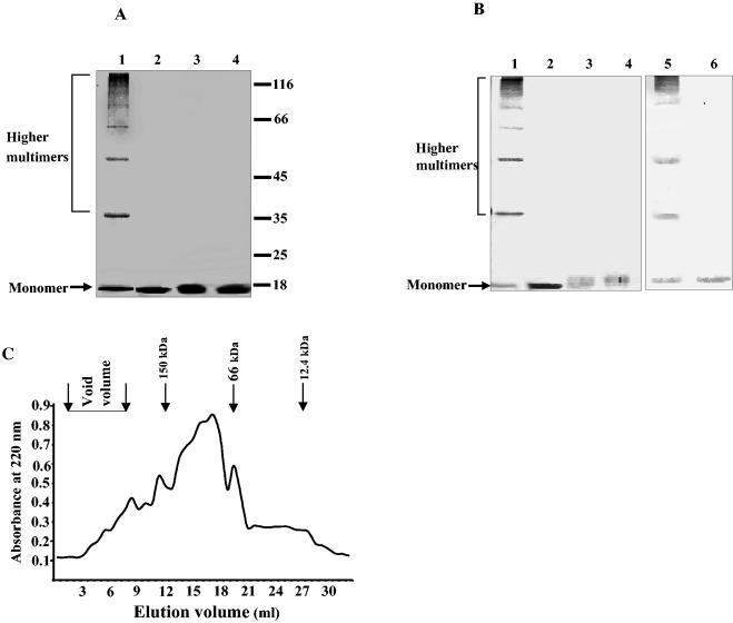
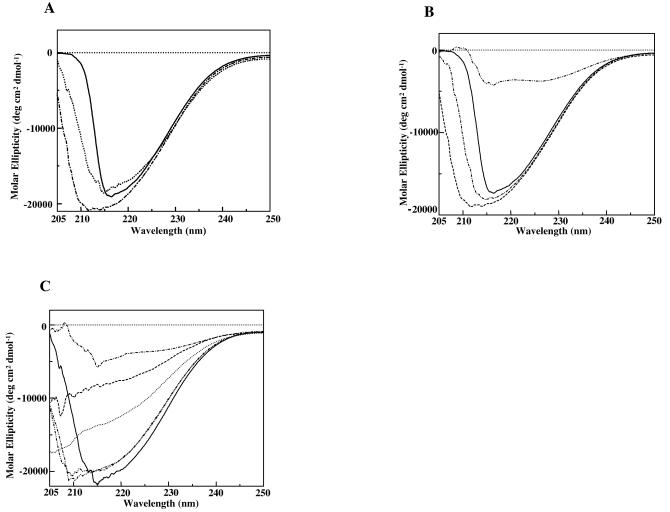
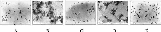
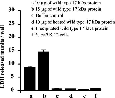
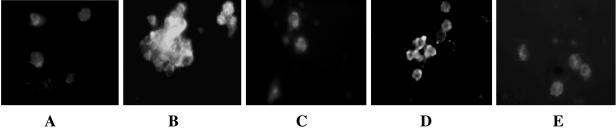
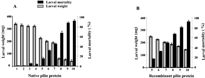
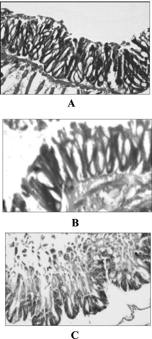
Xenorhabdus nematophila is an insect pathogen and produces protein toxins which kill the larval host. Previously, we characterized an orally toxic, large, outer membrane-associated protein complex from the culture medium of X. nematophila. Here, we describe the cloning, expression, and characterization of a 17-kDa pilin subunit of X. nematophila isolated from that protein complex. The gene was amplified by PCR, cloned, and expressed in Escherichia coli. The recombinant protein was refolded in vitro in the absence of its cognate chaperone by using a urea gradient. The protein oligomerized during in vitro refolding, forming multimers. Point mutations in the conserved N-terminal residues of the pilin protein greatly destabilized its oligomeric organization, demonstrating the importance of the N terminus in refolding and oligomerization of the pilin subunit by donor strand complementation. The recombinant protein was cytotoxic to cultured Helicoverpa armigera larval hemocytes, causing agglutination and subsequent release of the cytoplasmic enzyme lactate dehydrogenase. The agglutination of larval cells by the 17-kDa protein was inhibited by several sugar derivatives. The biological activity of the purified recombinant protein indicated that it has a conformation similar to that of the native protein. The 17-kDa pilin subunit was found to be orally toxic to fourth- or fifth-instar larvae of an important crop pest, H. armigera, causing extensive damage to the midgut epithelial membrane. To our knowledge, this is first report describing an insecticidal pilin subunit of a bacterium.


Figures

FIG. 1.
FIG. 1.
(A) X-ray crystallographic structure of PapE with donor strand complementing N-terminal peptide segment of PapK (indicated by sticks). Conserved deeply buried residues are indicated by arrows. An alignment of N-terminal sequences of various structural pilin proteins is also shown; conserved hydrophobic residues are indicated by boldface type. Sequences were aligned with the CLUSTALW program, followed by manual adjustments to minimize gaps within secondary structures. (B) Neighbor-joining tree showing the branching pattern of different pilin protein amino acid sequences and the phylogenic position of the Xenorhabdus pilin protein. Bootstrap resampling was done for 1,000 replicons. The numbers at the nodes in the consensus tree indicate the numbers of times that the subtree occurred in the 1,000 trees that were generated by NEIGHBOR. The designations of the proteins are followed by the accession numbers and names of the organisms in parentheses.
FIG. 2.
FIG. 2.
Purification of 17-kDa pilin protein from OMVs of X. nematophila. OMV proteins were solubilized in TENS buffer and applied to a Sephacryl S-300 column. The proteins were eluted with TENS buffer. Peak fractions were pooled and examined. Lanes 1 and 2, void volume fractions; lanes 3 to 7, subsequent column fractions. The proteins were resolved on an SDS—12% PAGE gel and were visualized by staining with Coomassie brilliant blue. Numbers on the right are molecular weight markers (in thousands).
FIG. 3.
FIG. 3.
(A) SDS-PAGE profile of purified recombinant 17-kDa protein. The proteins were resolved on an SDS—12% PAGE gel and were visualized by staining with Coomassie brilliant blue or blotted with antiserum. Lane 1, purified recombinant 17-kDa protein without histidine tag after DEAE ion-exchange purification; lane 2, Ni-NTA affinity column-purified 17-kDa protein; lane 3, Ni-NTA affinity column-purified triple-mutant protein (Val-Phe-Ile → Ala-Ala-Ala). Numbers on the left are molecular weight markers (in thousands). (B) Western blot of recombinant 17-kDa pilin protein of X. nematophila. Lanes 1, 2, and 3 were developed with monoclonal antibodies against the six-His tag. Lanes 4, 5, and 6 were developed with polyclonal antibodies against native 17-kDa protein of X. nematophila. Lane 1, cell lysate of uninduced culture of strain PK5; lane 2, cell lysate of induced culture of strain PK5; lanes 3 and 5, Ni-NTA-purified 17-kDa protein; lane 4, DEAE column-purified 17-kDa protein (without tag); lane 6, Ni-NTA-purified triple-mutant protein (Val-Phe-Ile → Ala-Ala-Ala). Numbers on the left are prestained markers.
FIG. 4.
FIG. 4.
Detection of oligomers of recombinant 17-kDa pilin subunit by SDS-PAGE. (A) Ni-NTA-purified wild-type 17-kDa protein and triple-mutant 17-kDa protein (Val-Phe-Ile → Ala-Ala-Ala) were incubated at 25 or 95°C for 5 min in SDS loading buffer containing 1.5% SDS prior to loading onto the gel. The proteins were resolved on an SDS—10% PAGE gel and were visualized by staining with Coomassie brilliant blue. Lane 1, wild-type 17-kDa protein incubated at 25°C; lane 2, wild-type protein incubated at 95°C; lane 3, triple-mutant protein incubated at 25°C; lane 4, triple-mutant protein incubated at 95°C. Numbers on the right are molecular weight markers (in thousands). (B) Detection of protein oligomers by Western blotting. The protein samples were prepared and resolved as described above. Lanes 1 and 2, wild-type 17-kDa protein incubated at 25 and 95°C, respectively; lanes 3 and 4, triple-mutant protein incubated at 25 and 95°C, respectively; lanes 5 and 6, recombinant 17-kDa protein (without His tag) incubated at 25 and 95°C, respectively. Lanes 1, 2, 3, and 4 were developed with antibodies against the six-His tag, and lanes 5 and 6 were developed with polyclonal antibodies against native 17-kDa protein. (C) Recombinant 17-kDa pilin protein (His tagged) analyzed for oligomer formation by gel filtration on a Sephadex G-200 column (35 by 1.2 cm; Pharmacia). Protein fractions were eluted with 20 mM sodium phosphate buffer (pH 8.0). Protein concentrations in the fractions were determined by measuring the absorbance at 220 nm. Peaks of standard molecular mass markers are indicated by arrows.
FIG. 5.
FIG. 5.
Comparision of far-UV CD spectra of the native and recombinant 17-kDa pilin proteins. Twenty spectra were averaged, and CD signals were converted to molar ellipticity. (A) CD spectra of both the native and recombinant 17-kDa pilin proteins at a concentration of 20 μM. Solid line, recombinant 17-kDa protein with six-His tag; dotted line, recombinant 17-kDa protein without tag; dotted and dashed line, purified native pilin protein. (B) CD spectra for different concentrations of recombinant 17-kDa protein. Solid line, 20 μM; dashed line with single dots, 30 μM; dashed line, 40 μM; dashed line with double dots, 20 μM recombinant 17-kDa pilin protein denatured with 8 M urea. (C) CD spectra at a protein concentration of 20 μM, showing relative CD signal intensities. Solid line, wild-type 17-kDa protein; dashed line with single dots, 17-kDa protein denatured with 8 M urea; dotted line, Val32Ala mutant; dashed lines with double dots, Phe34Ala and Ile38Ala mutants; dashed line, triple mutant (Val-Phe-Ile → Ala-Ala-Ala).
FIG. 6.
FIG. 6.
Agglutination of H. armigera fifth-instar larval hemocytes by recombinant 17-kDa protein (His tagged). Hemocytes obtained from H. armigera were suspended in Grace's insect medium. Protein samples were added, and the plate was incubated at 28°C for 2 h. Agglutination was observed by light microscopy. (A) Control (hemocytes in PBS); (B) 10 μg of recombinant 17-kDa protein; (C) recombinant 17-kDa protein preincubated with polyclonal antiserum (1:100) against native 17-kDa protein; (D) recombinant 17-kDa protein preincubated with preimmune serum; (E) 107 E. coli K-12 cells per ml.
FIG. 7.
FIG. 7.
Cytotoxicity for larval hemocytes of recombinant 17-kDa proteins (His tagged). The cells were incubated with the test proteins at 28°C for 4 to 5 h. The supernatants were separated, and LDH activity was determined.
FIG. 8.
FIG. 8.
Immunofluorescence, showing binding of recombinant 17-kDa protein (His tagged) on H. armigera fourth- or fifth-instar larval hemocytes. Hemocyte monolayers were incubated with protein samples for 30 min and fixed with 0.4% formaldehyde in PBS. Binding of the protein was detected by using polyclonal antibodies against native 17-kDa protein of X. nematophila, followed by fluorescein-labeled conjugate. (A) Control hemocytes with buffer; (B) recombinant 17-kDa protein; (C) recombinant 17-kDa proteins preincubated with polyclonal antiserum against native 17-kDa protein; (D) recombinant 17-kDa protein preincubated with preimmune serum before binding to the cells; (E) E. coli K-12 cells.
FIG. 9.
FIG. 9.
Bioassay of purified native (A) and wild-type (B) 17-kDa proteins of X. nematophila, showing larval mortality and growth inhibition. Each group contained 24 larvae, and mortality was recorded for the entire larval period, which varied from 12 and 15 days in the controls and the test groups, respectively. Bar 1, control; bar 2, 20 μg of BSA/cm3 of diet; bar 3, 20 μg of heat-inactivated recombinant 17-kDa protein/cm3 of diet; bar 4, E. coli K-12 cells; bars 5 to 10, 0.4, 2, 4, 6, 8, and 12 μg of 17-kDa protein/cm3 of diet, respectively.
FIG. 10.
FIG. 10.
Histopathological sections of gut epithelial membrane of H. armigera larvae treated with the 17-kDa protein. (A) Control; (B) larva fed E. coli K-12; (C) purified recombinant 17-kDa protein. (A and C) Magnification, ×10; (B) magnification, ×20.

Similar articles

Cited by

References

    1. Akhurst, R. J. 1982. Antibiotic activity of Xenorhabdus spp. bacteria symbiotically associated with insect pathogenic nematodes of the families Heterorhabditae and Steinernematidae. J. Gen. Microbiol. 128:3061-3065. - PubMed
    1. Akhurst, R. J., and G. B. Dunphy. 1993. Symbiotically associated entomopathogenic bacteria, nematodes and their insect hosts, p. 1-23. In N. Beckage, S. Thompson, and B. Federici (ed.), Parasites and pathogens of insects, vol. 2. Academic Press, Inc., New York, N.Y.
    1. Beveridge, T. J. 1999. Structures of Gram-negative cell walls and their derived membrane vesicles. J. Bacteriol. 181:4725-4733. - PMC - PubMed
    1. Boemare, N. E., and R. J. Akhrust. 1988. Biochemical and physiological characterization of colony form variants in Xenorhabdus ssp. ( Enteriobacteriaceae). J. Gen. Microbiol. 134:751-761.
    1. Bowen, D., T. A. Rocheleau, M. Blackburn, O. Andreev, E. Golubeva, R. Bhartia, and R. H. Ffrench-Constant. 1998. Insecticidal toxins from the bacterium Photorhabdus luminescens. Science 280:2129-2132. - PubMed

Publication types

Associated data

LinkOut - more resources