. 2004 Oct;186(19):6605-16.
doi: 10.1128/JB.186.19.6605-6616.2004.

Transcription regulation by the Mycobacterium tuberculosis alternative sigma factor SigD and its role in virulence

Affiliations

Transcription regulation by the Mycobacterium tuberculosis alternative sigma factor SigD and its role in virulence

Sahadevan Raman et al. J Bacteriol. 2004 Oct.

Abstract

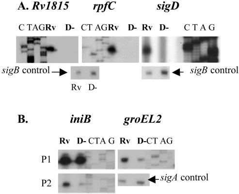
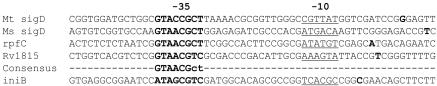
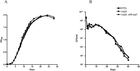
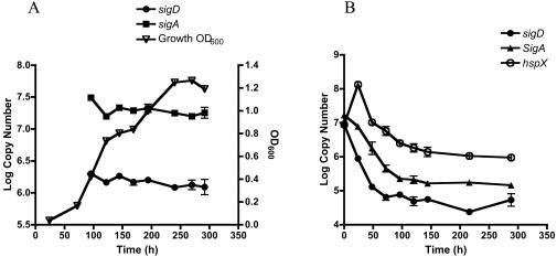
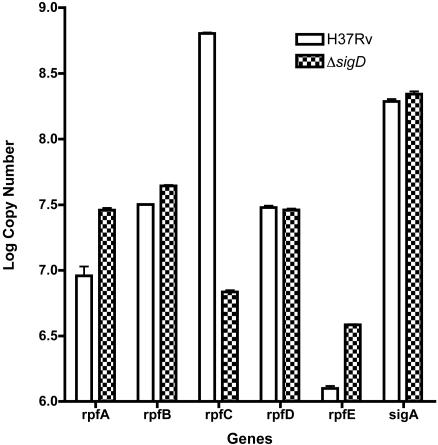
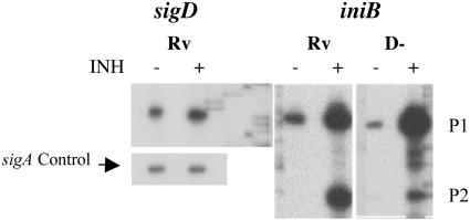
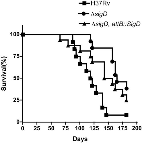
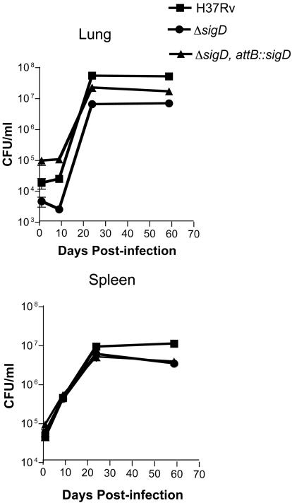
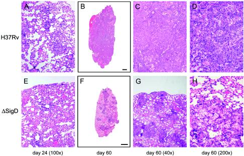
Mycobacterium tuberculosis, an obligate mammalian pathogen, adapts to its host during the course of infection via the regulation of gene expression. Of the regulators of transcription that play a role in this response, several alternative sigma factors of M. tuberculosis have been shown to control gene expression in response to stresses, and some of these are required for virulence or persistence in vivo. For this study, we examined the role of the alternative sigma factor SigD in M. tuberculosis gene expression and virulence. Using microarray analysis, we identified several genes whose expression was altered in a strain with a sigD deletion. A small number of these genes, including sigD itself, the gene encoding the autocrine growth factor RpfC, and a gene of unknown function, Rv1815, appear to be directly regulated by this sigma factor. By identifying the in vivo promoters of these genes, we have determined a consensus promoter sequence that is putatively recognized by SigD. The expression of several genes encoding PE-PGRS proteins, part of a large family of related genes of unknown function, was significantly increased in the sigD mutant. We found that the expression of sigD is stable throughout log phase and stationary phase but that it declines rapidly with oxygen depletion. In a mouse infection model, the sigD mutant strain was attenuated, with differences in survival and the inflammatory response in the lung between mice infected with the mutant and those infected with the wild type.


Figures

FIG. 1.
FIG. 1.
Primer extension analysis was performed with genes whose expression was increased in H37Rv relative to the ΔsigD strain to identify in vivo transcription start sites. RNAs were isolated from exponential-phase cultures of H37Rv (Rv) or the ΔsigD strain (D−) of M. tuberculosis. Sequencing ladders shown adjacent to each transcript were generated with the same primers as those used for RT. The results for promoters that were completely (A) or partially (B) SigD dependent are shown. Control experiments demonstrating sigA or sigB transcripts were performed with each RNA tested.
FIG. 2.
FIG. 2.
Alignment of promoter regions from the completely SigD-dependent in vivo promoters shown in Fig. 1 plus the experimentally defined SigD-dependent promoter of M. smegmatis (Ms-sigD). A strong consensus sequence in the −35 region is evident (bold letters), and an AT-rich −10 element is also present (underlined). Below the consensus line, the iniB P2 promoter, which is partially SigD dependent, is shown.
FIG. 3.
FIG. 3.
(A) Growth kinetics of M. tuberculosis H37Rv, the ΔsigD mutant, and the ΔsigD complemented strain, as determined in Middlebrook 7H9 broth supplemented with ADC and 0.05% Tween 80 incubated at 37°C. OD600 values were measured at serial time points for 4 weeks. (B) Survival of M. tuberculosis H37Rv, the ΔsigD mutant, and the ΔsigD complemented strain in an oxygen depletion system. Seventeen-milliliter samples of early-log-phase cultures (OD600 = 0.3) were placed in 25-ml sealed bottles to obtain a headspace ratio of 0.5. Aliquots were removed for CFU determinations and were plated in duplicate on Middlebrook 7H11 medium at serial time points for 40 days.
FIG. 4.
FIG. 4.
Expression of sigD mRNA in M. tuberculosis H37Rv, as measured by quantitative real-time RT-PCR during exponential growth and stationary phase (A) and during gradual oxygen depletion (B). Aliquots were removed at each time point, RNAs were isolated, and RT-PCRs were performed with 100 ng of RNA as described in Materials and Methods. Quantification of sigA mRNA was performed with an aliquot of the same RNA preparation at each time point. For the oxygen depletion experiments, hspX mRNA was quantified as a control for hypoxia. For each gene, the relative copy number was determined by use of a standard curve of H37Rv genomic DNA. Errors bars indicate standard errors of the means.
FIG. 5.
FIG. 5.
Expression of each of the five M. tuberculosis rpf gene mRNAs during exponential growth, as measured in H37Rv and the ΔsigD strain by quantitative real-time RT-PCR. RNAs were isolated, and RT-PCRs were performed with 100 ng of RNA as described in Materials and Methods. Quantification of sigA mRNA was performed on aliquots of the same RNA preparations. Errors bars indicate standard errors of the means.
FIG. 6.
FIG. 6.
Primer extension analysis of sigD and iniB promoters in response to INH exposure. Cultures of H37Rv (Rv) and ΔsigD (D−) were grown to mid-log phase and split into two aliquots, and INH was added to one aliquot to a final concentration of 1 μg/ml. The cells were harvested after 4 hours for RNA isolation, and primer extension experiments were performed with primers specific for sigD, iniB, or sigA (control).
FIG. 7.
FIG. 7.
Survival of mice infected with M. tuberculosis H37Rv, the ΔsigD mutant, and the ΔsigD complemented strain. BALB/c mice were infected by lateral tail vein injections with 106 CFU. Morbidity and weights were monitored throughout the experiment, and mice that became moribund were sacrificed. Twelve mice were infected in each group.
FIG. 8.
FIG. 8.
Replication and persistence of M. tuberculosis H37Rv, the ΔsigD mutant, and the ΔsigD complemented strain in mice. BALB/c mice were infected by lateral tail vein injections with 106 CFU, mice were sacrificed on days 1, 9, 24, and 59, and plating for bacterial burdens in the lungs and spleens was performed as described in Materials and Methods.
FIG. 9.
FIG. 9.
Histopathology of lungs of BALB/c mice infected with M. tuberculosis H37Rv or the ΔsigD strain. The images show hematoxylin and eosin staining of lung sections from mice infected with H37Rv (A to D) or the ΔsigD mutant (E to H). The time points at which the mice were sacrificed are indicated below the columns of panels.

Similar articles

Cited by

References

    1. Alland, D., A. J. Steyn, T. Weisbrod, K. Aldrich, and W. R. Jacobs, Jr. 2000. Characterization of the Mycobacterium tuberculosis iniBAC promoter, a promoter that responds to cell wall biosynthesis inhibition. J. Bacteriol. 182:1802-1811. - PMC - PubMed
    1. Banu, S., N. Honore, B. Saint-Joanis, D. Philpott, M. C. Prevost, and S. T. Cole. 2002. Are the PE-PGRS proteins of Mycobacterium tuberculosis variable surface antigens? Mol. Microbiol. 44:9-19. - PubMed
    1. Betts, J. C., P. T. Lukey, L. C. Robb, R. A. McAdam, and K. Duncan. 2002. Evaluation of a nutrient starvation model of Mycobacterium tuberculosis persistence by gene and protein expression profiling. Mol. Microbiol. 43:717-731. - PubMed
    1. Brennan, M. J., G. Delogu, Y. Chen, S. Bardarov, J. Kriakov, M. Alavi, and W. R. Jacobs, Jr. 2001. Evidence that mycobacterial PE_PGRS proteins are cell surface constituents that influence interactions with other cells. Infect. Immun. 69:7326-7333. - PMC - PubMed
    1. Chen, P., R. E. Ruiz, Q. Li, R. F. Silver, and W. R. Bishai. 2000. Construction and characterization of a Mycobacterium tuberculosis mutant lacking the alternate sigma factor gene sigF. Infect. Immun. 68:5575-5580. - PMC - PubMed

Publication types

LinkOut - more resources