. 2004 Oct;186(19):6667-70.
doi: 10.1128/JB.186.19.6667-6670.2004.

Channel-forming (Porin) activity in Herpetosiphon aurantiacus Hp a2

Affiliations

Channel-forming (Porin) activity in Herpetosiphon aurantiacus Hp a2

Rainer Harwardt et al. J Bacteriol. 2004 Oct.

Abstract

Detergent extracts of cell envelopes of the gliding bacterium Herpetosiphon aurantiacus formed channels in lipid bilayers. Fast protein liquid chromatography across a HiTrap-Q cation-exchange column demonstrated that a 45-kDa protein forms the channel. The observation of a channel-forming protein suggests that Herpetosiphon aurantiacus Hp a2 has a permeability barrier on its surface.


Figures
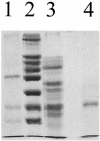
FIG. 1.
FIG. 1.
SDS-PAGE (7% polyacrylamide), performed according to Laemmli (14), as part of the purification procedure for the 45-kDa channel-forming protein of H. aurantiacus Hp a2. The gel was stained with Coomassie brilliant blue, and all protein samples were treated for 10 min at 100°C. Lane 1, low-molecular-mass markers of 36, 45, and 66 kDa; lane 2, high-molecular-mass markers of 36, 45, 55, 66, 84, 97, 116, and 205 kDa; lane 3, 15 μl of the 0.4% LDAO extract of the cell envelope fraction dissolved in 10 μl of sample buffer; lane 4, 5 μg of protein of fraction 32 of the Hitrap-Q FPLC column dissolved in 15 μl of sample buffer.
FIG. 2.
FIG. 2.
Single-channel recording of a PC/n-decane membrane in the presence of pure 45-kDa protein of H. aurantiacus Hp a2. The aqueous phase contained 1 M KCl (pH 6) and 10-ng/ml protein. The applied membrane potential was 20 mV (temperature of 20°C).
FIG. 3.
FIG. 3.
Single-channel recording of a PC/n-decane membrane in the presence of pure 45-kDa protein of H. aurantiacus Hp a2. The aqueous phase contained 1 M NH4Cl (pH 5) and 10-ng/ml protein. The applied membrane potential was 10 mV (temperature of 20°C). Note that the channels were transient under these conditions and open and closed in a single step, indicating that the channel is formed by a protein monomer.

Similar articles

Cited by

References

    1. Behlau, M., D. J. Mills, H. Quader, W. Kühlbrand, and J. Vonck. 2001. Projection structure of the monomeric porin OmpG at 6 Å resolution. J. Mol. Biol. 305:71-77. - PubMed
    1. Benz, R. 2001. Porins—structure and function, p. 227-246. In G. Winkelmann (ed.), Microbial transport systems. Wiley-VCH Verlag GmbH, Weinheim, Germany.
    1. Benz, R., K. Janko, W. Boos, and A. Läuger. 1978. Formation of large, ion-permeable membrane channels by the matrix protein (porin) of Escherichia coli. Biochim. Biophys. Acta 511:305-319. - PubMed
    1. Benz, R., K. Janko, and P. Läuger. 1979. Ionic selectivity of pores formed by the matrix protein (porin) of Escherichia coli. Biochim. Biophys. Acta 551:238-247. - PubMed
    1. Benz, R., A. Schmid, and R. E. W. Hancock. 1985. Ion selectivity of gram-negative bacterial porins. J. Bacteriol. 162:722-727. - PMC - PubMed

Publication types