. 2004 Nov 19;279(47):48993-9002.
doi: 10.1074/jbc.M409776200. Epub 2004 Sep 16.

Endonuclease-mediated mRNA decay requires tyrosine phosphorylation of polysomal ribonuclease 1 (PMR1) for the targeting and degradation of polyribosome-bound substrate mRNA

Affiliations

Endonuclease-mediated mRNA decay requires tyrosine phosphorylation of polysomal ribonuclease 1 (PMR1) for the targeting and degradation of polyribosome-bound substrate mRNA

Feng Yang et al. J Biol Chem. .

Abstract

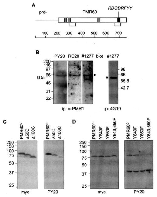
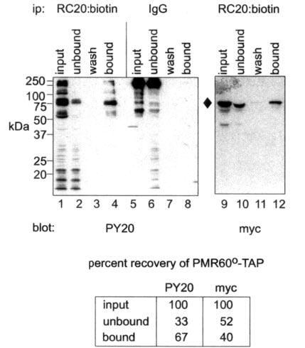
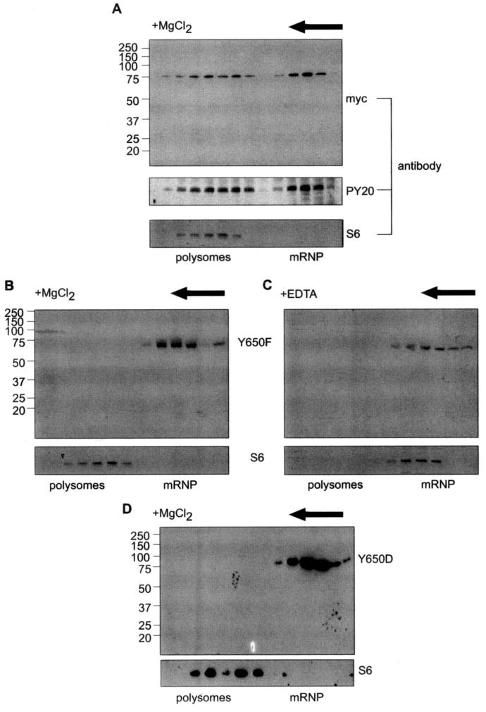
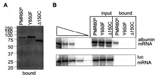
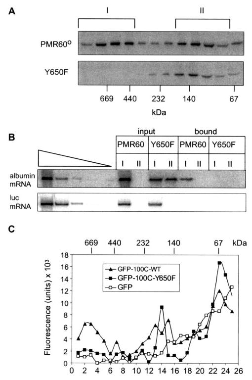
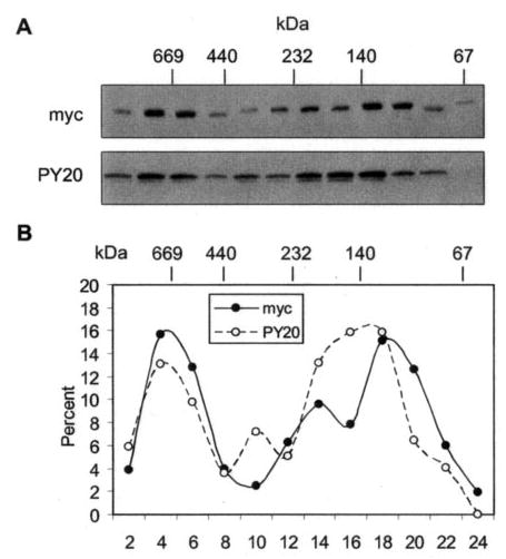
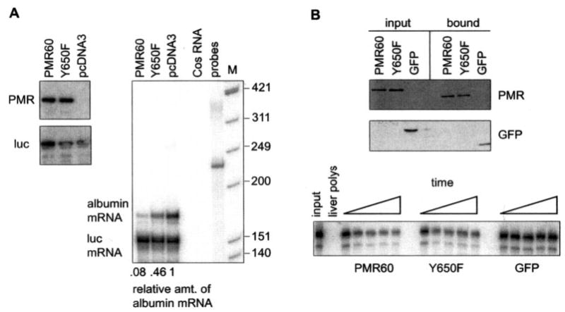
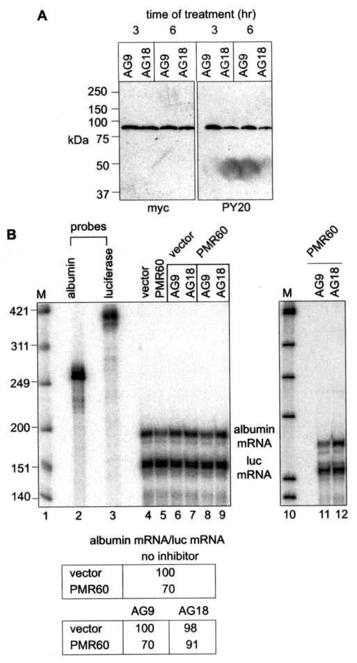
PMR1 is an endonuclease that is activated by estrogen to degrade Xenopus albumin mRNA. A previous report showed that the functional unit of endonuclease-mediated mRNA decay is a approximately 680-kDa polysome-bound complex that contains both PMR1 and substrate mRNA. PMR1 contains two domains involved in endonuclease targeting to polysomes, an N-terminal domain that lies between residues 200 and 250, and a C-terminal domain that lies within the last 100 residues. Loss of either domain inactivated PMR1 targeting to polysomes and stabilized albumin mRNA. The current study identified a phosphorylated tyrosine residue within the C-terminal polysome-targeting domain and showed that this modification is required for PMR1-mediated mRNA decay. Changing this tyrosine to phenylalanine inactivated the targeting of PMR1 to polysomes, blocked binding of PMR1 to the functional complex containing its substrate mRNA, prevented the targeting of a green fluorescent protein fusion protein to this complex, and stabilized albumin mRNA to degradation by PMR1 in vivo. A general tyrosine kinase inhibitor inhibited the phosphorylation of PMR1, which in turn inhibited PMR1-catalyzed degradation of albumin mRNA. These results indicate that one or more tyrosine kinases functions as a regulator of endonuclease-mediated mRNA decay.


Figures

Fig. 1
Fig. 1. Mapping the tyrosine phosphorylation site on PMR1
A, a schematic of the 80-kDa unprocessed PMR1 is shown with the mature 60-kDa form (PMR60) indicated, sequences with potential for binding to SH3 domains shown in gray boxes, a potential tyrosine kinase phospho-rylation site in the black box, and the locations of the two polysome targeting domains in brackets. B, a 0.4 M KCl extract of Xenopus liver polysomes was immunoprecipitated (ip) with a rabbit polyclonal antibody to PMR1 (three left panels) or with the phosphotyrosine mono-clonal antibody 4G10 (right panel), and recovered protein was separated by SDS-PAGE. Individual lanes of protein recovered with the PMR1 antibody were probed with the phosphotyrosine monoclonal antibodies PY20 or RC20:biotin or with a peptide-specific antibody to PMR1, 1277. The position of PMR1 is indicated with a filled circle (•). The lower band seen with the latter is IgG heavy chain. The blot of protein recovered with 4G10 was probed with the PMR1 antibody 1277. C, to map the general location of the tyrosine phosphorylation site Cos-1 cells were transfected with plasmids expressing TAP-tagged forms of the full-length protein (PMR60o) or deletions lacking 50 (~50C) or 100 (~100C) amino acids from the C terminus. Protein recovered on IgG-Sepharose was extracted with SDS sample buffer and analyzed by Western blot with antibody to the N-terminal Myc tag on PMR60oor PY20. D, the tyrosine residues at positions 649 (Y649F), 650 (Y650F), or both sites (Y649,650F) in PMR60o-TAP were mutated to phenylalanine, and the phosphorylation state of each protein expressed in transfected cells was determined as in C.
Fig. 2
Fig. 2. Estimating the population of PMR1 that is tyrosine-phosphorylated
Cytoplasmic extracts from cells transfected with PMR60o -TAP were incubated with RC20:biotin or nonimmune IgG before binding to immobilized avidin. Input, unbound, wash and bound fractions eluted with SDS sample buffer were separated by SDS-PAGE and analyzed by Western blot with PY20 or antibody to the Myc tag on PMR60o -TAP. The blot in the left panel probed with PY20 (lanes 1–8) determined the specificity and efficiency for recovering tyrosine-phosphorylated protein, and the blot in the right panel probed with Myc antibody (lanes 9–12) determined the efficiency of recovering PMR60o -TAP. The data quantified by scanning densitometry are shown beneath the figure. The recovery of Myc staining protein on avidin resin was normalized to the efficiency of recovery for tyrosine-phosphorylated PMR60o -TAP determined with PY20. ip, immunoprecipitate.
Fig. 3
Fig. 3. Mutating Y650 blocks the targeting PMR1 to polysomes
A, Cos-1 cells were transfected with PMR60o -TAP, and cytoplasmic extracts were separated on a 10–40% sucrose gradient that contained MgCl2 to maintain ribosome integrity. Even-numbered fractions were treated with EDTA to dissociate polysome-bound complexes, applied to IgG-Sepharose, and eluted with SDS sample buffer. The recovered proteins were analyzed by Western blot with antibody to the Myc tag or with PY20. The sedimentation of polysomes was determined by Western blot of unselected gradient fractions with a monoclonal antibody to ribosomal protein S6. B, Cos-1 cells with a plasmid expressing PMR60o with the Y650F mutation and fractionating cytoplasmic extract on a 10–40% sucrose gradient. Even-numbered fractions were analyzed by Western blot with antibody to the Myc tag or with antibody to S6 to map the sedimentation of polysomes on the gradient. C, the sample analyzed in B was sedimented on a gradient containing 5 mM EDTA, and even-numbered fractions were analyzed as in B with antibodies to the Myc tag or S6. D, cells were trans-fected with PMR60o, in which Tyr-650 was changed to aspartic acid (Y650D), and the sedimentation of PMR1 was analyzed on a gradient containing MgCl2 as in B.
Fig. 4
Fig. 4. The Y650F mutation prevents recovery of substrate mRNA by PMR1
Full-length PMR60o -TAP, PMR60o-TAP with the Y650F mutation (Y650F), or PMR60o -TAP deleted of the C-terminal 150 amino acids (Δ150C) were transfected into Cos-1 cells together with plasmids expressing albumin and luciferase mRNA. Extracts from puromycin-treated cells were bound to IgG-Sepharose and eluted by cleavage with Tev protease. Protein recovered in the bound complexes was analyzed by Western blot with antibody to the Myc tag (left panel), and RNA extracted from the bound complexes was analyzed by quantitative RT-PCR and phosphorimaging analysis (19) (right panel).
Fig. 5
Fig. 5. The Y650F mutation blocks targeting of PMR1 to the functional ~680-kDa complex
A, Cos-1 cells transfected with PMR60o or PMR60o with the Y650F mutation plus plasmids expressing albumin and luciferase mRNA were treated with puromycin before harvest, and cytoplasmic extracts were separated on 10–40% glycerol gradients. Even-numbered fractions were analyzed by Western blot with antibody to the Myc tag. B, the fractions identified with brackets in A were pooled and identified as complex I and complex II. Half of each pooled complex was bound to IgG-Sepharose and eluted by cleavage with Tev protease, and RNA extracted from the bound and unbound complexes was analyzed by quantitative RT-PCR as in Fig. 4. C, the C-terminal 100 amino acids of PMR1 bearing the native sequence or the Y650F mutation were fused to the C terminus of GFP. Cytoplasmic extracts from Cos-1 cells transfected with these or GFP alone were fractionated on a 10–40% glycerol gradient, and individual fractions were assayed for fluorescence. WT, wild type.
Fig. 6
Fig. 6. Total and tyrosine-phosphorylated PMR1 have overlapping but not identical sedimentation profiles
A, cytoplasmic extracts prepared from cells with PMR60o-TAP were bound to IgG-Sepharose, and complexes recovered by Tev protease cleavage were fractionated on a 10–40% glycerol gradient. Gradient fractions were analyzed by Western blot with antibody to the Myc tag to detect the total distribution of PMR1 and PY20 to detect the distribution of tyrosine-phosphorylated PMR1. B, the relative amount of protein determined with each antibody was determined by scanning densitometry and plotted as the percent present in each fraction of the gradient.
Fig. 7
Fig. 7. The Y650F mutation stabilizes albumin mRNA to degradation by PMR1
A, Cos-1 cells were transfected with pcDNA3, pcDNA expressing catalytically active PMR60, or pcDNA3 expressing PMR60 with the Y650F mutation together with plasmids expressing albumin and luciferase mRNA. The relative expression each protein was determined by Western blot (left panel), and their impact on steady-state levels of albumin and luciferase mRNA was determined by RNase protection (right panel). The relative amount of albumin mRNA normalized to luciferase mRNA determined by phosphorimaging analysis is shown beneath the autoradiogram. B, the catalytic activity of PMR60 and PMR60 with the Y650F mutation was determined by assaying the degradation of 5′32P-labeled albumin mRNA by PMR60-TAP, PMR60-TAP with the Y650F mutation, or GFP-TAP that were expressed in transfected cells and recovered on IgG-Sepharose. The upper panel is a Western blot of input protein and protein recovered from IgG-Sepharose Tev protease cleavage, and the lower panel is a time course of 0, 5, 10, 20, and 30 min of incubation with substrate RNA. M, Mr standards.
Fig. 8
Fig. 8. Tyrosine kinase inhibitor stabilizes albumin mRNA to degradation by PMR1
A, PMR60o-TAP was cloned into the tetracy-cline-regulated plasmid pTRE-myc and transfected into Cos-1 (tTA) cells in medium containing doxycycline. Fifteen hours later doxycycline was removed, and AG9 or AG18 were added to the medium. Cytoplasmic extracts prepared after 3 and 6 h were bound to IgG-Sepharose, eluted with SDS sample buffer, and analyzed by Western blot with antibody to the Myc tag or PY20. B, Cos-1 (tTA) cells were transfected in doxycycline-containing medium with plasmids expressing albumin and luciferase mRNA and either pTRE-myc (vector) or pTRE-PMR60. Doxycycline was removed 15 h later, and cells were incubated for 6 h with no additions (lanes 4 and 5), with AG9 (lanes 6, 8, and 11), or with AG18 (lanes 7, 9, and 12). The recovered RNA was analyzed by RNase protection assay, and albumin mRNA normalized to luciferase mRNA is presented in the table. The right panel (lanes 11 and 12) is an independent repeat of the data in lanes 8 and 9. M, Mr standards.

Similar articles

Cited by

References

    1. Wilusz CJ, Wormington M, Peltz SW. Nat Rev Mol Cell Biol. 2001;2:237–246. - PubMed
    1. Tharun S, Parker R. Mol Cell. 2001;8:1075–1083. - PubMed
    1. Sheth U, Parker R. Science. 2003;300:805–808. - PMC - PubMed
    1. Eystathioy T, Jakyymiw A, Chan EKL, Seraphin B, Cougot N, Fritzler MJ. RNA (N Y) 2003;9:1171–1173. - PMC - PubMed
    1. van Hoof A, Staples RR, Baker RE, Parker R. Mol Cell Biol. 2000;20:8230–8243. - PMC - PubMed

Publication types

MeSH terms

LinkOut - more resources