. 2004 Nov 1;560(Pt 3):627-38.
doi: 10.1113/jphysiol.2004.069054. Epub 2004 Sep 16.

Inhibitory modulation of distal C-terminal on protein kinase C-dependent phospho-regulation of rat TRPV1 receptors

Affiliations

Inhibitory modulation of distal C-terminal on protein kinase C-dependent phospho-regulation of rat TRPV1 receptors

Beiying Liu et al. J Physiol. .

Abstract

The vanilloid receptor TRPV1, previously known as VR1, has been implicated in pain sensation under both physiological and pathological conditions. The channel is highly expressed in sensory ganglion neurones and is activated by a range of noxious stimuli including irritant chemicals, acids and heat. In order to understand the structural basis underlying this polymodal activation and the regulation by intracellular signalling pathways, we have investigated the functional roles of the cytoplasmic C-terminal of rat TRPV1. A mutant with the maximal truncation of the distal C-terminal encompassing the last 88 residues was constructed. Of interest, this mutant exhibited a Ca(2+)-dependent functional loss; it was irresponsive to capsaicin in the presence of extracellular Ca(2+), but fully functional otherwise. Further studies of this construct revealed that extracellular Ca(2+) alone could activate the channel, and that the activation required protein kinase C (PKC) phosphorylation at S502, an event that was up-regulated by external Ca(2+) entry. We compared the truncation mutant with wild-type TRPV1 and demonstrated that it had a significantly increased sensitivity to PKC phosphorylation. These results suggest the distal C-terminal of TRPV1 can inhibit phosphorylation-induced potentiation of the wild-type channel. They also call into question some established functions of the distal C-terminal of TRPV1, including its roles in agonist binding and functional desensitization. We suggest that the functional loss of the truncation mutant, in the presence of extracellular Ca(2+), was not due to disruption of agonist binding or gating, but rather to desensitization promoted by unstimulated extracellular Ca(2+) entry.


Figures

Figure 1
Figure 1. Functional domains of distal C-terminal of TRPV1
The distal C-terminal of TRPV1 has been implicated in a variety of functions ranging from ligand binding to functional regulations. The molecular regions identified for these functions are indicated. Of them, 726–740 forms a Walker motif for ATP binding, S800 and T704 for PKC phosphorylation, S774 and S820 for PKA phosphorylation, E761 for ligand recognition, 777–820 for PIP2 binding and 767–801 for CaM association. The distal half of the terminal also confers specific thermal sensitivity. The truncation mutants that were studied here include TRPV1-Δ88 and TRPV1-Δ40, involving deletions of the last 88 and 40 residues, respectively. The TRPV1-Δ88 mutant represents a functional channel with the maximal truncation.
Figure 2
Figure 2. The truncated mutants retain normal activity at the whole-cell level in Ca2+-free solutions
A and B, whole-cell currents evoked by capsaicin, low pH and the combination of both for TRPV1-Δ88 and TRPV1-Δ40. Large and robust ionic currents were observed for both truncation mutants. C and D, heat responses of the mutant channels as a function of temperature. Each curve represents a recording from an individual cell. The insets show the time course of the elevation of the solution temperature. E, maximal currents of wild-type and mutant channels tested in the three conditions, 1 μm capsaicin, pH 5.5 and the combination of the two. Currents were normalized by membrane capacitance to account for variations in cell sizes. The number of cells in each trial is indicated over each bar. F, dose–response curves of capsaicin for the wild-type and the mutant channels. Each point describes mean ±s.e.m. from WT (n = 6), TRPV1-Δ40 (n = 5) and TRPV1-Δ88 (n = 10) experiments. The currents were normalized by the maximal responses obtained at 10 μm capsaicin in each experiment. The smooth curves are the best fits by the Hill equation, with wild-type: EC50, 0.6 ± 0.1; Hill coefficient (nH), 1.8 ± 0.5; TRPV1-Δ88: EC50, 0.6 ± 0.04; nH, 2.1 ± 0.3; TRPV1-Δ40: EC50, 0.7 ± 0.1; nH, 2.7 ± 0.4. All recordings were made from transiently transfected HEK293 cells in Ca2+-free solutions at a holding potential of −60 mV. Cap, capsaicin; WT, wild-type; Δ40, TRPV1-Δ40; Δ88, TRPV1-Δ88.
Figure 3
Figure 3. The truncated mutants retain near normal single-channel activity
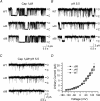
AC, single-channel currents from wild-type and truncation mutants evoked by different stimuli. Downward deflection represents channel opening. D, unitary current–voltage relationship activated by 1 μm capsaicin at normal pH. The TRPV1-Δ88 mutant exhibited a reduced unitary current at positive membrane potentials, while TRPV1-Δ40 was approximately the same as the wild-type. Data were summarized from three to four patches. All single-channel currents were recorded from Xenopus oocytes at +60 mV in the outside-out configuration. Cap, capsaicin; WT, wild-type; Δ40, TRPV1-Δ40; Δ88, TRPV1-Δ88.
Figure 4
Figure 4. Functional loss of TRPV1-Δ88 in the presence of extracellular Ca2+
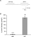
A, current response recorded from a HEK293 cell expressing TRPV1-Δ88 in a bath solution containing 1.8 mm Ca2+. The construct was nearly irresponsive to either capsaicin or low pH. B, current density of the wild-type and TRPV1-Δ88 measured in the presence of extracellular Ca2+. Capsaicin at 1 μm was used as the stimulus. The mutant channel retained only a portion (< 3%) of the wild-type current. Holding potential (Vh), −60 mV. Cap, capsaicin; WT, wild-type; Δ88, TRPV1-Δ88.
Figure 5
Figure 5. Extracellular Ca2+ activates TRPV1-Δ88
A and B, when cells were bathed in the Ca2+-free solution, application of extracellular Ca2+ alone induced a current in the absence of agonist. The Ca2+-evoked current was substantial as compared to that of 1 μm capsaicin. C and D, inhibition of Ca2+-evoked currents by intracellular Ca2+ chelation. Dialysis of cells with 10 mm BAPTA was sufficient to abolish the Ca2+ response. E, leak current detected in the whole-cell mode. Cells were bathed in the recording medium for > 5 min before recordings. All data were recorded from transiently transfected HEK293 cells at a Vh of −60 mV. Cap, capsaicin; Czp, capsazepine.
Figure 6
Figure 6. PKC phosphorylation at S502 underlies Ca2+-activation of TRPV1-Δ88
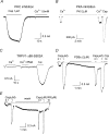
A and B, effects of protein kinase inhibitors on Ca2+-evoked currents. For PKC inhibition, cells were pre-incubated with BIM (2 μm) for 1 h. For PKA inhibition, cells were perfused for 10 min with 2 μm PKI 14–22, which is a membrane-permeable PKA inhibitor. C, mutation of S502 to alanine in TRPV1-Δ88 abolished the Ca2+-evoked current. D and E, leak current and potentiation of capsaicin responses (0.1 μm) induced by application of 2 μm PDBu (D) and intracellular dialysis of 140 nm free Ca2+ through the pipette solution containing 10 mm EGTA and 8 mm Ca2+ (E). All recordings were made at Vh of −60 mV. Cap, capsaicin; RR, ruthenium red.
Figure 7
Figure 7. Distal C-terminal modulates the potentiation effect induced by PKC phosphorylation
A, application of 1 μm PMA, with or without Ca2+, produced no detectable currents on HEK293 cells transiently transfected with the wild-type TRPV1. But subsequent application of low capsaicin (0.1 μm) elicited a large response comparable to that of 10 μm capsaicin, suggesting that the channel was phosphorylated prior to capsaicin application. B and C, current responses to application of capsaicin alone and together with 0.5 μm PMA for wild-type (B) and TRPV1-Δ88 (C). D, ratios of the increase of the current after PMA treatment. TRPV1-Δ88 showed a higher level of potentiation by PMA, at both 0.1 and 0.5 μm concentrations, than the wild-type. The capsaicin concentration was 0.1 μm. Cap, capsaicin; WT, wild-type.
Figure 8
Figure 8. Desensitization of TRPV1-Δ88
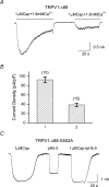
A, current responses of TRPV1-Δ88 to two repeated applications of 1 μm capsaicin in the presence of 1.8 mm Ca2+. Cells were bathed in Ca2+-free solutions. B, plot of the normalized peak current density during the two consecutive capsasaicin applications. C, current responses to capsaicin and pH recorded from cells expressing the construct TRPV1-Δ88-S502 in Ca2+-containing solution. The mutation S502A rescued the channel activity of the truncation mutant in Ca2+-containing bath solutions. The perfusion solutions contained no Ca2+. Currents were recorded at Vh of −60 mV.

Similar articles

Cited by

References

    1. Bhave G, Hu HJ, Glauner KS, Zhu W, Wang H, Brasier DJ, Oxford GS, Gereau RW. Protein kinase C phosphorylation sensitizes but does not activate the capsaicin receptor transient receptor potential vanilloid 1 (TRPV1) Proc Natl Acad Sci U S A. 2003;100:12480–12485. - PMC - PubMed
    1. Bhave G, Zhu W, Wang H, Brasier DJ, Oxford GS, Gereau RW. cAMP-dependent protein kinase regulates desensitization of the capsaicin receptor (VR1) by direct phosphorylation. Neuron. 2002;35:721–731. - PubMed
    1. Bonnington JK, McNaughton PA. Signalling pathways involved in the sensitisation of mouse nociceptive neurones by nerve growth factor. J Physiol. 2003;551:433–446. - PMC - PubMed
    1. Caterina MJ, Leffler A, Malmberg AB, Martin WJ, Trafton J, Petersen-Zeitz KR, Koltzenburg M, Basbaum AI, Julius D. Impaired nociception and pain sensation in mice lacking the capsaicin receptor. Science. 2000;288:306–313. - PubMed
    1. Caterina MJ, Schumacher MA, Tominaga M, Rosen TA, Levine JD, Julius D. The capsaicin receptor: a heat-activated ion channel in the pain pathway. Nature. 1997;389:816–824. 10.1038/39807. - DOI - PubMed

Publication types