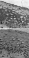
Blood group related antigens in ocular cicatricial pemphigoid
- PMID: 15377543
- PMCID: PMC1772371
- DOI: 10.1136/bjo.2003.039784
Blood group related antigens in ocular cicatricial pemphigoid
Abstract
Aim: To study the MUC5AC and the blood group related antigen expression in ocular cicatricial pemphigoid (OCP) according to the distribution of Lewis and secretor phenotypes in OCP patients compared to normal subjects.
Methods: Immunostaining was performed on conjunctival biopsy specimens from 22 consecutive patients suffering from OCP, using monoclonal antibodies (Mabs) directed against the peptidic core MUC5AC mucin (anti-M1/MUC5AC Mabs) and against the saccharide moieties (anti-blood group related antigens). These latter included anti-Le(a), anti-Le(b), anti-sialyl Le(a), and H type 2 Mabs, which immunoreact with Lewis positive and non-secretor (Le(a)), Lewis positive and secretor (Le(b)), Lewis positive (sialyl Le(a)), and secretor (H type 2) phenotypes respectively. Serological tests were also performed to confirm the phenotype of each patient. The immunohistopathological patterns and the distribution of Lewis and secretor phenotypes were compared with the results of a previous study in normal individuals.
Results: (1) In OCP patients compared to the normal population, anti-M1 immunoreactivity of goblet cells was unchanged, whereas anti-Le(a), anti-Le(b), and anti-sialyl Le(a) immunoreactivities of epithelial and/or goblet cells were markedly decreased. (2) 41% of OCP patients had a non-secretor phenotype, which is statistically significantly more than the estimated incidence of the same phenotype in the French population (20%) (p approximately 0.04).
Conclusions: Mucins in OCP patients showed a decreased expression of blood group related antigens whereas the MUC5AC peptidic core detected by anti-M1 Mab remained unchanged. These results also seem to indicate that OCP may be associated with a non-secretor phenotype.
Figures


References
-
- Mondino BJ, Brown SI. Ocular cicatricial pemphigoid. Ophthalmology 1981;88:95–100. - PubMed
-
- Wright P. Cicatrizing conjunctivitis. Trans Ophthalmol Soc UK 1986;105:1–17. - PubMed
-
- Hoang-Xuan T, Robin H, Demers PE, et al. Pure ocular cicatricial pemphigoid. A distinct immunopathologic subset of cicatricial pemphigoid. Ophthalmology 1999;106:355–61. - PubMed
-
- Rice BA, Foster CS. Immunopathology of cicatricial pemphigoid affecting the conjunctiva. Ophthalmology 1990;97:1476–83. - PubMed
MeSH terms
Substances
LinkOut - more resources
Full Text Sources
Medical
