

Transplantation of cultivated autologous oral mucosal epithelial cells in patients with severe ocular surface disorders
- PMID: 15377551
- PMCID: PMC1772364
- DOI: 10.1136/bjo.2003.038497
Transplantation of cultivated autologous oral mucosal epithelial cells in patients with severe ocular surface disorders
Abstract
Background/aims: To determine outcomes of transplants of cultivated autologous oral epithelial cells in patients with severe ocular surface disorders.
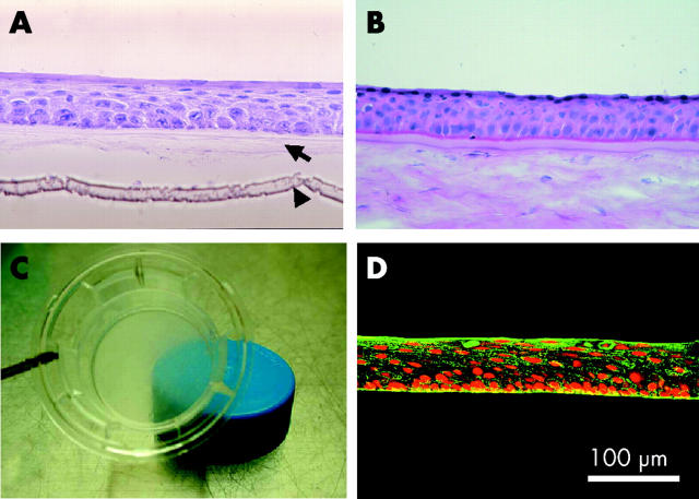
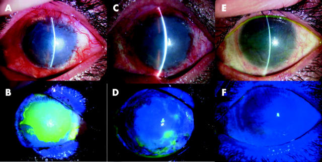
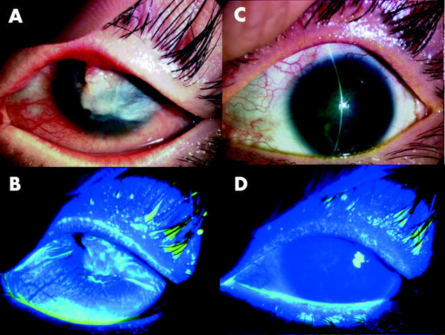
Methods: The eyes (n = 6) of four patients with Stevens-Johnson syndrome (three eyes) or chemical burns (three eyes) were studied. Autologous oral epithelial cells, grown for 2-3 weeks on a denuded amniotic membrane carrier in the presence of 3T3 fibroblasts, were air lifted. The resultant sheet was transplanted onto the damaged eye, and acceptance of the sheet by the corneal surface was confirmed 48 hours after surgery. The success of ocular surface reconstruction, graft survival, changes in visual acuity, and postoperative complications were assessed and the quality of the cultivated oral epithelial sheet was evaluated histologically.
Results: At 48 hours after transplant, the entire corneal surface of all six eyes was free of epithelial defects indicating complete survival of the transplanted oral epithelium. Visual acuity was improved in all eyes. During follow up (mean 13.8 (SD 2.9) months), the corneal surface remained stable, although all eyes manifested mild peripheral neovascularisation.
Conclusions: Autologous oral epithelial cells grown on denuded amniotic membrane can be transplanted to treat severe ocular surface disorders.
Figures
References
-
- Thoft RA, Friend J. Biochemical transformation of regenerating ocular surface epithelium. Invest Ophthalmol Vis Sci 1977;16:14–20. - PubMed
-
- Tsai RJF, Sun TT, Tseng SCG. Comparison of limbal and conjunctival autograft transplantation in corneal surface reconstruction in rabbits. Ophthalmology 1990;97:446–55. - PubMed
-
- Wei ZG, Wu RL, Lavker LM, et al. In vitro growth and differentiation of rabbit bulbar, fornix, and palpebral conjunctival epithelia: implications on conjunctival epithelial transdifferentiation and stem cells. Invest Ophthalmol Vis Sci 1993;34:1814–28. - PubMed
-
- Shapiro MS, Friend J, Thoft RA. Corneal re-epithelialization from the conjunctiva. Invest Ophthalmol Vis Sci 1981;21:135–42. - PubMed
-
- Dua H, Forrester JV. The corneoscleral limbus in human corneal epithelial wound healing. Am J Ophthalmol 1990;110:646–56. - PubMed
Publication types
MeSH terms
LinkOut - more resources
Full Text Sources
Other Literature Sources
Medical