. 2004 Oct;16(10):2614-28.
doi: 10.1105/tpc.104.024588. Epub 2004 Sep 17.

A classical arabinogalactan protein is essential for the initiation of female gametogenesis in Arabidopsis

Affiliations

A classical arabinogalactan protein is essential for the initiation of female gametogenesis in Arabidopsis

Gerardo Acosta-García et al. Plant Cell. 2004 Oct.

Abstract

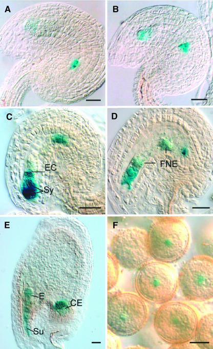
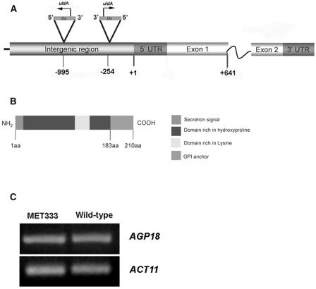
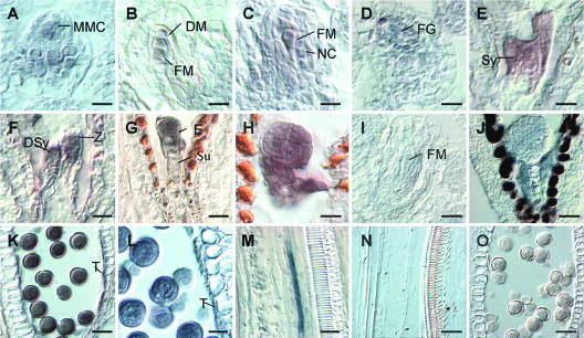
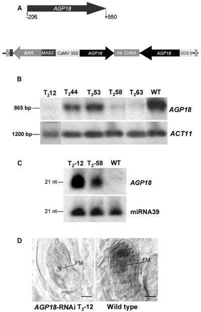
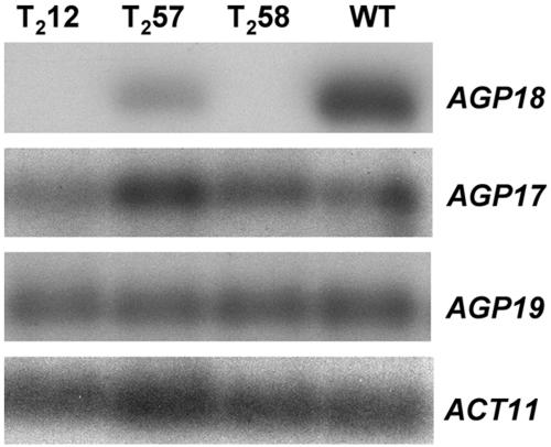
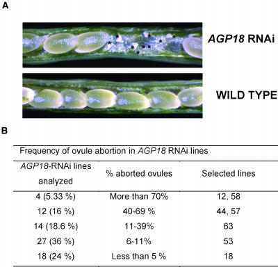
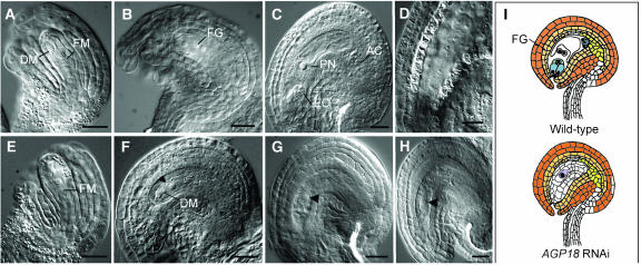
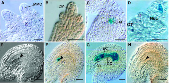
Classical arabinogalactan proteins (AGPs) are an abundant class of cell surface proteoglycans widely distributed in flowering plants. We have used a combination of enhancer detection tagging and RNA interference (RNAi)-induced posttrancriptional silencing to demonstrate that AGP18, a gene encoding a classical arabinogalactan protein, is essential for female gametogenesis in Arabidopsis thaliana. AGP18 is expressed in cells that spatially and temporally define the sporophytic to gametophytic transition and during early stages of seed development. More than 75% of the T1 transformants resulted in T2 lines showing reduced seed set during at least three consecutive generations but no additional developmental defects. AGP18-silenced T2 lines showed reduced AGP18 transcript levels in female reproductive organs, the presence of 21-bp RNA fragments specific to the AGP18 gene, and the absence of in situ AGP18 mRNA localization in developing ovules. Reciprocal crosses to wild-type plants indicate that the defect is female specific. The genetic and molecular analysis of AGP18-silenced plants containing a single T-DNA RNAi insertion suggests that posttranscriptional silencing of AGP18 is acting both at the sporophytic and gametophytic levels. A cytological analysis of all defective AGP18-RNAi lines, combined with the analysis of molecular markers acting at key stages of female gametogenesis, showed that the functional megaspore fails to enlarge and mitotically divide, indicating that AGP18 is essential to initiate female gametogenesis in Arabidopsis. Our results assign a specific function in plant development to a gene encoding a classical AGP.


Figures

Figure 1.
Figure 1.
Pattern of GUS Expression in the Enhancer Detector Line MET333. (A) Female gametophyte at four-nucleate stage. (B) Cellularized female gametophyte. (C) Mature female gametophyte before fertilization. (D) Female gametophyte after fertilization. (E) Embryo at four-cell stage. (F) Mature pollen with GUS expression associated with the vegetative nucleus. Sy, synergids; EC, egg cell; E, embryo; FNE, free nuclear endosperm; Su, suspensor; CE, chalazal endosperm. Bars in (A) to (E) = 20 μm; bar in (F) = 10 μm.
Figure 2.
Figure 2.
Genomic Structure and Protein Organization of AGP18. The enhancer detector line MET333 has two DsE elements inserted in the 5′ regulatory region of AGP18. (A) Genomic structure of AGP18. The arrows show the direction of transcription of uidA (GUS). (B) Predicted protein structure of AGP18. aa, amino acids. (C) RT-PCR analysis shows that the levels of transcription of AGP18 are identical in MET333 and wild-type plants.
Figure 3.
Figure 3.
Localization of AGP18 mRNA by in Situ Hybridization. (A) MMC stage. Bar = 9 μm. (B) Female meiosis stage. Bar = 8 μm. (C) Functional megaspore stage with young nucellus. Bar = 8.5 μm. (D) Two-nucleate stage female gametophyte. Bar = 14 μm. (E) Mature female gametophyte. Bar = 8 μm. (F) Zygote stage. Bar = 9μm. (G) Embryo four-cell stage. Bar = 15 μm. (H) Embryo at early globular stage. Bar = 11 μm. (I) Functional megaspore and young nucellus, sense probe. Bar = 15 μm. (J) Embryo at early globular stage, sense probe. Bar = 20 μm. (K) Mature pollen. Bar = 24 μm. (L) Anther showing the tapetum. Bar = 20 μm. (M) Longitudinal section of a stem. Bar = 20 μm. (N) Longitudinal section of a stem, sense probe. Bar = 20 μm. (O) Mature pollen, sense probe. Bar = 20 μm. (A) to (H) and (K) to (M) hybridizations with antisense probe; (I), (J), (N), and (O) hybridizations with sense probe. NC, nucellar cells; FM, functional megaspore; DM, degenerating megaspores; Sy, synergids; DSy, degenerating synergid; FG, female gametophyte; Z, zygote; E, embryo; Su, suspensor; T, tapetum.
Figure 4.
Figure 4.
Accumulation of AGP18 Transcript, Presence of 21-bp Small RNAs, and Absence of AGP18 Expression in the Gynoecium of AGP18-RNAi T2 Lines. (A) Schematic diagram of the vector used to posttrancriptionally silence AGP18. The arrow indicates the sequence cloned in the RNAi silencing vector. Numbers indicate nucleotide positions with respect to initiation of the AGP18 mRNA. (B) Expression analysis of four AGP18-RNAi T2 lines and a wild-type control. RNA was isolated from mature gynoecia in both silenced and wild-type plants. A portion of the AGP18 cDNA was used as a probe. RNA gel blots were subsequently rehybridized with a specific actin probe (ACT11) as a loading control. (C) A polyacrylamide gel of 100 μg of low molecular weight RNA extracted from gynoecia of AGP18-RNAi T2 lines and wild-type plants was blotted and hybridized with a portion of the AGP18 cDNA. The blot was rehybridized with a probe specific to the constitutively expressed microRNA 39 (miR39) as a control. nt, nucleotides. (D) Localization of AGP18 mRNA in developing ovules at the functional megaspore stage. In situ hybridization with specific AGP18 digoxygenin-labeled antisense probes was performed on gynoecia of both silenced (AGP18-RNAi T2-12; bar = 8.5 μm) and wild-type plants (bar = 10 μm). FM, functional megaspore.
Figure 5.
Figure 5.
Posttranscriptional Gene Silencing Is Specific to AGP18. RNA extracted from developing gynoecia of selected AGP18-RNAi T2 lines (T2-12, T2-57, and T2-58) was used for cDNA synthesis. PCR amplification was performed with primers specific to AGP17, AGP18, or AGP19 (Schultz et al., 2002) using as a template samples corresponding to the same cDNA synthesis. Agarose gels were blotted on nitrocellulose membranes and probed with a corresponding AGP probe. Wild-type cDNA and amplification of ACT11 were used as positive controls.
Figure 6.
Figure 6.
Siliques of AGP18-RNAi Lines Show Aborted Ovules. (A) Micrographs of AGP18-RNAi and wild-type siliques. The asterisks indicate the aborted ovules observed in AGP18-RNAi lines. (B) An average of 250 ovules was scored for each AGP18-RNAi line. A χ2 statistical analysis showed that lines having >5% ovule abortion were significantly different from the wild type (2.5% ovule abortion).
Figure 7.
Figure 7.
Female Gametophyte Development Is Defective in Ovules of AGP18-RNAi Lines. Wild-type and AGP18-RNAi T2 gynoecia were fixed, cleared, whole mounted, and viewed under Nomarsky optics. (A) to (D) Development of wild-type ovules. (A) Functional megaspore in a developing ovule. (B) Female gametophyte at the two-nucleate stage. (C) Mature female gametophyte. (D) Young embryo at the two-cellular stage. (E) to (H) Development of AGP18-RNAi T2-12 defective ovules. (E) Functional megaspore in the developing T2-12 ovule. (F) Arrested cell in defective T2-12 mature ovule (arrowhead); normal ovules in the same gynoecium are at the two-nucleate stage. (G) Arrested cell in defective T2-12 mature ovule (arrowhead); normal ovules in the same gynoecium contain a mature female gametophyte. (H) Arrested cell in T2-12 mature ovule; normal ovules in the same gynoecium contain seeds undergoing early stages of embryogenesis. (I) Schematic representation compares a mature wild-type ovule to a mature AGP18-RNAi T2-12 defective ovule. FM, functional megaspore; DM, degenerating megaspore cells; EC, egg cell; AC, antipodal cells; FG, female gametophyte; PN, polar nuclei. Arrowheads indicate the presence of an arrested cell at the one-nucleate stage. Bars = 20 μm.
Figure 8.
Figure 8.
The Functional Megaspore Does Not Initiate Female Gametogenesis in AGP18-RNAi Ovules. Ovules of F1 plants resulting from crosses of line AGP18-RNAi T2-12 with individuals homozygous either for the ET499 or for ET2209 were either fixed and whole-mount cleared or processed for histochemical localization of GUS activity. Shown are patterns of GUS expression in ET499 ([A] to [C]), ET4127 (D), F1 plants resulting from the cross of AGP18-RNAi T2-12 and homozygous ET499 plants ([E] and [F]), ET2209 (G), and F1 plants resulting from the cross of AGP18-RNAi T2-12 and homozygous ET2209 plants (H). (A) Ovules of ET499 at MMC not showing GUS expression. (B) Ovules of ET499 showing absence of expression in all three dying megaspores. (C) Functional megaspore showing GUS expression in ET499. (D) Ovule of ET4127 showing GUS expression in a dying megaspore. (E) Whole-mounted cleared ovule showing the phenotype observed in defective ET499;AGP18-RNAi T2-12 F1 ovules. (F) GUS expression in the arrested functional megaspore of defective ET499;AGP18-RNAi T2-12 F1 ovules. The arrowhead shows the position of the arrested functional megaspore. (G) Pattern of GUS expression in the mature female gametophyte of ET2209. (H) Absence of GUS expression in defective ET2209;AGP18-RNAi T2-12 F1 ovules. FM, functional megaspore; IT, inner integument; OT, outer integument; Nuc, nucellus; DM, dying megaspore; EC, egg cell; Sy, synergid cells; CC, central cell. The arrowheads indicate the arrested cells. Bars = 20 μm.

References

    1. Bajon, C., Horlow, C., Motamayor, J.C., Sauvanet, A., and Robert, D. (1999). Megasporogenesis in Arabidopsis thaliana L.: An ultrastructural study. Sex. Plant Reprod. 12, 99–109.
    1. Bechtold, N., Jaudeau, B., Jolivet, S., Maba, B., Vezon, D., Voisin, R., and Pelletier, G. (2000). The maternal chromosome set is the target of the T-DNA in the in planta transformation of Arabidopsis thaliana. Genetics 155, 1875–1887. - PMC - PubMed
    1. Bell, P.R. (1989). The alternation of generations. Adv. Bot. Res. 16, 55–93.
    1. Bell, P.R. (1995). Incompatibility in flowering plants: Adaptation of an ancient response. Plant Cell 7, 5–16. - PMC - PubMed
    1. Bellen, H.J. (1999). Ten years of enhancer detection: Lessons from the fly. Plant Cell 11, 2271–2282. - PMC - PubMed

Publication types

Associated data