Comparative Study
. 2004 Sep 28;101(39):14045-50.
doi: 10.1073/pnas.0405274101. Epub 2004 Sep 17.

The channel architecture of aquaporin 0 at a 2.2-A resolution

Affiliations
Comparative Study

The channel architecture of aquaporin 0 at a 2.2-A resolution

William E C Harries et al. Proc Natl Acad Sci U S A. .

Abstract

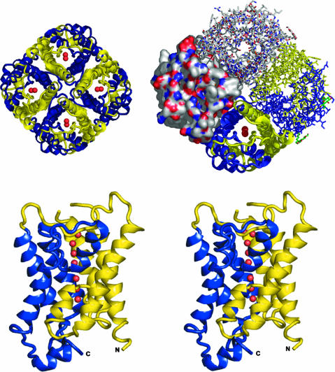
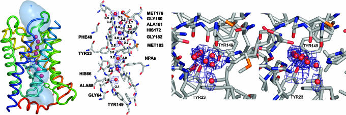
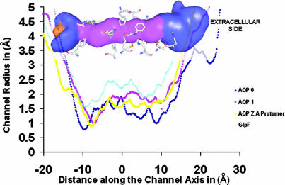
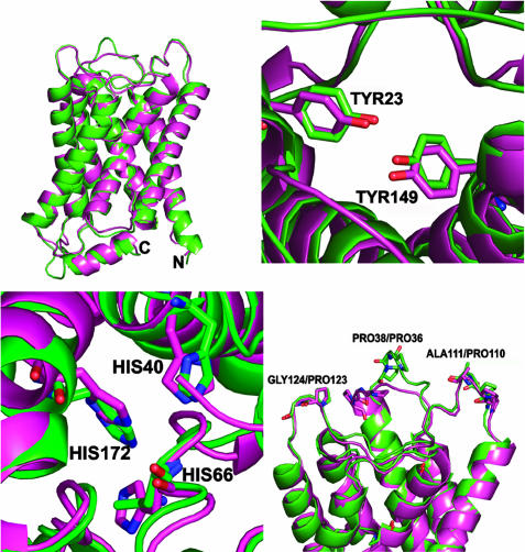
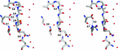
We determined the x-ray structure of bovine aquaporin 0 (AQP0) to a resolution of 2.2 A. The structure of this eukaryotic, integral membrane protein suggests that the selectivity of AQP0 for water transport is based on the identity and location of signature amino acid residues that are hallmarks of the water-selective arm of the AQP family of proteins. Furthermore, the channel lumen is narrowed only by two, quasi-2-fold related tyrosine side chains that might account for reduced water conductance relative to other AQPs. The channel is functionally open to the passage of water because there are eight discreet water molecules within the channel. Comparison of this structure with the recent electron-diffraction structure of the junctional form of sheep AQP0 at pH 6.0 that was interpreted as closed shows no global change in the structure of AQP0 and only small changes in side-chain positions. We observed no structural change to the channel or the molecule as a whole at pH 10, which could be interpreted as the postulated pH-gating mechanism of AQP0-mediated water transport at pH >6.5. Contrary to the electron-diffraction structure, the comparison shows no evidence of channel gating induced by association of the extracellular domains of AQP0 at pH 6.0. Our structure aids the analysis of the interaction of the extracellular domains and the possibility of a cell-cell adhesion role for AQP0. In addition, our structure illustrates the basis for formation of certain types of cataracts that are the result of mutations.


Figures

Fig. 1.
Fig. 1.
Tetramer and monomer structure of bAQP0. (Upper Left) Cartoon of the bAQP0 tetramer looking down the z axis from the extracellular side of the protein. Yellow and blue indicate structures derived from each of the two gene-duplicated portions of the primary sequence. (Upper Right) Cartoon showing the same view as in Upper Left, with each monomer shown in a different representation. (Lower) Cartoon of an bAQP0 monomer in a side view, with the uppermost extracellular side in crossed eye stereo. All images were made with pymol (DeLano Scientific, San Carlos, CA).
Fig. 2.
Fig. 2.
Monomer channel views of bAQP0. (Left) Side view of the monomer and water molecules (red spheres) in the channel. The channel luminal surface is shown in light blue. Each helix is colored in order of the rainbow. (Center) Side view looking from the midmembrane plane toward the monomer channel residues and water molecules in the channel. Hydrogen bonds to the channel waters are shown as dotted lines. Electron density around waters is shown in a composite omit 2Fo–Fc map contoured at 0.5 σ for clarity. (Right) Stereo view from the extracellular side of the channel. Electron density around waters and Tyr-23 and Tyr-149 are contoured at 0.5 σ. All images were made with pymol.
Fig. 3.
Fig. 3.
Channel radius profile plot. Channel radius profiles of AQPs of known structure with corresponding structural elements are shown (22, 23). The AQPZ “A” protomer was used for radius calculations for AQPZ. The distance along the channel axis is calculated by using a point midway between the Asn-Pro-Ala sequences (NPAs) as the zero point. Radii were calculated with hole (39). Channel volume is shown in the background, with major channel-forming residues. The pink central region has a diameter of <2.5 Å, the blue region has a diameter of >2.5 Å and <10 Å. All images were made with pymol.
Fig. 4.
Fig. 4.
Comparisons of the x-ray and electron crystal structures. (Upper Left) Structures overlaid with x-ray structure are shown in purple, and the electron-diffraction structure is shown in green. (Upper Right) View down the monomer channel z axis from the extracellular side showing the positions of Tyr-23 and Tyr-149. (Lower Left) View down the monomer channel z axis from the extracellular side showing the positions of the three histidines that are close to the channel and vestibules. (Lower Right) Side view. The extracellular residues involved in cell-to-cell contacts are highlighted and labeled.
Fig. 5.
Fig. 5.
Diagrams of the region surrounding the E134G and T138R cataract mutations. Overall, bovine and human amino acid sequences share 94% identity, and with regard to the amino acids shown in Fig. 6, all are conserved. Therefore, parallels can be made regarding their effect on channel permeability. The figures were produced with moloc and the mutation structure underwent minimization by using the molecular mechanics force field of cns. Possible hydrogen-bonding pairs are connected by yellow dotted lines.

Similar articles

Cited by

References

    1. Kinoshita, J. H., Kador, P. & Catiles, M. (1981) J. Am. Med. Assoc. 246, 257–261. - PubMed
    1. Trokel, S. (1962) Invest. Ophthalmol. 1, 493–501. - PubMed
    1. Kinoshita, J. H. & Merola, L. O. (1964) Invest. Ophthalmol. 47, 577–584. - PubMed
    1. Bloemendal, H. (1977) Science 197, 127–138. - PubMed
    1. Rink, H. (1982) Biophys. Struct. Mech. 9, 95–101. - PubMed

Publication types

Associated data

LinkOut - more resources