. 2004 Sep 28;43(38):12113-22.
doi: 10.1021/bi0494166.

Fetal Alz-50 clone 1 interacts with the human orthologue of the Kelch-like Ech-associated protein

Affiliations

Fetal Alz-50 clone 1 interacts with the human orthologue of the Kelch-like Ech-associated protein

Gordon D Strachan et al. Biochemistry. .

Abstract

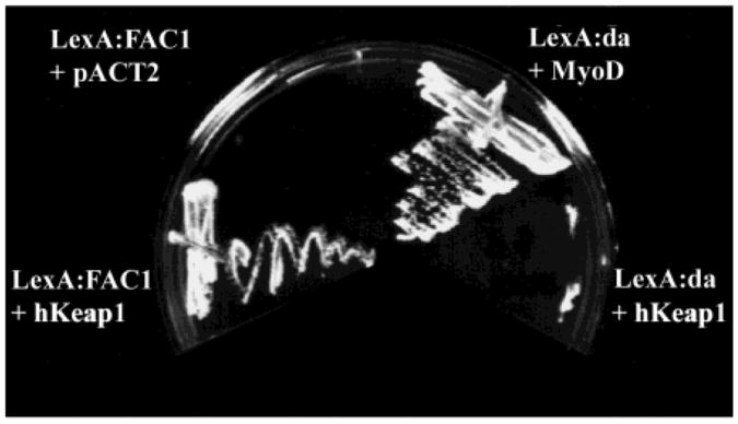
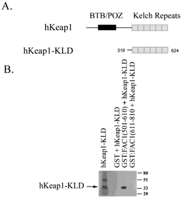
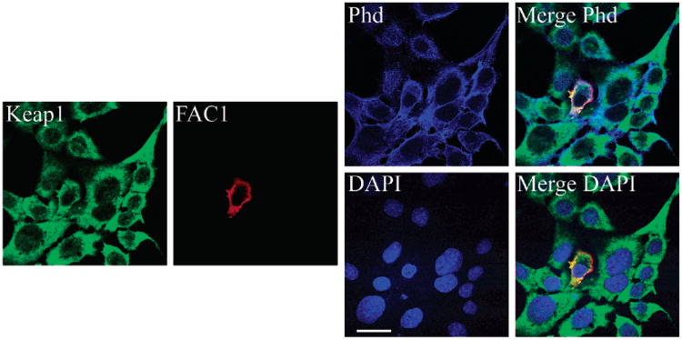
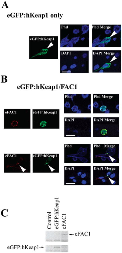
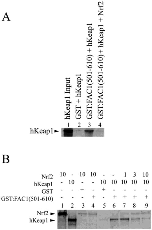
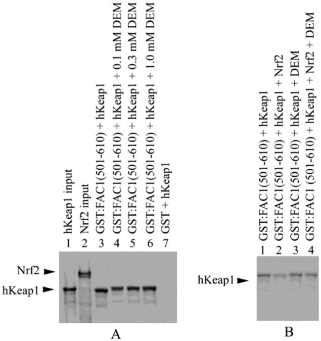
The fetal Alz-50 reactive clone 1 (FAC1) protein exhibits altered expression and subcellular localization during neuronal development and neurodegenerative diseases such as Alzheimer's disease. Using the yeast two-hybrid screen, the human orthologue of Keap1 (hKeap1) was identified as a FAC1 interacting protein. Keap1 is an important regulator of the oxidative stress response pathway through its interaction with the Nrf family of transcription factors. An interaction between full-length FAC1 and hKeap1 proteins has been demonstrated, and the FAC1 binding domain of hKeap1 has been identified as the Kelch repeats. In addition, FAC1 colocalizes with endogenous Keap1 within the cytoplasm of PT67 cells. Exogenously introduced eGFP:hKeap1 fusion protein redistributed FAC1 to colocalize with eGFP:hKeap1 in perinuclear, spherical structures. The interaction between FAC1 and hKeap1 is reduced by competition with the Nrf2 protein. However, competition by Nrf2 for hKeap1 is reduced by diethylmaleate (DEM), a known disrupter of the Nrf2:Keap1 interaction. DEM does not affect the ability of FAC1 to bind hKeap1 in our assay. These results suggest that hKeap1 regulates FAC1 in addition to its known role in control of Nrf2. Furthermore, the observed competition between FAC1 and Nrf2 for binding hKeap1 indicates that the interplay between these three proteins has important implications for neuronal response to oxidative stress.


Figures

Figure 1
Figure 1
FAC1 interacts with hKeap1 in vivo as assayed by the dihybrid yeast screen. Four cultures of the L40 yeast strain were transformed with (1) pLexA:FAC1(438–810) and hKeap1 (amino acids 42–624 as identified in the screen), (2) pLexA:FAC1 and pACT2 (library vector only), (3) LexA:da and MyoD (positive control for functioning interactions), or (4) LexA:da and hKeap1 (control for nonspecific interaction with LexA or an unrelated protein). Colonies were streaked on plates lacking the amino acid histidine on which only yeast containing interacting fusion proteins will grow.
Figure 2
Figure 2
hKeap1 interacts with a region of FAC1 containing a putative PEST domain. (A) GST:FAC1 fusion proteins shown here were used to make affinity columns. GST is shown in black, whereas the remainder contains the indicated FAC1 domains. FAC1 deletion mutants containing the putative PEST domain, nuclear localization sequence (NLS), and a nuclear export sequence (NES) are shown. Relative binding of each FAC1 domain to hKeap1 is indicated on the right. In (B) 35S-labeled, in vitro translated hKeap1 (42–624) (hKeap1; lane 1) was incubated with the following affinity columns: GST (lane 2), GST:FAC1(438–810) (lane 3), GST:FAC1(611–810) (lane 4), GST:FAC1(401–500) (lane 5), and GST:FAC1(501–610). Column-bound proteins were fractionated by size on a 10% SDS–polyacrylamide gel, which was transferred to PVDF. The autoradiograph of the transferred gel is shown in (B).
Figure 3
Figure 3
FAC1 binds the Kelch-like domain of hKeap1. (A) hKeap1 has two conserved motifs, the BTB/POZ domain and the Kelch-like repeat domain (KLD). In (B) 35S-labeled, in vitro translated hKeap1-KLD (lane 1) was incubated with the following affinity columns: GST (lane 2), GST:FAC1(501–610) (lane 3), and GST:FAC1(611–810) (lane 4). Column-bound proteins were fractionated by size on a 10% SDS–polyacrylamide gel, which was transferred to PVDF. The autoradiograph of the transferred gel is shown in (B).
Figure 4
Figure 4
FAC1 colocalizes with endogenous Keap1 and actin in mouse fibroblasts. PT67 cells were transfected with epitope tagged FAC1. Quadruple-label immunofluorescent confocal microscopy for endogenous murine Keap1 (green), FAC1 (red), actin via phalloidin (Phd, blue), and nuclear DNA via DAPI (DAPI, blue) demonstrated colocalization between FAC1 and Keap1 (Merge Phd and Merge DAPI). FAC1 and Keap1 also colocalized with actin (Merge Phd; green, red, and blue colocalization appears white). However, neither FAC1 nor Keap1 colocalizes with DAPI (FAC1 and Keap1 appear yellow to orange, indicating colocalization of the red and green, but not the blue). Bar= 20 μM.
Figure 5
Figure 5
Coexpression of hKeap1 with FAC1 redistributes FAC1 within the cytoplasm. PT67 murine fibroblasts were transfected with eGFP:hKeap1 alone (A) or with eGFP:hKeap1 and epitope tagged FAC1 (B). Transfected cells were stained for FAC1 by immunohistochemistry, actin by TRITC conjugated phalloidin, and DNA by DAPI and analyzed by quadruple-label immunofluorescent laser confocal microscopy. For panels A and B, FAC1 is shown in red, eGFP:hKeap1 is shown in green, actin is labeled with phalloidin and shown as blue in the Phd panel, and DAPI labeling nuclei are shown as blue in the DAPI panel. Panel B demonstrates FAC1 colocalization with eGFP:hKeap1. In panel C, western analysis of cells transfected with eGFP:hKeap1 (lane 2) and epitope-tagged FAC1 (eFAC1; lane 3) or untransfected (lane 1) demonstrates the levels of eGFP:hKeap1 and eFAC1 upon transfection. Bar = 20 μM.
Figure 6
Figure 6
FAC1 binding to hKeap1 is dramatically reduced by competition with NRF2. In (A), 35S-labeled hKeap1 (lane 1) was incubated with GST (lane 2) or GST:FAC1(501–610) in the presence (lane 4) or absence (lane 3) of in vitro translated Nrf2. Bound proteins were electrophoresed on a 10% SDS–polyacryl-amide gel, transferred to PVDF, and visualized by autoradiography. In (B), 35S-labeled, in vitro translated Nrf2 (lane 1) was incubated with GST affinity column (lane 3) or GST:FAC1(501–610) (lane 4). Shown also is the 35S-labeled, in vitro translated hKeap1 (lane 2), which is in equal amounts to Nrf2 (lane 1). A constant amount of hKeap1 (10 μL) was added to each reaction indicated. GST and hKeap1 alone (lane 5) did not interact. GST:FAC1(501–610) bound hKeap1 (lane 6) as shown in Figure 2. However, when Nrf2 was added to the GST:FAC1(501–610) binding reaction (lanes 7–9), we saw a reduction in bound hKeap1 in a dose-dependent manner. Nrf2 retention on GST:FAC1(501–610) was the same as GST control (lanes 7–9 compared to lane 3). At equimolar amounts of hKeap1 and Nrf2, >90% of the hKeap1 no longer bound FAC1.
Figure 7
Figure 7
Nrf2 does not outcompete FAC1 for hKeap1 as efficiently in the presence of diethylmaleate (DEM). In (A), 35S-labeled hKeap1 (lane 1) was incubated with GST (lane 7) or GST: FAC1(501–610) in the presence of increasing concentrations of DEM (lane 4–6) or in the absence of DEM (lane 3). Bound proteins were electrophoresed on a 10% SDS–polyacrylamide gel, transferred to PVDF, and visualized by autoradiography. Also shown is the in vitro translated 35S-labeled Nrf2 used in part B (lane 2). In (B), 35S-labeled, in vitro translated hKeap1 alone (lanes 1 and 3) or equal amounts of 35S-labled in vitro translated hKeap1 and Nrf2 (lanes 2 and 4) were incubated with GST:FAC1(501–610) in the absence (lanes 1 and 2) or presence of 0.2 μM DEM (lanes 3 and 4). GST:FAC1(501–610) bound hKeap1 (lane 1) as shown in Figures 2, 6A, and 7A. However, when Nrf2 was added to the GST:FAC1(501–610) binding reaction (lane 2), we saw a reduction in bound hKeap1 as seen in Figure 6. In the presence of 0.2 μM DEM, hKeap1 binding to GST:FAC1(501–610) was not altered (lane 3) as shown in Figure 7A. However, Nrf2 was not able to compete as efficiently with GST:FAC1(501–610) for hKeap1 binding in the presence of 0.2 μM DEM (lane 4).

References

    1. Jordan-Sciutto KL, Dragich JM, Bowser R. DNA binding activity of the fetal Alz-50 clone 1 (FAC1) protein is enhanced by phosphorylation. Biochem Biophys Res Commun. 1999;260:785–789. - PubMed
    1. Styren SD, Bowser R, Dekosky ST. Expression of fetal ALZ-50 reactive clone 1 (FAC1) in dentate gyrus following entorhinal cortex lesion. J Comp Neurol. 1997;386:555–561. - PubMed
    1. Mu X, Springer JE, Bowser R. FAC1 expression and localization in motor neurons of developing, adult, and amyotrophic lateral sclerosis spinal cord. Exp Neurol. 1997;146:17–24. - PubMed
    1. Schoonover S, Davies P, Bowser R. Immunolocalization and redistribution of the FAC1 protein in Alzheimer's disease. J Neuropathol Exp Neurol. 1996;55:444–455. - PubMed
    1. Bowser R, Giambrone A, Davies P. FAC1, a novel gene identified with the monoclonal antibody Alz50, is develop-mentally regulated in human brain. Dev Neurosci. 1995;17:20–37. - PubMed

Publication types

MeSH terms

LinkOut - more resources