

Identification of subpopulations with characteristics of mesenchymal progenitor cells from human osteoarthritic cartilage using triple staining for cell surface markers
- PMID: 15380042
- PMCID: PMC546281
- DOI: 10.1186/ar1210
Identification of subpopulations with characteristics of mesenchymal progenitor cells from human osteoarthritic cartilage using triple staining for cell surface markers
Abstract
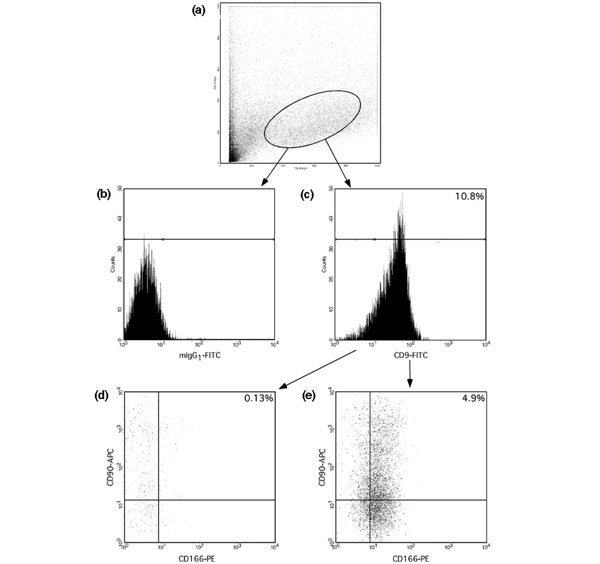
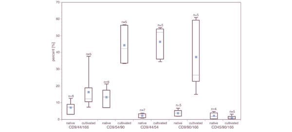
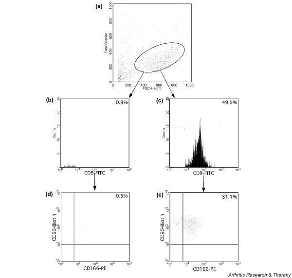
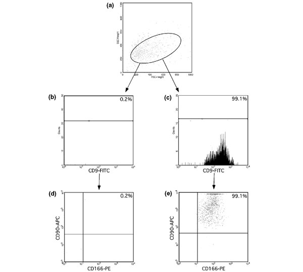
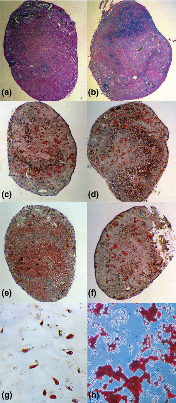
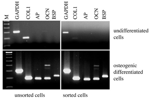
We first identified and isolated cellular subpopulations with characteristics of mesenchymal progenitor cells (MPCs) in osteoarthritic cartilage using fluorescence-activated cell sorting (FACS). Cells from osteoarthritic cartilage were enzymatically isolated and analyzed directly or after culture expansion over several passages by FACS using various combinations of surface markers that have been identified on human MPCs (CD9, CD44, CD54, CD90, CD166). Culture expanded cells combined and the subpopulation derived from initially sorted CD9+, CD90+, CD166+ cells were tested for their osteogenic, adipogenic and chondrogenic potential using established differentiation protocols. The differentiation was analyzed by immunohistochemistry and by RT-PCR for the expression of lineage related marker genes. Using FACS analysis we found that various triple combinations of CD9, CD44, CD54, CD90 and CD166 positive cells within osteoarthritic cartilage account for 2-12% of the total population. After adhesion and cultivation their relative amount was markedly higher, with levels between 24% and 48%. Culture expanded cells combined and the initially sorted CD9/CD90/CD166 triple positive subpopulation had multipotency for chondrogenic, osteogenic and adipogenic differentiation. In conclusion, human osteoarthritic cartilage contains cells with characteristics of MPCs. Their relative enrichment during in vitro cultivation and the ability of cell sorting to obtain more homogeneous populations offer interesting perspectives for future studies on the activation of regenerative processes within osteoarthritic joints.
Figures
Similar articles
-
Identification, quantification and isolation of mesenchymal progenitor cells from osteoarthritic synovium by fluorescence automated cell sorting.Osteoarthritis Cartilage. 2003 Nov;11(11):790-800. doi: 10.1016/s1063-4584(03)00167-5. Osteoarthritis Cartilage. 2003. PMID: 14609532
-
Progenitor cells from cartilage--no osteoarthritis-grade-specific differences in stem cell marker expression.Biotechnol Prog. 2013 Jan-Feb;29(1):206-12. doi: 10.1002/btpr.1668. Epub 2012 Dec 20. Biotechnol Prog. 2013. PMID: 23172745
-
Effects of oxygen and culture system on in vitro propagation and redifferentiation of osteoarthritic human articular chondrocytes.Cell Tissue Res. 2012 Mar;347(3):649-63. doi: 10.1007/s00441-011-1193-7. Epub 2011 Jun 4. Cell Tissue Res. 2012. PMID: 21638206
-
Immunohistochemistry in the study of normal and osteoarthritic articular cartilage.Prog Histochem Cytochem. 1998;33(2):93-165. doi: 10.1016/s0079-6336(98)80004-1. Prog Histochem Cytochem. 1998. PMID: 10319375 Review. No abstract available.
-
Histochemistry as a unique approach for investigating normal and osteoarthritic cartilage.Eur J Histochem. 2014 Apr 15;58(2):2371. doi: 10.4081/ejh.2014.2371. Eur J Histochem. 2014. PMID: 24998926 Free PMC article. Review.
Cited by
-
Chondroitin sulfate sulfation motifs as putative biomarkers for isolation of articular cartilage progenitor cells.J Histochem Cytochem. 2008 Feb;56(2):125-38. doi: 10.1369/jhc.7A7320.2007. Epub 2007 Oct 15. J Histochem Cytochem. 2008. PMID: 17938280 Free PMC article.
-
Targeting of chondrocyte plasticity via connexin43 modulation attenuates cellular senescence and fosters a pro-regenerative environment in osteoarthritis.Cell Death Dis. 2018 Dec 5;9(12):1166. doi: 10.1038/s41419-018-1225-2. Cell Death Dis. 2018. PMID: 30518918 Free PMC article.
-
Cell surface markers for mesenchymal stem cells related to the skeletal system: A scoping review.Heliyon. 2023 Feb 10;9(2):e13464. doi: 10.1016/j.heliyon.2023.e13464. eCollection 2023 Feb. Heliyon. 2023. PMID: 36865479 Free PMC article.
-
Senolytic activity of small molecular polyphenols from olive restores chondrocyte redifferentiation and promotes a pro-regenerative environment in osteoarthritis.Aging (Albany NY). 2020 Aug 3;12(16):15882-15905. doi: 10.18632/aging.103801. Epub 2020 Aug 3. Aging (Albany NY). 2020. PMID: 32745074 Free PMC article.
-
Complement C3a and C5a modulate osteoclast formation and inflammatory response of osteoblasts in synergism with IL-1β.J Cell Biochem. 2011 Sep;112(9):2594-605. doi: 10.1002/jcb.23186. J Cell Biochem. 2011. PMID: 21598302 Free PMC article.
References
-
- Wickham MQ, Erickson GR, Gimble JM, Vail TP, Guilak F. Multipotent stromal cells derived from the infrapatellar fat pad of the knee. Clin Orthop. 2003;412:196–212. - PubMed
Publication types
MeSH terms
Substances
LinkOut - more resources
Full Text Sources
Other Literature Sources
Medical
Research Materials
Miscellaneous
