. 2004 Sep 21;32(16):4915-24.
doi: 10.1093/nar/gkh837. Print 2004.

Conserved tertiary base pairing ensures proper RNA folding and efficient assembly of the signal recognition particle Alu domain

Affiliations

Conserved tertiary base pairing ensures proper RNA folding and efficient assembly of the signal recognition particle Alu domain

Laurent Huck et al. Nucleic Acids Res. .

Abstract

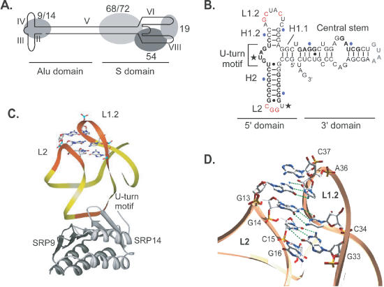
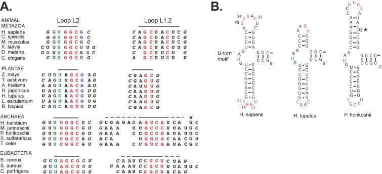
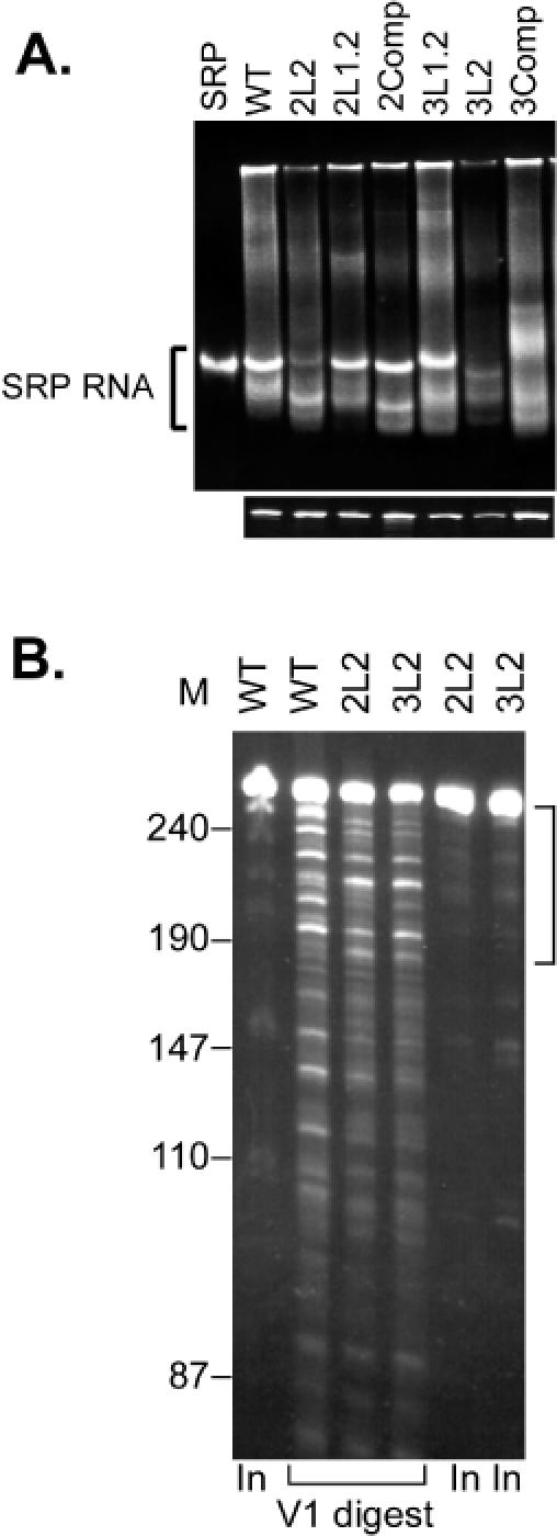
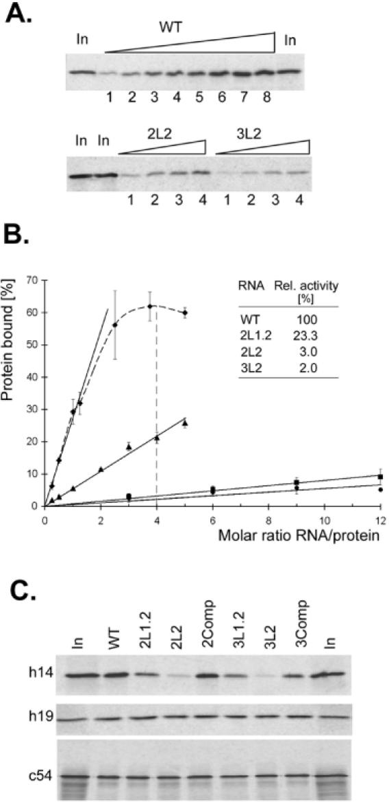
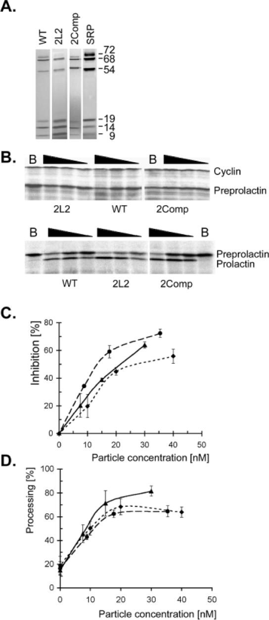
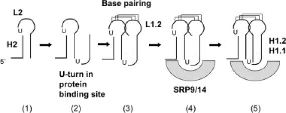
Proper folding of the RNA is an essential step in the assembly of functional ribonucleoprotein complexes. We examined the role of conserved base pairs formed between two distant loops in the Alu portion of the mammalian signal recognition particle RNA (SRP RNA) in SRP assembly and functions. Mutations disrupting base pairing interfere with folding of the Alu portion of the SRP RNA as monitored by probing the RNA structure and the binding of the protein SRP9/14. Complementary mutations rescue the defect establishing a role of the tertiary loop-loop interaction in RNA folding. The same mutations in the Alu domain have no major effect on binding of proteins to the S domain suggesting that the S domain can fold independently. Once assembled into a complete SRP, even particles that contain mutant RNA are active in arresting nascent chain elongation and translocation into microsomes, and, therefore, tertiary base pairing does not appear to be essential for these activities. Our results suggest a model in which the loop-loop interaction and binding of the protein SRP9/14 play an important role in the early steps of SRP RNA folding and assembly.


Figures

Figure 1
Figure 1
Structure of SRP and the SRP Alu domain. (A) Schematic representation of SRP, (B) secondary structure of the minimal Alu RNA that still binds 9/14 efficiently. Stems and loops are named according to the topological nomenclature. Bases in the loops that form tertiary base pairs are highlighted in red. Protein footprints are shown in boldface. U-turns are marked with asterisks and stretches of 10 nt are marked in blue. The U-turn motif marks a highly conserved sequence that represents the major protein-binding site. (C) Structure of the SRP Alu 5′ domain. SRP9 and SRP14 are displayed as dark and light grey ribbons, respectively. The RNA is shown as a yellow ribbon with the loop sequences and the U-turn motif shown in orange. Nucleotides from loops L2 and L1.2 that are involved in tertiary interactions between the loops are shown as wireframe. (D) Detailed view of the tertiary base pairing between loops. Bases G13, G14 and C15 form hydrogen bonds with C37, C34 and G33, respectively (dotted green lines). One base from each loop, G16 and A36, are positioned to extend the stack formed by the three base pairs.
Figure 2
Figure 2
Structure-based alignment of loop L1.2 and L2 sequences in SRP RNAs of animal metazoans, of plants and of eubacterial and archaeal species. (A) Nucleotides proposed to base pair are shown in red, sheared G-G pairs in blue and U-turns in green. First nucleotides in stems are shown in italic. Black bars delineate the loop sequences. For each species, only one representative is shown. Sequences were obtained from SRPDB. (B) Helix diagrams of mammalian, plant and archaeal Alu RNA 5′ domains. Asterisks highlight a conserved nucleotide in the archaeal RNAs. The helix diagram of a eubacterial SRP RNA resembles most closely the one of archaeal SRP RNAs.
Figure 3
Figure 3
Analysis of wild-type and mutated synthetic SRP RNAs. (A) Native (upper panel) and denaturing (lower panel) 6% PAGE. Equal amounts of RNA were loaded on both gels. The RNAs were visualized by staining with Gelstar®. SRP RNA was extracted from purified canine SRP. The synthetic RNAs are labelled as shown in Table 1. (B) Limited V1 ribonuclease digestion experiments. The digestion products were displayed by 10% denaturing PAGE and the RNA fragments visualized with ethidium bromide staining. The bracket highlights the region that contains RNAs obtained by single cleavages in the Alu portion of SRP RNA. In, 50% of the RNA used in the experiments.
Figure 4
Figure 4
Binding of human SRP9/14 to mutated SRP RNAs. (A) Titration experiments with synthetic WT; 2L2 and 3L2 biotinylated RNAs. The binding reactions contained 40 nM h9/14 and tracer amounts of 35S-labelled h14 in complex with recombinant h9. WT RNA concentrations in lanes 1–8: 10, 20, 30, 40, 50, 100, 150 and 200 nM. 2L2 and 3L2 RNA concentrations in lanes 1–4: 60, 120, 240 and 480 nM. In, 50% of total protein used in the experiment. The bound protein was displayed by SDS–PAGE followed by autoradiography. (B) Quantitative analysis of the titration experiments. WT (diamonds), 2L1.2 (triangles), 2L2 (squares) and 3L2 (dots) RNAs. (C) Protein binding with the mutated synthetic SRP RNAs. The protein and RNA concentrations were 40 and 160 nM, respectively, in the binding reactions. The binding reactions of c54 also contained 40 nM recombinant h19. The quantification of the results is shown in Table 2.
Figure 5
Figure 5
Elongation arrest and translocation activities of particles reconstituted with mutated SRP RNAs. (A) Fractions enriched in complete SRP that was reconstituted in vitro with all SRP proteins and WT, 2L2 and 2Comp synthetic RNAs. (B) Elongation arrest (upper panel) and translocation (lower panel) assays with 2, 1, 0.5 and 0 μl (B, Buffer) of the fractions shown in (A). Translocation assays contain SRP-depleted microsomes. B, buffer. (C and D) Quantification of the elongation arrest and translocation assays. The ratio of preprolactin to cyclin in the buffer sample was taken as 0% inhibition in the elongation arrest assay. In the absence of exogenous SRP, the membranes have a residual translocation activity of about 20%. Values represent the average of at least two independent experiments. WT SRP (diamonds), 2L2 SRP (triangles) and 2Comp SRP (dots).
Figure 6
Figure 6
Model for the early steps of SRP RNA folding. Step (1) formation of the first very stable hairpin structure; step (2) the central U-turn ensures orientation of the emerging strand to allow base pairing with loop L2 [step (3)]; SRP9/14 recognizes the correctly folded central U-turn region and by binding to it stabilizes the fold [step (4)]; formation and alignment of H1.2 and H1.2 ensues [step (5)]. It is possible that the protein may bind the U-turn before base pairing occurs.

References

    1. Weeks K.M. (1997) Protein-facilitated RNA folding. Curr. Opin. Struct. Biol., 7, 336–342. - PubMed
    1. Woodson S.A. (2000) Recent insights on RNA folding mechanisms from catalytic RNA. Cell Mol. Life Sci., 57, 796–808. - PMC - PubMed
    1. Walter P. and Johnson,A.E. (1994) Signal sequence recognition and protein targeting to the endoplasmic reticulum membrane. Annu. Rev. Cell Biol., 10, 87–119. - PubMed
    1. Johnson A.E. and van Waes,M.A. (1999) The translocon: a dynamic gateway at the ER membrane. Annu. Rev. Cell Dev. Biol., 15, 799–842. - PubMed
    1. Keenan R.J., Freymann,D.M., Stroud,R.M. and Walter,P. (2001) The signal recognition particle. Annu. Rev. Biochem., 70, 755–775. - PubMed

Publication types