. 2004 Sep 28;101(39):14126-31.
doi: 10.1073/pnas.0405899101. Epub 2004 Sep 21.

Targeted disruption of the Walker-Warburg syndrome gene Pomt1 in mouse results in embryonic lethality

Affiliations

Targeted disruption of the Walker-Warburg syndrome gene Pomt1 in mouse results in embryonic lethality

Tobias Willer et al. Proc Natl Acad Sci U S A. .

Abstract

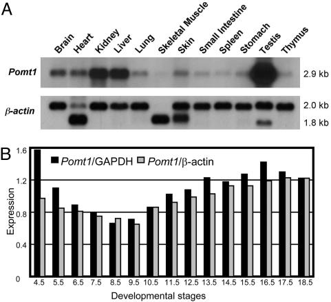
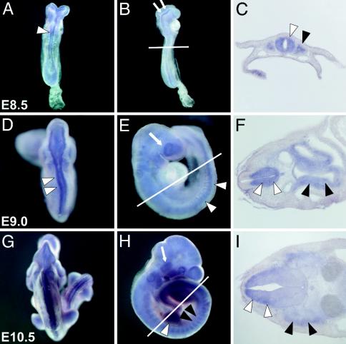
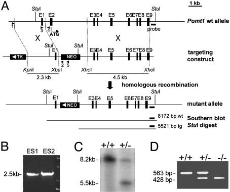
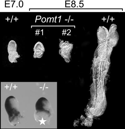
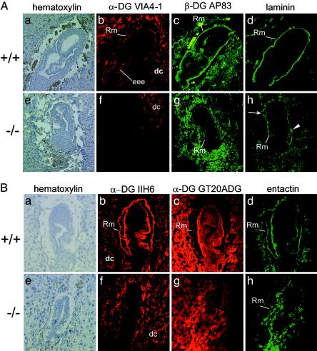
O-mannosylation is an important protein modification in eukaryotes that is initiated by an evolutionarily conserved family of protein O-mannosyltransferases. The first mammalian protein O-mannosyltransferase gene described was the human POMT1. Mutations in the hPOMT1 gene are responsible for Walker-Warburg syndrome (WWS), a severe recessive congenital muscular dystrophy associated with defects in neuronal migration that produce complex brain and eye abnormalities. During embryogenesis, the murine Pomt1 gene is prominently expressed in the neural tube, the developing eye, and the mesenchyme. These sites of expression correlate with those in which the main tissue alterations are observed in WWS patients. We have inactivated a Pomt1 allele by gene targeting in embryonic stem cells and produced chimeras transmitting the defect allele to offspring. Although heterozygous mice were viable and fertile, the total absence of Pomt1(-/-) pups in the progeny of heterozygous intercrosses indicated that this genotype is embryonic lethal. An analysis of the mutant phenotype revealed that homozygous Pomt1(-/-) mice suffer developmental arrest around embryonic day (E) 7.5 and die between E7.5 and E9.5. The Pomt1(-/-) embryos present defects in the formation of Reichert's membrane, the first basement membrane to form in the embryo. The failure of this membrane to form appears to be the result of abnormal glycosylation and maturation of dystroglycan that may impair recruitment of laminin, a structural component required for the formation of Reichert's membrane in rodents. The targeted disruption of mPomt1 represents an example of an engineered deletion of a known glycosyltransferase involved in O-mannosyl glycan synthesis.


Figures

Fig. 1.
Fig. 1.
Expression of mouse Pomt1 gene. (A) Northern blot analysis of adult tissues hybridized with cDNA probes: Pomt1 (2.9 kb) and β-actin (2.0 and 1.8 kb). (B) Pomt1 expression during mouse embryogenesis. Original Northern blot is shown in Fig. 6C. Northern blot was normalized by using GAPDH (black columns) and β-actin (gray columns).
Fig. 2.
Fig. 2.
Whole-mount expression of Pomt1 in mouse embryos. Whole-mount in situ hybridizations (A, B, D, E, G, and H) and representative cryosections (C, F, and I) through the anterior neural tube at the level of the forelimb bud (indicated by a white line in B, E, and H). (AC) At E8.5, strong Pomt1 expression is found along the neural tube (white arrowhead) and in the dorsal aspects of the neural fold (arrows). Expression was also detected in the somites (black arrowhead). (DF) At E9.0, strong expression was seen in the ventral part of the neural tube (white arrowheads), in the developing eye (white arrow), and in the gut endoderm (black arrowheads). (G and H) At E10.5, high levels of Pomt1 mRNA were detected in the somites (black arrowheads), limb buds (white arrowhead), and trigeminal ganglion (white arrow). (I) Pronounced Pomt1 expression in the mantle layer of the dorsal neural tube (white arrowheads), as well as in the dermomyotome (black arrowheads), was verified in the E10.5 section.
Fig. 3.
Fig. 3.
Targeted disruption of the Pomt1 gene. (A) Schematic representation of the targeting strategy: the genomic locus, the targeting construct, and the expected mutant Pomt1 allele after homologous recombination. Selectable markers: herpes simplex virus thymidine kinase gene (TK) and neomycin gene (NEO). The short (2.3-kb) and long (4.5-kb) arms for homologous recombination are represented. PCR primers are represented by arrows. (B) Primers 1 and 2 were used to identify two targeted embryonic stem clones after homologous recombination. (C) Southern blot analysis of genomic DNA from mouse tail tissue. Endogenous (8.2-kb) and targeted (5.5-kb) Pomt1 alleles. (D) PCR genotyping of embryos from timed matings. Primers 3 and 4 identify the endogenous allele, whereas primers 3 and 5 identify the targeted allele.
Fig. 4.
Fig. 4.
Morphology of wild-type and Pomt1–/– embryos. Representative littermates from an E8.5 Pomt1 heterozygous intercross are shown. PCR genotyping confirmed Pomt1–/– null mutants. Pomt1-deficient embryos display severe growth retardation presumably due to a developmental block in E6–7. For size comparison, an additional E7.0 wild-type embryo is presented. (Inset) Whole-mount in situ hybridization with the gastrulation marker brachyury. The asterisk indicates a missing part of the extraembryonic ectoderm used for PCR genotypization.
Fig. 5.
Fig. 5.
Immunohistochemical characterization of extracellular matrix components in Pomt1–/– mutant embryos. Sagittal sections of paraffin-embedded E7.5 (A) or frozen E6.5 (B) embryos were stained with hematoxylin and analyzed with anti-α-DG antibodies directed against a glycoepitope (VIA4–1, IIH6) or the protein core (GT20ADG) and anti-β-DG (AP83), anti-laminin, and anti-nidogen/entactin antibodies, as indicated. Wild-type embryos (A and B ad) and independent Pomt1 null mutants (A and Beh) are shown. In Pomt1–/– embryos, the glycoepitope is missing in all embryo-derived cellular structures, although it is still present in the decidual cells. Discontinuous (arrows) and patchy (arrowhead) laminin and nidogen/entactin staining of the mutant embryos indicates a defect in the formation of Reichert's membrane. Genotypes were determined by PCR genotyping of laser-captured material. Rm, Reichert's membrane; eee, extraembryonic ectoderm; dc, maternal decidual cells.

References

    1. Strahl-Bolsinger, S., Gentzsch, M. & Tanner, W. (1999) Biochim. Biophys. Acta 1426, 297–307. - PubMed
    1. Willer, T., Valero, M. C., Tanner, W., Cruces, J. & Strahl, S. (2003) Curr. Opin. Struct. Biol. 13, 621–630. - PubMed
    1. Endo, T. & Toda, T. (2003) Biol. Pharmacol. Bull. 26, 1641–1647. - PubMed
    1. Michele, D. E. & Campbell, K. P. (2003) J. Biol. Chem. 278, 15457–15460. - PubMed
    1. Muntoni, F., Brockington, M., Torelli, S. & Brown, S. C. (2004) Curr. Opin. Neurol. 17, 205–209. - PubMed

Publication types

MeSH terms

Associated data