

Fast and effective prediction of microRNA/target duplexes
- PMID: 15383676
- PMCID: PMC1370637
- DOI: 10.1261/rna.5248604
Fast and effective prediction of microRNA/target duplexes
Abstract
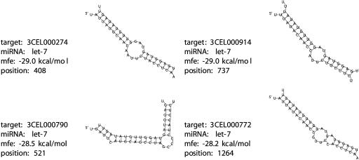
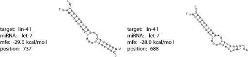
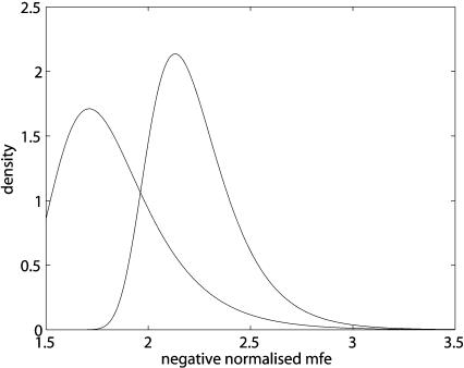
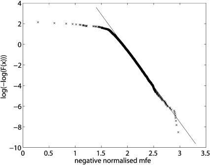
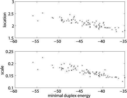
MicroRNAs (miRNAs) are short RNAs that post-transcriptionally regulate the expression of target genes by binding to the target mRNAs. Although a large number of animal miRNAs has been defined, only a few targets are known. In contrast to plant miRNAs, which usually bind nearly perfectly to their targets, animal miRNAs bind less tightly, with a few nucleotides being unbound, thus producing more complex secondary structures of miRNA/target duplexes. Here, we present a program, RNA-hybrid, that predicts multiple potential binding sites of miRNAs in large target RNAs. In general, the program finds the energetically most favorable hybridization sites of a small RNA in a large RNA. Intramolecular hybridizations, that is, base pairings between target nucleotides or between miRNA nucleotides are not allowed. For large targets, the time complexity of the algorithm is linear in the target length, allowing many long targets to be searched in a short time. Statistical significance of predicted targets is assessed with an extreme value statistics of length normalized minimum free energies, a Poisson approximation of multiple binding sites, and the calculation of effective numbers of orthologous targets in comparative studies of multiple organisms. We applied our method to the prediction of Drosophila miRNA targets in 3'UTRs and coding sequence. RNAhybrid, with its accompanying programs RNAcalibrate and RNAeffective, is available for download and as a Web tool on the Bielefeld Bioinformatics Server (http://bibiserv.techfak.uni-bielefeld.de/rnahybrid/).
Copyright 2004 RNA Society
Figures
Similar articles
-
Prediction of microRNA targets.Methods Mol Biol. 2006;342:87-99. doi: 10.1385/1-59745-123-1:87. Methods Mol Biol. 2006. PMID: 16957369
-
Analysis of microRNA-target interactions by a target structure based hybridization model.Pac Symp Biocomput. 2008:64-74. Pac Symp Biocomput. 2008. PMID: 18232104
-
Computational analysis of microRNA targets in Caenorhabditis elegans.Gene. 2006 Jan 3;365:2-10. doi: 10.1016/j.gene.2005.09.035. Epub 2005 Dec 13. Gene. 2006. PMID: 16356665
-
Prediction of human microRNA targets.Methods Mol Biol. 2006;342:101-13. doi: 10.1385/1-59745-123-1:101. Methods Mol Biol. 2006. PMID: 16957370 Review.
-
Prediction of microRNA targets.Drug Discov Today. 2007 Jun;12(11-12):452-8. doi: 10.1016/j.drudis.2007.04.002. Epub 2007 Apr 26. Drug Discov Today. 2007. PMID: 17532529 Review.
Cited by
-
The effect of Jiedu Huoxue decoction on rat model of experimental nonbacterial prostatitis via regulation of miRNAs.Pharm Biol. 2020 Dec;58(1):745-759. doi: 10.1080/13880209.2020.1797124. Pharm Biol. 2020. PMID: 32758035 Free PMC article.
-
MiR-124a Regulates Extracellular Vesicle Release by Targeting GTPase Rabs in Lung Cancer.Front Oncol. 2020 Aug 20;10:1454. doi: 10.3389/fonc.2020.01454. eCollection 2020. Front Oncol. 2020. PMID: 32974168 Free PMC article.
-
Unraveling the plasticity of translation initiation in prokaryotes: Beyond the invariant Shine-Dalgarno sequence.PLoS One. 2024 Jan 11;19(1):e0289914. doi: 10.1371/journal.pone.0289914. eCollection 2024. PLoS One. 2024. PMID: 38206950 Free PMC article.
-
A conserved RpoS-dependent small RNA controls the synthesis of major porin OmpD.Nucleic Acids Res. 2012 Apr;40(8):3623-40. doi: 10.1093/nar/gkr1156. Epub 2011 Dec 17. Nucleic Acids Res. 2012. PMID: 22180532 Free PMC article.
-
Computational Screening to Predict MicroRNA Targets in the Flavivirus 3' UTR Genome: An Approach for Antiviral Development.Int J Mol Sci. 2024 Sep 21;25(18):10135. doi: 10.3390/ijms251810135. Int J Mol Sci. 2024. PMID: 39337625 Free PMC article.
References
-
- Abrahante, J.E., Daul, A.L., Li, M., Volk, M.L., Tennessen, J.M., Miller, E.A., and Rougvie, A.E. 2003. The Caenorhabditis elegans hunchback-like gene lin-57/hbl-1 controls developmental time and is regulated by microRNAs. Dev. Cell 4: 625–637. - PubMed
-
- Brennecke, J., Hipfner, D.R., Stark, A., Russell, R.B., and Cohen, S.M. 2003. bantam encodes a developmentally regulated microRNA that controls cell proliferation and regulates the proapoptotic gene hid in Drosophila. Cell 113: 25–36. - PubMed
-
- Butler, M.J., Jacobsen, T.L., Cain, D.M., Jarman, M.G., Hubank, M., Whittle, J.R.S., Phillips, R., and Simcox, A. 2003. Discovery of genes with highly restricted expression patterns in the Drosophila wing disc using DNA oligonucleotide microarrays. Development 130: 659–670. - PubMed
MeSH terms
Substances
LinkOut - more resources
Full Text Sources
Other Literature Sources
Molecular Biology Databases