Comparative Study
. 2004 Oct;72(10):5799-806.
doi: 10.1128/IAI.72.10.5799-5806.2004.

Differential expression of Toll-like receptors 2 and 4 in tissues of the human female reproductive tract

Affiliations
Comparative Study

Differential expression of Toll-like receptors 2 and 4 in tissues of the human female reproductive tract

Patricia A Pioli et al. Infect Immun. 2004 Oct.

Abstract

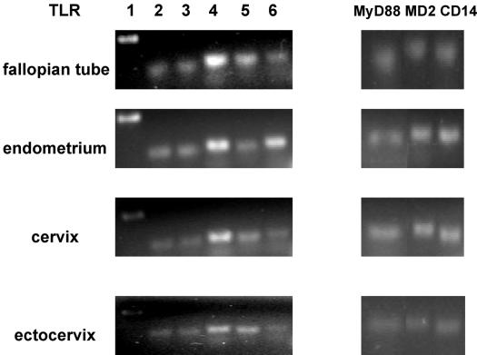
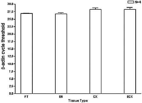
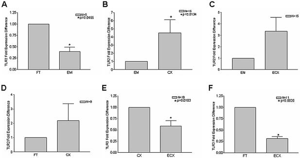
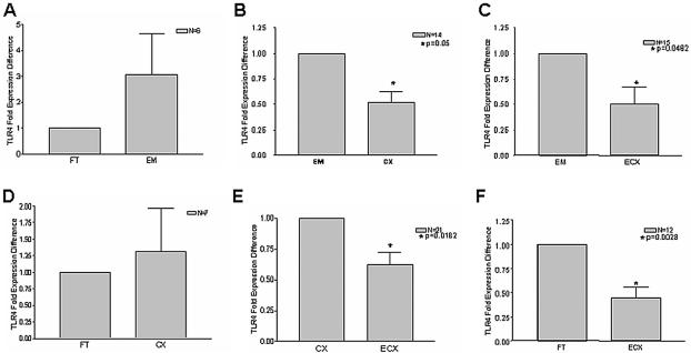
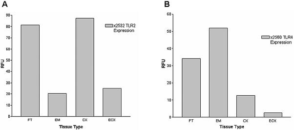
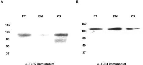
Toll-like receptor (TLR) signal transduction is a central component of the innate immune response to pathogenic challenge. Although recent studies have begun to elucidate differences in acquired immunity in tissues of the human female reproductive tract, there is a relative paucity of work regarding innate defense mechanisms. We investigated TLR mRNA and protein expression in tissues of the human female reproductive tract. Constitutive mRNA expression of TLRs 1 to 6 was observed in fallopian tubes, uterine endometrium, cervix, and ectocervix. Furthermore, transcripts of the signaling adapter MyD88 and the accessory molecule CD14 were also detected in all tissues assayed. Quantitative analysis of TLR2 mRNA levels revealed highest expression of this molecule in fallopian tube and cervical tissues, followed by endometrium and ectocervix. In contrast to TLR2, TLR4 expression declined progressively along the tract, with highest expression in the upper tissues (fallopian tubes and endometrium), followed by cervix and ectocervix. In addition to mRNA, protein expression of TLR2 and TLR4 was also documented in these tissues. These data suggest that TLRs are differentially expressed in distinct compartments of the female reproductive tract and may provide insight regarding the regulation of inflammation and immunity within the tract.


Figures

FIG. 1.
FIG. 1.
mRNA expression of TLRs, MyD88, MD-2, and CD14 in tissues of the female reproductive tract. Total RNA was extracted from fallopian tube, endometrial, cervical and ectocervical tissues, reverse transcribed, and amplified with the primers described in Table 1 for expression of pattern recognition receptors. For each tissue, the data shown are representative of gene expression observed in 10 different patient samples.
FIG. 2.
FIG. 2.
Validation of β-actin as a normalization control. Total RNA was extracted from each female reproductive tract tissue, reverse transcribed, and analyzed by TaqMan PCR with validated β-actin primers and probe (as described in the text). Comparisons were made with tissue samples derived from the same patient to maintain internal consistency; therefore, four individual patients from whom fallopian tube (FT), endometrium (EM), cervix (CX), and ectocervix (ECX) samples were obtained are represented in this study. All reactions were performed in triplicate.
FIG. 3.
FIG. 3.
TLR2 mRNA is most highly expressed in fallopian tubes and cervix. Total RNA was isolated from the fallopian tube (FT), endometrium (EM), cervix (CX), and ectocervix (ECX) as described in the text, and reverse transcription was performed with random hexamers. Comparisons among tissue types were made with paired tissue samples derived from the same patients. The sample size for each comparison is indicated in the upper right quadrant of each figure. Real-time PCR analysis of relative TLR2 mRNA expression was performed in duplicate with TaqMan TLR2- and β-actin-specific primers and probes. Samples incubated in the absence of reverse transcriptase were analyzed as well to control for genomic DNA contamination.
FIG. 4.
FIG. 4.
TLR4 mRNA expression declines along tissues of the female reproductive tract. cDNA was synthesized by reverse transcription from total RNA, and relative TLR4 mRNA levels were assessed by real-time PCR analysis with normalization to β-actin controls. Each reaction was performed in duplicate. Comparisons among tissue types were made with paired tissue samples of the fallopian tube (FT), endometrium (EM), cervix (CX), and ectocervix (ECX) derived from the same patients. The sample size for each comparison is indicated in the upper right quadrant of each panel.
FIG. 5.
FIG. 5.
Representative analysis of TLR2 and TLR4 expression profiles. (A) Total RNA was extracted from fallopian tube (FT), endometrium (EM), cervix (CX), and ectocervix (ECX) samples of patient 2532 and reverse transcribed with random hexamers. TLR2 levels were quantified by real-time analysis with Taqman TLR2 primers and probes as in Fig. 3. (B) TLR4 mRNA levels were quantified in fallopian tube (FT), endometrium (EM), cervix (CX), and ectocervix (ECX) samples derived from patient 2560 by real-time Taqman PCR as described for Fig. 4. Reactions were standardized against β-actin and are expressed in relative fluorescence units (RFU). Values shown are the means of duplicate PCR runs, which never varied by more than 3%.
FIG. 6.
FIG. 6.
Detection of TLR2 and TLR4 proteins in upper and lower tissues of the female reproductive tract. Whole-cell lysates were generated from total tissue of the fallopian tube (FT), endometrium (EM), cervix (CX), and ectocervix (ECX) and quantified with the BCA kit. One milligram of each of these lysates was immunoprecipitated with either (A) anti-TLR2 polyclonal antibody (H-175) or (B) mouse monoclonal anti-TLR4 antibody (HTA 125), and subsequently probed with (A) goat polyclonal antibody N-17 for detection of TLR2 or (B) rabbit polyclonal anti-TLR4 antibody H-80. Data shown are representative of three separate immunoprecipitations from three different tissues.

References

    1. Asai, Y., Y. Ohyama, K. Gen, and T. Ogawa. 2001. Bacterial fimbriae and their peptides activate human gingival epithelial cells through Toll-like receptor 2. Infect. Immun. 69:7387-7395. - PMC - PubMed
    1. Barlow, R. E., I. D. Cooke, O. Odukoya, M. Heatley, J. Jenkins, G. Narayansingh, S. S. Ramsewak, and A. Eley. 2001. The prevalence of Chlamydia trachomatis in fresh tissue specimens from patients with ectopic pregnancy or tubal factor infertility as determined by PCR and in-situ hybridisation. J. Med. Microbiol. 50:902-908. - PubMed
    1. Bowie, A., and L. A. O'Neill. 2000. The interleukin-1 receptor/Toll-like receptor superfamily: signal generators for pro-inflammatory interleukins and microbial products. J. Leukoc. Biol. 67:508-514. - PubMed
    1. Darville, T., J. M. O'Neill, C. W. Andrews, Jr., U. M. Nagarajan, L. Stahl, and D. M. Ojcius. 2003. Toll-like receptor-2, but not toll-like receptor-4, is essential for development of oviduct pathology in chlamydial genital tract infection. J. Immunol. 171:6187-6197. - PubMed
    1. Eggert-Kruse, W., Reimann-Andersen, J., G. Rohr, S. Pohl, W. Tilgen, and B. Runnebaum. 1995. Clinical relevance of sperm morphology assessment using strict criteria and relationship with sperm-mucus interaction in vivo and in vitro. Fertil. Steril. 63:612-624. - PubMed

Publication types

MeSH terms