. 2004 Oct;72(10):5832-9.
doi: 10.1128/IAI.72.10.5832-5839.2004.

Bacteroides fragilis enterotoxin induces intestinal epithelial cell secretion of interleukin-8 through mitogen-activated protein kinases and a tyrosine kinase-regulated nuclear factor-kappaB pathway

Affiliations

Bacteroides fragilis enterotoxin induces intestinal epithelial cell secretion of interleukin-8 through mitogen-activated protein kinases and a tyrosine kinase-regulated nuclear factor-kappaB pathway

Shaoguang Wu et al. Infect Immun. 2004 Oct.

Abstract

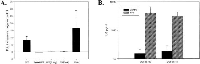
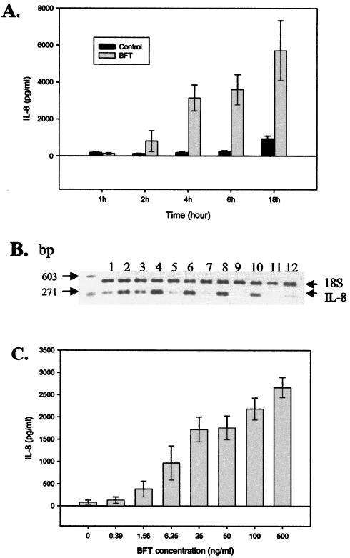
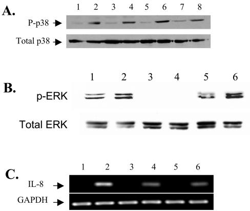
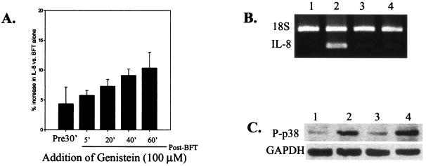
Enterotoxigenic Bacteroides fragilis (ETBF) secretes a 20-kDa metalloprotease toxin termed B. fragilis toxin (BFT). ETBF disease in animals is associated with an acute inflammatory response in the intestinal mucosa, and lethal hemorrhagic colitis may occur in rabbits. In this study, we confirmed recent reports (J. M. Kim, Y. K. Oh, Y. J. Kim, H. B. Oh, and Y. J. Cho, Clin. Exp. Immunol. 123:421-427, 2001; L. Sanfilippo, C. K. Li, R. Seth, T. J. Balwin, M. J. Menozzi, and Y. R. Mahida, Clin. Exp. Immunol. 119:456-463, 2000) that purified BFT stimulates interleukin-8 (IL-8) secretion by human intestinal epithelial cells (HT29/C1 cells) and demonstrate that stimulation of IL-8 production is dependent on biologically active BFT and independent of serum. Induction of IL-8 mRNA expression occurs rapidly and ceases by 6 h after BFT treatment, whereas IL-8 secretion continues to increase for at least 18 h. Our data suggest that BFT-stimulated IL-8 secretion involves tyrosine kinase-dependent activation of nuclear factor-kappaB (NF-kappaB) as well as activation of the mitogen-activated protein kinases (MAPKs), p38 and extracellular signal-related kinase. Simultaneous activation of NF-kappaB and MAPKs appears necessary for secretion of IL-8 by HT29/C1 cells treated with BFT.


Figures

FIG. 1.
FIG. 1.
BFT specifically stimulates IL-8 secretion by HT29/C1 cells that is not serum dependent. (A) Subconfluent HT29/C1 cells were treated with BFT (100 ng/ml, for 18 h), boiled BFT, B. fragilis LPS (5 μg/ml), E. coli LPS (5 μg/ml), or PMA (1 μg/ml) in DMEM with 2% FBS. Secreted IL-8 was measured in the culture medium as described in Materials and Methods. P < 0.01, BFT versus untreated control HT29/C1 cells (n = 8 experiments); P < 0.01, PMA versus untreated control HT29/C1 cells (n = 3 experiments). (B) HT29/C1 cells were treated with BFT (100 ng/ml, for 4 h) or BFT buffer alone, and the level of IL-8 was measured in the culture medium as described in Materials and Methods. P ≤ 0.05 or 0.01, BFT versus control in the presence or absence of 2% FBS, respectively (N = 3 to 7 experiments per condition).
FIG. 2.
FIG. 2.
Time course and concentration dependence of BFT-induced IL-8 production by HT29/C1 cells. (A) Subconfluent HT29/C1 cells were treated with BFT (100 ng/ml), and the level of secreted IL-8 was measured in the culture medium (DMEM with 2% FBS) at the indicated times, as described in Materials and Methods. P < 0.01 at 4, 6, and 18 h, BFT versus untreated control HT29/C1 cells (n = 3 experiments). (B) The time course of IL-8 expression was assessed by semiquantitative RT-PCR after BFT (100 ng/ml) treatment of subconfluent HT29/C1 cells. Results for untreated control cells are given in lanes 1 (1 h), 3 (2 h), 5 (3 h), 7 (4 h), 9 (6 h), and 11 (18 h). Results for BFT-treated cells are given in lanes 2 (1 h), 4 (2 h), 6 (3 h), 8 (4 h), 10 (6 h), and 12 (18 h). Synthesis of B. fragilis 18S rRNA was used as an internal control. (C) The concentration dependence of BFT-induced IL-8 production was assessed by treating HT29/C1 cells with various concentrations of BFT for 4 h. Cell supernatants were then assessed for IL-8 by ELISA. BFT at 1.56 ng/ml significantly stimulated IL-8 secretion compared to the control (P < 0.02), with a maximal effect at 100 ng/ml. n = 3 to 4 experiments per BFT concentration.
FIG. 3.
FIG. 3.
BFT activates p38 and ERK MAPK. (A) Subconfluent HT29/C1 cells were treated with BFT (100 ng/ml) for various periods and assessed for phosphorylated p38 kinase by Western blotting. Results for untreated control cells are in lanes 1 (30 min), 3 (1 h), 5 (2 h), and 7 (3 h). Results for BFT-treated cells are in lanes 2 (30 min), 4 (1 h), 6 (2 h), and 8 (3 h). Total p38 protein served as an internal control for protein loading. (B) Subconfluent HT29/C1 cells were treated with BFT for 1 h and assessed for phosphorylated ERK kinase by Western blotting. Lanes: 1, control; 2, BFT (100 ng/ml); 3, U126 (10 μM) alone; 4, U126 plus BFT; 5, genistein (100 μM) alone; 6, Genistein plus BFT. Total ERK served as an internal control for protein loading. (C) IL-8 expression was assessed by semiquantitative RT-PCR after BFT treatment of subconfluent HT29/C1 cells for 4 h in the presence or absence of p38 (SB203580) or ERK (U126) MAPK inhibitors. Lanes: 1, untreated control cells; 2, BFT (100 ng/ml); 3, SB203580 (10 μM) alone; 4, SB203580 plus BFT; 5, U126 (10 μM) alone; 6, U126 plus BFT. Synthesis of GAPDH mRNA was used as an internal benchmark control.
FIG. 4.
FIG. 4.
The tyrosine kinase inhibitor, genistein, inhibits BFT-induced IL-8 secretion. (A) Subconfluent HT29/C1 cells treated with BFT (100 ng/ml, for 4 h) in serum-free DMEM. Genistein (100 μM) was added either 30 min before or at different times (5 to 60 min) after BFT. Data are presented as the percentage of IL-8 secreted compared to that in BFT-treated cells in the absence of genistein (n = 3 to 4 experiments per time point). P ≤ 0.01, BFT alone versus genistein plus BFT at any time point. (B) IL-8 expression was assessed by semiquantitative RT-PCR after BFT treatment (100 ng/ml, for 4 h) of subconfluent HT29/C1 cells in the presence or absence of genistein (100 μM). Genistein was added 30 min prior to treatment of the cells with BFT. Lanes: 1, control HT29/C1 cells; 2, BFT treatment only; 3, genistein treatment only; 4, BFT plus genistein. Synthesis of B. fragilis 18S rRNA was used as an internal control. (C) The activation of p38 MAPK was analyzed by Western blotting, as described in Materials and Methods, in the presence or absence of BFT (100 ng/ml, for 4 hs) or genistein (100 μM). Genistein was added 30 min prior to treatment of the cells with BFT. Lanes: 1, control HT29/C1 cells; 2, BFT treatment only; 3, genistein treatment only; 4, BFT plus genistein. GAPDH was used as a benchmark control for protein loading.
FIG. 5.
FIG. 5.
BFT induces IL-8 secretion that is dependent on activation of MAPKs, tyrosine kinase(s), and NF-κB. Confocal analysis of the location of NF-κB in HT29/C1 monolayers in the presence or absence of BFT treatment and/or inhibitors was performed. (A to D) are xy plane confocal sections; (E to H) z plane confocal sections. (A and E). Untreated control HT29/C1 cells. (B and F) BFT-treated (100 ng/ml, for 3 hs) HT29/C1 cells. Note the coalescence of NF-κB in a supranuclear position (arrow) of some, but not all, cells. (C and G). HT29/C1 cells treated with the p38 inhibitor, SB203580 (10 μM), for 30 min prior to BFT treatment (100 ng/ml, for 3 h). Note that the redistribution of NF-κB (arrow) is similar to that in cells treated only with BFT. (D and H) HT29/C1 cells treated with the tyrosine kinase inhibitor, genistein (100 μM), for 30 min prior to BFT treatment (100 ng/ml, for 3 h). Note that the distribution of NF-κB is similar to that in control cells for the majority of BFT-treated cells. The observed differences from control cells are secondary to the cell shape changes stimulated by BFT, which are associated, in some cells, with loss of the basal location of the HT29/C1 cell nuclei (4), resulting in a less uniform pattern on xy confocal sections compared to control cells. Treatment of HT29/C1 cells with any of the inhibitors in the absence of BFT did not alter the cellular location of NF-κB compared to that in control HT29/C1 cells (data not shown). The data shown are representative of three experiments.

References

    1. Banks, C., A. Bateman, R. Payne, P. Johnson, and N. Sheron. 2003. Chemokine expression in IBD. Mucosal chemokine expression is unselectively increased in both ulcerative colitis and Crohn's disease. J. Pathol. 199:28-35. - PubMed
    1. Blaecke, A., Y. Delneste, N. Herbault, P. Jeannin, J. Y. Bonnefoy, A. Beck, and J. P. Aubry. 2002. Measurement of nuclear factor-kappa B translocation on lipopolysaccharide-activated human dendritic cells by confocal microscopy and flow cytometry. Cytometry 48:71-79. - PubMed
    1. Cao, W., C. Bao, and C. J. Lowenstein. 2003. Inducible nitric oxide synthase expression inhibition by adenovirus E1A. Proc. Natl. Acad. Sci. USA 100:7773-7778. - PMC - PubMed
    1. Chambers, F. G., S. S. Koshy, R. F. Saidi, D. P. Clark, R. D. Moore, and C. L. Sears. 1997. Bacteroides fragilis toxin exhibits polar activity on monolayers of human intestinal epithelial cells (T84 cells) in vitro. Infect. Immun. 65:3561-3570. - PMC - PubMed
    1. Craig, R., A. Larkin, A. M. Mingo, D. J. Thuerauf, C. Andrews, P. M. McDonough, and C. C. Glembotski. 2000. p38 MAPK and NF-kappa B collaborate to induce interleukin-6 gene expression and release. Evidence for a cytoprotective autocrine signaling pathway in a cardiac myocyte model system. J. Biol. Chem. 275:23814-23824. - PubMed

Publication types

MeSH terms