. 2004 Oct;72(10):5892-902.
doi: 10.1128/IAI.72.10.5892-5902.2004.

A new cruzipain-mediated pathway of human cell invasion by Trypanosoma cruzi requires trypomastigote membranes

Affiliations

A new cruzipain-mediated pathway of human cell invasion by Trypanosoma cruzi requires trypomastigote membranes

Isabela M Aparicio et al. Infect Immun. 2004 Oct.

Abstract

The intracellular protozoan Trypanosoma cruzi causes Chagas' disease, a chronic illness associated with cardiomyopathy and digestive disorders. This pathogen invades mammalian cells by signaling them through multiple transduction pathways. We previously showed that cruzipain, the main cysteine protease of T. cruzi, promotes host cell invasion by activating kinin receptors. Here, we report a cruzipain-mediated invasion route that is not blocked by kinin receptor antagonists. By testing different strains of T. cruzi, we observed a correlation between the level of cruzipain secreted by trypomastigotes and the capacity of the pathogen to invade host cells. Consistent with a role for cruzipain, the cysteine protease inhibitor N-methylpiperazine-urea-Phe-homophenylalanine-vinylsulfone-benzene impaired the invasion of human smooth muscle cells by strains Dm28c and X10/6 but not by the G isolate. Cruzipain-rich supernatants of Dm28c trypomastigotes enhanced the infectivity of isolate G parasites twofold, an effect which was abolished by the cysteine protease inhibitor l-trans-epoxysuccinyl-leucylamido-(4-guanidino)butane and by thapsigargin, a drug that induces depletion of the intracellular Ca(2+) stores. The enhancement due to Dm28 supernatants was abolished upon cruzipain immunodepletion, and the activity was restored by purified cruzipain. In contrast, supernatants from isolate G trypomastigotes (with low levels of cruzipain) or supernatants from Dm28c epimastigotes or purified cruzipain alone did not enhance parasite invasion, indicating that the protease is required but not sufficient to engage this invasion pathway. We provide evidence that activation of this pathway requires cruzipain-mediated processing of a trypomastigote molecule associated with parasite-shed membranes. Our results couple cruzipain to host cell invasion through a kinin-independent route and further suggest that high-level cruzipain expression may contribute to parasite infectivity.


Figures
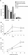
FIG. 1.
FIG. 1.
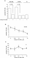
Differential infectivity of T. cruzi isolates. (A) Tissue culture trypomastigotes were incubated with monolayers containing different cell types at a parasite/host cell ratio of 3:1 in the appropriate medium (see Materials and Methods for details) supplemented with 0.1% BSA for 3 h at 37°C. (B and C) Invasion of human smooth muscle cells by the Dm28c and G isolates as described above for panel A at different interaction times (B) or at different parasite/host cell ratios (C). The data are representative of the data from three independent experiments.
FIG. 2.
FIG. 2.
Highly infective T. cruzi isolates depend on functional CP for host cell invasion. (A) Invasion of human smooth muscle cells by Dm28c, X10/6, or G trypomastigotes in DMEM containing 0.1% BSA for 3 h at 37°C at a parasite/cell ratio of 3:1 in the presence of 1% DMSO and in the presence of 1% DMSO and 10 μM N-Pip-F-hF-VSPh. (B and C) Invasion of human smooth muscle cells by strain Dm28c (B) or by strain G (at a 10:1 parasite/host cell ratio) (C) in the presence of different concentrations of N-Pip-F-hF-VSPh. The data are representative of the data from three independent experiments.
FIG. 3.
FIG. 3.
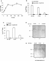
Highly infective T. cruzi isolates contain higher levels of functional CP. (A) Time course of CP release by trypomastigotes. Dm28c trypomastigotes were incubated in HBSS at 37°C. Aliquots (200 μl) were collected at different times and processed as described in Materials and Methods, and the peptidase activity was detected by using 10 μM ɛ-NH2-(Cap)Leu-(SBzl)Cys-AMC as a substrate. (B) Quantification of CP released by Dm28c, X10/6, or G trypomastigotes. Supernatants were obtained after incubation of parasites in HBSS at 37°C for 2 h. The peptidase activity was determined as described above for panel A. (C) Quantification of intracellular CP in Dm28c, X10/6, or G trypomastigotes. Trypomastigote lysates (30 μg) were used in enzymatic assays as described above for panel A. (D) Cruzipain accumulation in trypomastigotes: Western blot of trypomastigote lysates (50 μg) obtained by using anti-cruzipain antiserum at a 1:1,000 dilution. The numbers below the blot indicate the values obtained by densitometry of the bands. Cruzipain purified from epimastigotes was included as a control. (E) Quantification of OPB in trypomastigotes: Western blot of trypomastigote lysates (50 μg) obtained by using anti-OPB antiserum at a 1:1,000 dilution. The densitometry values are indicated below the blot. MW, molecular mass.
FIG. 4.
FIG. 4.
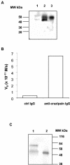
Cruzipain is the CP released by trypomastigotes. (A) Western blot of T. cruzi Dm28c lysates obtained by using anti-cruzipain antiserum. Lane 1, trypomastigotes; lane 2, epimastigotes; lane 3, purified cruzipain. (B) Supernatants were subjected to immunoprecipitation with anti-cruzipain or control IgG, and the peptidase activity bound to the beads was determined by using 10 μM ɛ-NH2-(Cap)Leu-(SBzl)Cys-AMC as a substrate. (C) Supernatants (lane 1) or purified cruzipain (lane 2) were supplemented with 5 mM DTT and incubated with 10 μM biotin-N-Pip-F-hF-VSPh at 37°C for 1 h. The samples were subsequently subjected to immunoprecipitation with anti-cruzipain IgG and visualized by Western blotting after incubation of the membrane with alkaline phosphatase-streptavidin conjugates. ctrl, control; MW, molecular mass.
FIG. 5.
FIG. 5.
CP released by trypomastigotes mediates host cell invasion. (A) Invasion of human smooth muscle cells by G trypomastigotes at a parasite/cell ratio of 4:1 in DMEM containing 0.1% BSA for 3 h at 37°C. The invasion assays were performed in the presence or absence of Dm28c trypomastigote supernatants supplemented with 2.5 mM DTT or with the reducing agent and 30 μM E-64, as indicated. The controls received HBSS containing 2.5 mM DTT. (B) Invasion of human smooth muscle cells by G trypomastigotes at parasite/host cell ratios 4:1, 10:1, and 20:1. The data are representative of the data from three independent experiments.
FIG. 6.
FIG. 6.
Extracellular cruzipain is required for host cell invasion: smooth muscle cell invasion by G trypomastigotes at a parasite/cell ratio of 4:1 in DMEM containing 0.1% BSA for 3 h at 37°C. Assays were performed in the presence (striped bar) or in the absence (open bar) of supernatant from Dm28c trypomastigotes or in the presence of supernatant subjected to immunoprecipitation with anti-cruzipain IgG (solid bars) or with control IgG (gray bar) as described in Materials and Methods. In some experiments, purified cruzipain was supplemented with 2.5 mM DTT, incubated for 5 min at 37°C, and added to the monolayers at a concentration of 5 nM. E-64 was added at a concentration of 30 μM. The data are representative of the data from two independent experiments.
FIG. 7.
FIG. 7.
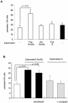
Cruzipain-mediated invasion requires a trypomastigote-associated factor. (A) Host cell invasion by G trypomastigotes at a parasite/cell ratio of 20:1 in DMEM containing 0.1% BSA for 3 h at 37°C in the presence or in the absence of supernatants from Dm28c or G trypomastigotes (Tryp), Dm28c epimastigotes (Epi), obtained in the same way, or in the presence of purified cruzipain at a final concentration of 5 nM (solid bar). The enzyme was supplemented with 2.5 mM DTT and incubated for 5 min at 37°C for activation prior to addition to the assay mixture. (B) Assays performed as described above for panel A in the presence or in the absence (open bar) of supernatants from Dm28c trypomastigotes (solid bars) or supernatants from G trypomastigotes (gray bars). The processed samples consisted of Dm28c supernatants (solid bar, processed) incubated with 2.5 mM DTT for 3 h at 37°C and subsequently supplemented with 30 μM E-64 prior to addition to the assay mixture or supernatants from G trypomastigotes (gray bar, + cruzipain) supplemented with 5 nM purified cruzipain and 2.5 mM DTT.
FIG. 8.
FIG. 8.
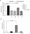
Cruzipain-mediated invasion requires trypomastigote membranes. (A) Host cell invasion by G trypomastigotes at a parasite/cell ratio of 20:1 in DMEM containing 0.1% BSA for 3 h at 37°C in the presence or in the absence (open bar) of supernatants from Dm28c trypomastigotes. Untreated supernatants were added to the invasion assay mixture in full (solid bar) or after fractionation by centrifugation at 100,000 × g (gray bars). S, soluble fraction; M, membrane fraction. Processed supernatants (striped bars) were incubated with 2.5 mM DTT for 3 h at 37°C and subsequently supplemented with 30 μM E-64, followed by fractionation by centrifugation at 100,000 × g prior to addition to the invasion assay mixture. S, soluble fraction; M, membrane fraction. (Inset) Peptidase activity of unprocessed supernatants (100 μl) from Dm28c trypomastigotes after fractionation by centrifugation at 100,000 × g, as determined with 10 μM carbobenzoxy-phenylalanyl-arginyl-7-amido-4-methylcoumarin. The graph shows the initial velocity. S, soluble fraction; M, membrane fraction. The data are representative of the data from two independent experiments. (B) Host cell invasion by G trypomastigotes at a parasite/cell ratio of 4:1 in DMEM containing 0.1% BSA for 3 h at 37°C. Supernatants from Dm28c trypomastigotes were fractionated by centrifugation at 100,000 × g, and the membrane fraction was incubated with 2.5 mM DTT and 5 nM purified cruzipain for 3 h at 37°C and subsequently supplemented with 30 μM E-64 before addition to the assay mixtures. Open bar, saline; solid bar, untreated fraction; gray bar, membrane fraction treated with cruzipain; striped bar, membrane fraction treated with cruzipain which was preinactivated by 30 μM E-64.
FIG. 9.
FIG. 9.
Increase in infectivity induced by Dm28c trypomastigote supernatants is inhibited by thapsigargin: host cell invasion by G trypomastigotes at a parasite/cell ratio of 50:1 in DMEM containing 0.1% BSA for 1 h at 37°C in the presence or in the absence of supernatants from Dm28c trypomastigotes. Where indicated, the host cells were pretreated with 0.5 μM thapsigargin in DMEM containing 10% fetal calf serum for 40 min at 37°C. The coverslips were washed three times with HBSS before addition of trypomastigotes. The low level of infectivity resulted from the short time interval used in the assay (1 h). The data are representative of the data from three independent experiments.

Similar articles

Cited by

References

    1. Burleigh, B. A., E. V. Caler, P. Webster, and N. W. Andrews. 1997. A cytosolic serine endopeptidase from Trypanosoma cruzi is required for the generation of Ca2+ signaling in mammalian cells. J. Biol. Chem. 136:609-620. - PMC - PubMed
    1. Burleigh, B. A., and A. M. Woolsey. 2002. Cell signaling and Trypanosoma cruzi invasion. Cell. Microbiol. 4:701-711. - PubMed
    1. Caler, E. V., S. Vaena de Avalos, P. A. Haynes, N. W. Andrews, and B. A. Burleigh. 1998. Oligopeptidase B-dependent signaling mediates host cell invasion by Trypanosoma cruzi. EMBO J. 17:4975-4986. - PMC - PubMed
    1. Caler, E. V., S. Chakrabarti, K. T. Fowler, S. Rao, and N. W. Andrews. 2001. The exocytosis-regulatory protein synaptotagmin VII mediates cell invasion by Trypanosoma cruzi. J. Exp. Med. 193:1097-1104. - PMC - PubMed
    1. Campetella, O., J. Henriksson, L. Aslund, A. C. C. Frasch, U. Pettterson, and J. J. Cazzulo. 1992. The major cysteine proteinase (cruzipain) from Trypanosoma cruzi is encoded by multiple polymorphic tandemly organized genes located on different chromosomes. Mol. Biochem. Parasitol. 50:225-234. - PubMed

Publication types

LinkOut - more resources