

Modulation of airway inflammation by immunostimulatory CpG oligodeoxynucleotides in a murine model of allergic aspergillosis
- PMID: 15385513
- PMCID: PMC517601
- DOI: 10.1128/IAI.72.10.6087-6094.2004
Modulation of airway inflammation by immunostimulatory CpG oligodeoxynucleotides in a murine model of allergic aspergillosis
Abstract
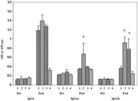
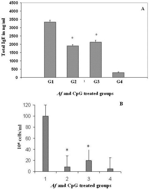
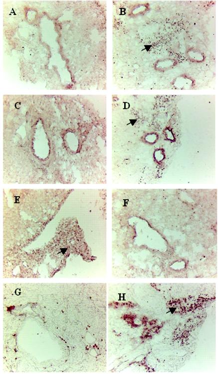
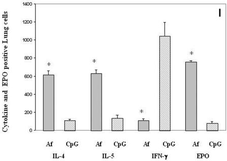
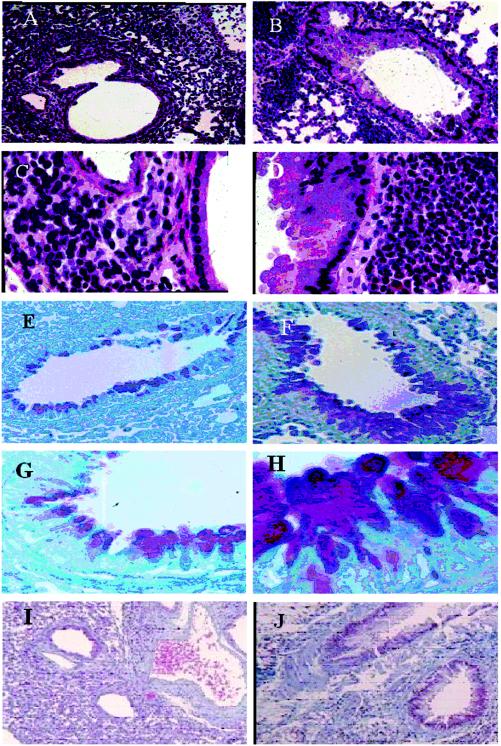
Allergic aspergillosis is a Th2 T-lymphocyte-mediated pulmonary complication in patients with atopic asthma and cystic fibrosis. Therefore, any therapeutic strategy that selectively inhibits Th2 T-cell activation may be useful in downregulating allergic lung inflammation in asthma. In the present study, we developed a CpG oligodeoxynucleotide (ODN)-based immune intervention of allergic inflammation in a mouse model of allergic aspergillosis. Four different groups of mice were used in a short-term immunization protocol. Three experimental groups of animals (groups 1 to 3) were sensitized with Aspergillus fumigatus antigens. Animals in group 1 were immunized with A. fumigatus antigen alone, while those in group 2 were treated with CpG-ODN 1 day before the first antigen immunization, and the animals in group 3 received the first CpG-ODN administration between the antigen treatments. The animals in group 4 served as controls and were given phosphate-buffered saline. Allergen-specific serum immunoglobulins and total immunoglobulin E in different groups of animals were measured by enzyme-linked immunosorbent assay, while airway remodeling and cytokine production were studied by immunohistochemistry. The results demonstrated that CpG-ODN administration either before (group 2) or between (group 3) antigen treatments resulted in reduced total immunoglobulin E levels and peripheral blood eosinophil numbers compared to A. fumigatus allergen-sensitized group 1 animals. Similarly, treatment with CpG-ODN also downregulated inflammatory cell infiltration, goblet cell hyperplasia, and basement membrane thickening compared to A. fumigatus-sensitized mice. The distinct reduction in peripheral blood eosinophilia and airway remodeling in CpG-ODN-treated mice emphasized its usefulness as an immunomodulating agent for allergic fungal diseases.
Figures
Similar articles
-
Intranasal administration of CpG oligodeoxynucleotides reduces lower airway inflammation in a murine model of combined allergic rhinitis and asthma syndrome.Int Immunopharmacol. 2015 Sep;28(1):390-8. doi: 10.1016/j.intimp.2015.06.028. Epub 2015 Jul 9. Int Immunopharmacol. 2015. PMID: 26163938
-
Aspergillus antigen induces robust Th2 cytokine production, inflammation, airway hyperreactivity and fibrosis in the absence of MCP-1 or CCR2.Respir Res. 2004 Sep 15;5(1):12. doi: 10.1186/1465-9921-5-12. Respir Res. 2004. PMID: 15377395 Free PMC article.
-
Modulation of murine allergic rhinosinusitis by CpG oligodeoxynucleotides.Laryngoscope. 2002 Oct;112(10):1819-26. doi: 10.1097/00005537-200210000-00021. Laryngoscope. 2002. PMID: 12368622
-
Immunology of allergic bronchopulmonary aspergillosis.Indian J Chest Dis Allied Sci. 2000 Oct-Dec;42(4):225-37. Indian J Chest Dis Allied Sci. 2000. PMID: 15597669 Review.
-
Allergic bronchopulmonary mycosis complicating cystic fibrosis.Semin Respir Infect. 1992 Sep;7(3):179-92. Semin Respir Infect. 1992. PMID: 1475542 Review.
Cited by
-
A protective allergy vaccine based on CpG- and protamine-containing PLGA microparticles.Pharm Res. 2007 Oct;24(10):1927-35. doi: 10.1007/s11095-007-9318-0. Epub 2007 May 31. Pharm Res. 2007. PMID: 17541735
-
Immune response modulation by curcumin in a latex allergy model.Clin Mol Allergy. 2007 Jan 25;5:1. doi: 10.1186/1476-7961-5-1. Clin Mol Allergy. 2007. PMID: 17254346 Free PMC article.
-
Prophylactic administration of bacterially derived immunomodulators improves the outcome of influenza virus infection in a murine model.J Virol. 2010 Mar;84(6):2983-95. doi: 10.1128/JVI.01805-09. Epub 2010 Jan 6. J Virol. 2010. PMID: 20053748 Free PMC article.
-
Eat dirt: CpG DNA and immunomodulation of asthma.Proc Am Thorac Soc. 2007 Jul;4(3):283-8. doi: 10.1513/pats.200701-019AW. Proc Am Thorac Soc. 2007. PMID: 17607014 Free PMC article. Review.
-
Intranasal CpG therapy attenuated experimental fungal asthma in a TLR9-dependent and -independent manner.Int Arch Allergy Immunol. 2010;152(2):98-112. doi: 10.1159/000265531. Epub 2009 Dec 16. Int Arch Allergy Immunol. 2010. PMID: 20016192 Free PMC article.
References
-
- Bozza, S., R. Gaziano, G. B. Lipford, C. Montagnoli, A. Bacci, P. Di Francesco, V. P. Kurup, H. Wagner, and L. Romani. 2002. Vaccination of mice against invasive aspergillosis with recombinant Aspergillus proteins and CpG oligodeoxynucleotides as adjuvants. Microbes Infect. 4:1281-1290. - PubMed
-
- Broide, D., J. Schwarze, H. Tighe, T. Gifford, M. D. Nguyen, S. Malek, J. Van Uden, E. Martin-Orozco, and E. W. Gelfand. 1999. Immunostimulatory DNA sequences inhibit IL-5, eosinophilic inflammation, and airway hyperresponsiveness in mice. J. Immunol. 161:7054-7062. - PubMed
-
- Greenberger, P. A. 2003. Allergic bronchopulmonary aspergillosis, p. 1353-1371. In N. F. Adkinson, Jr., J. W. Yunginger, W. W. Busse, B. S. Bochner, S. T. Holgate, and F. E. R. Simons (ed.), Middleton's allergy principles and practice. Mosby, Philadelphia, Pa.
-
- Kline, J. N., T. J. Waldschmidt, T. R. Businga, J. E. Lemish, J. V. Weinstock, P. S. Thorne, and A. M. Krieg. 1998. Modulation of airway inflammation by CpG oligodeoxynucleotides in a murine model of asthma. J. Immunol. 160:2555-2559. - PubMed
-
- Krieg, A. M. 2002. From A to Z on CpG. Trends Immunol. 23:64-65. - PubMed
Publication types
MeSH terms
Substances
LinkOut - more resources
Full Text Sources
Other Literature Sources
