. 2004 Dec 17;279(51):53435-41.
doi: 10.1074/jbc.M409421200. Epub 2004 Sep 22.

Eukaryotic CTR copper uptake transporters require two faces of the third transmembrane domain for helix packing, oligomerization, and function

Affiliations

Eukaryotic CTR copper uptake transporters require two faces of the third transmembrane domain for helix packing, oligomerization, and function

Stephen G Aller et al. J Biol Chem. .

Abstract

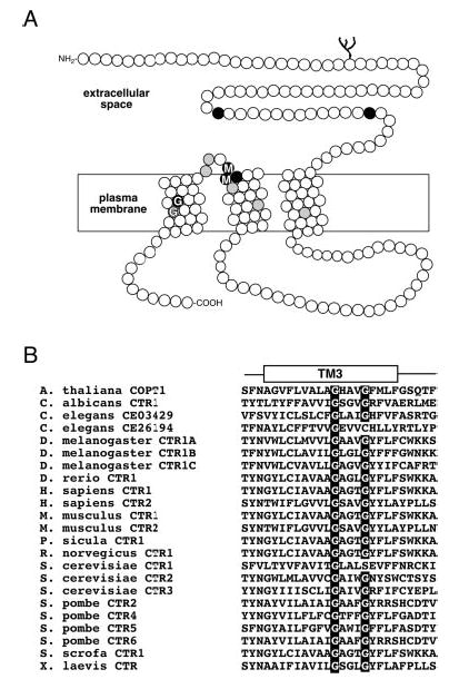
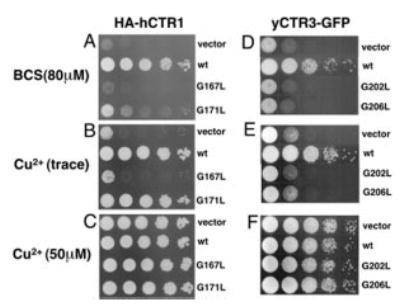
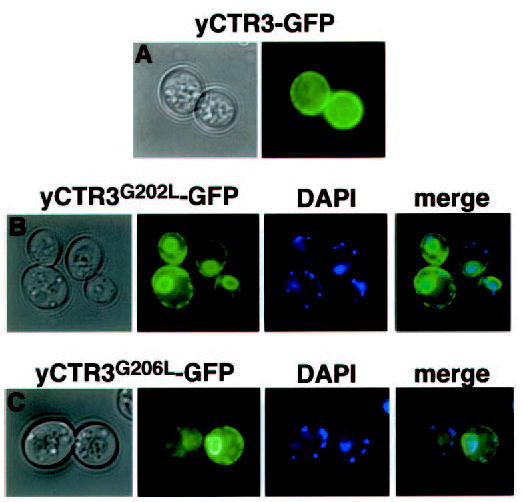
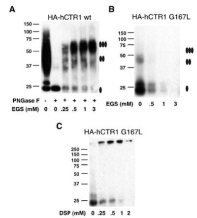
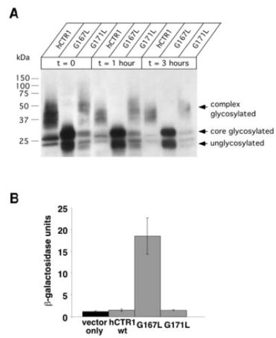
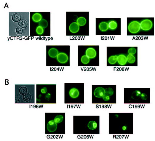
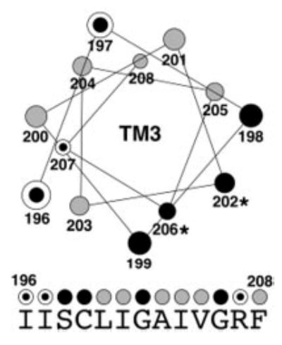
Members of the copper uptake transporter (CTR) family from yeast, plants, and mammals including human are required for cellular uptake of the essential metal copper. Based on biochemical data, CTRs have three transmembrane domains and have been shown to oligomerize in the membrane. Among individual members of the family, there is little amino acid sequence identity, raising questions as to how these proteins adopt a common fold, oligomerize, and participate in copper transport. Using site-directed mutagenesis, tryptophan scanning, genetic complementation, subcellular localization, chemical cross-linking, and the yeast unfolded protein response, we demonstrated that at least half of the third transmembrane domain (TM3) plays a vital role in CTR structure and function. The results of our analysis showed that TM3 contains two functionally distinct faces. One face bears a highly conserved Gly-X-X-X-Gly (GG4) motif, which we showed to be essential for CTR oligomerization. Moreover, we showed that steric constraints reach past the GG4-motif itself including amino acid residues that are not conserved throughout the CTR family. A second face of TM3 contains three amino acid positions that, when mutated to tryptophan, cause predominantly abnormal localization but are still partially functional in growth complementation experiments. These mutations cluster on the face opposite to the GG4-bearing face of TM3 where they may mediate interactions with the remaining two transmembrane domains. Taken together, our data support TM3 as being buried within trimeric CTR where it plays an essential role in CTR assembly.


Figures

Fig. 1
Fig. 1. Topology of human CTR1 and highly conserved Gly-X-X-X-Gly motif
A, diagram of a human CTR1 monomer shows the extracellular N terminus, three putative transmembrane domains, and an intracellular C terminus. Amino acid positions represented as black circles are invariantly conserved in CTRs from yeast, plants, and metazoans, and those in gray are >90% identical in the family. Two conserved amino acid motifs in the entire family of CTR copper uptake proteins, Met-X-X-X-Met in TM2 and Gly-X-X-X-Gly (GG4) motif in TM3, are indicated. B, multiple amino acid sequence alignment of the third TM domain reveals a nearly invariant conservation of the GG4 motif in all of the known members of the CTR copper uptake family.
Fig. 2
Fig. 2. Effect of GG4 mutations on human CTR1 and yeast CTR3 function
Yeast deficient in high affinity copper uptake (Δctr1,3) were transformed with vector only, HA-tagged wild type human CTR1 (wt), and GG4 mutants HA-hCTR1G167L (G167L) and HA-hCTR1G171L (G171L), wild type yeast CTR3-GFP fusion wt, and the corresponding yeast CTR3 GG4 mutants G202L and G206L. Yeast transformants were grown in minimal dextrose medium before two washes with double distilled water. All of the cultures were then adjusted to A600 = 1.0 and plated in serial 1:10 dilutions on plates containing glycerol as the sole non-fermentable carbon source. A, copper depletion for HA-hCTR1 was acheived with 80 μm copper chelator BCS. B, neither copper nor BCS was added to the plates. C, excess copper (50 μm). D, E, and F, same as A, B, and C, respectively, but with yCTR3-GFP.
Fig. 3
Fig. 3. Fluorescence localization of wild type and GG4 mutants G202L and G206L of yeast CTR3-GFP
Colonies of yeast transformants of the same constructs used in complementation experiments were spotted directly on glass coverslips for fluorescence microscopy. Bright-field and corresponding fluorescence images are shown. A, plasma membrane localization of wild type yCTR3-GFP fusion protein. GG4 mutants G202L (panel B) and G206L (panel C) are trapped in a perinuclear intracellular compartment as revealed by DAPI staining of nuclear DNA and a ring at the periphery of the cell. Accumulation of GFP-tagged mutant proteins in both regions is indicative of ER localization in yeast. Smaller DAPI-stained points represent mitochondrial DNA.
Fig. 4
Fig. 4. Chemical cross-linking of human CTR1 wild type and GG4 mutant G167L
Membranes prepared from yeast overexpressing HA-tagged human CTR1 wild type (wt) (A) or HA-hCTR1G167L mutant (B) were solubilized with Triton X-100 detergent and subjected to chemical cross-linking. Solubilized material was incubated in increasing concentrations of the ethylene glycol succinimidylsuccinimate (EGS) cross-linker before being subjected to Western blot using an anti-HA primary antibody. HA-tagged wild-type human CTR1 required removal of N-linked glycosylation by treatment with PNGase F prior to exposure to EGS to clearly resolve oligomeric states. Partially purified HA-hCTR1G167L was concentrated before exposure to increasing concentrations of the slightly lower molecular weight cross linker dithiobis succinimidylpropionate (DSP) (panel C). Filled ovals displayed to the right of the images indicate the expected migration positions for monomer, dimer, and trimer species of hCTR1.
Fig. 5
Fig. 5. Processing of GG4 mutants and induction of the unfolded protein response
A, yeast cultures expressing the indicated constructs were grown to mid-log phase before protein synthesis was halted with cycloheximide. Samples were removed from the culture at the indicated time points, and total protein extracts were subjected to Western blot using an anti-HA primary antibody. Note that a nearly overexposed blot demonstrates the lack of any higher order glycosylation for the G167L mutant compared with wild type and G171L. The doublet of bands observed for the G167L mutant corresponds to unglycosylated and core-glycosylated species, respectively. Recall in Fig. 4 that complex glycosylation (seen as high molecular mass smearing) as well as core glycosylation (as a distinct band that migrated at ~28 kDa), could be reduced to a single band (~23 kDa) upon exposure to PNGase F. B, yeast transformed with both pSZ1-UPRE-LacZ and an expression plasmid carrying either wild type G167L or G171L were subjected to β-galactosidase assay to determine the extent of induction of the unfolded protein response (n = 3–4 randomly selected colonies/condition).
Fig. 6
Fig. 6. Fluorescence localization of tryptophan-scanning mutants of the third transmembrane domain of yeast CTR3-GFP fusion
Thirteen separate constructs were synthesized by subcloning the wild-type yCTR3 into the p423GPD vector and performing site-directed mutagenesis to replace each amino acid position from 196 to 208 with tryptophan. Mutants were transformed, picked from minimal dextrose selective plates, and subjected to fluorescence microscopy. A, class of six mutants exhibited localization that was identical to wild type. B, second class of mutants revealed abnormal localization to intracellular compartments.
Fig. 7
Fig. 7. Helical wheel diagram summarizing the results of tryptophan-scanning mutagenesis and complementation experiments
A helical wheel was generated for the middle 13 positions of TM3 (amino acid residues 196–208). Gray circles represent amino acid positions where introduction of a tryptophan residue did not interfere with localization to the plasma membrane and function of the mutant protein in the complementation assay. Three dotted circles identify amino acid positions that caused mislocalization when mutated to tryptophan but were partially functional in the complementation assay. Black circles represent positions where introduction of tryptophan resulted in mislocalization and completely abolished function in complementation experiments. Under the helical wheel, the native sequence of yCTR3 is displayed. The same color-coded circles summarizing the fluorescence localization and complementation results obtained for each tryptophan mutant are placed above each amino acid letter.

Similar articles

Cited by

References

    1. Linder, M. C. (1991) Biochemistry of Copper, Plenum Press, New York
    1. Pena MM, Lee J, Thiele DJ. J Nutr. 1999;129:1251–1260. - PubMed
    1. DiDonato M, Sarkar B. Biochim Biophys Acta. 1997;1360:3–16. - PubMed
    1. Schaefer M, Gitlin JD. Am J Physiol. 1999;276:G311–G314. - PubMed
    1. Strausak D, Mercer JF, Dieter HH, Stremmel W, Multhaup G. Brain Res Bull. 2001;55:175–185. - PubMed

Publication types

MeSH terms

LinkOut - more resources