Evidence that each S cone in macaque fovea drives one narrow-field and several wide-field blue-yellow ganglion cells
- PMID: 15385619
- PMCID: PMC6729688
- DOI: 10.1523/JNEUROSCI.1063-04.2004
Evidence that each S cone in macaque fovea drives one narrow-field and several wide-field blue-yellow ganglion cells
Abstract
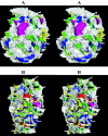
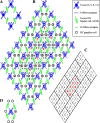
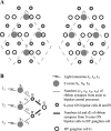
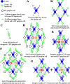
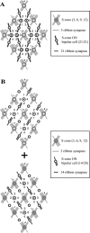
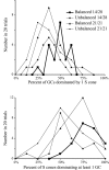
A rule of retinal wiring is that many receptors converge onto fewer bipolar cells and still fewer ganglion cells. However, for each S cone in macaque fovea, there are two S-cone ON bipolar cells and two blue-yellow (BY) ganglion cells. To understand this apparent rule reversal, we reconstructed synaptic patterns of divergence and convergence and determined the basic three-tiered unit of connectivity that repeats across the retina. Each foveal S cone diverges to four S-cone ON bipolar cells but contacts them unequally, providing 1-16 ribbon synapses per cell. Next, each bipolar cell diverges to two BY ganglion cells and also contacts them unequally, providing approximately 14 and approximately 28 ribbon synapses per cell. Overall, each S cone diverges to approximately six BY ganglion cells, dominating one and contributing more modestly to the others. Conversely, of each pair of BY ganglion cells, one is dominated by a single S cone and one is diffusely driven by several. This repeating circuit extracts blue/yellow information on two different spatiotemporal scales and thus parallels the circuits for achromatic, spatial vision, in which each cone dominates one narrow-field ganglion cell (midget) and contributes some input to several wider-field ganglion cells (parasol). Finally, because BY ganglion cells have coextensive +S and -(L+M) receptive fields, and each S cone contributes different weights to different BY ganglion cells, the coextensive receptive fields must be already present in the synaptic terminal of the S cone. The S-cone terminal thus constitutes the first critical locus for BY color vision.
Figures



References
-
- Ahmad KM, Klug K, Herr S, Sterling P, Schein S (2003) Cell density ratios in a foveal patch in macaque retina. Vis Neurosci 20: 189–209. - PubMed
-
- Ahnelt P, Kolb H (1994a) Horizontal cells and cone photoreceptors in primate retina: a Golgi-light microscopic study of spectral connectivity. J Comp Neurol 343: 387–405. - PubMed
-
- Ahnelt P, Kolb H (1994b) Horizontal cells and cone photoreceptors in human retina: a Golgi-electron microscopic study of spectral connectivity. J Comp Neurol 343: 406–427. - PubMed
-
- Bumsted K, Hendrickson A (1999) Distribution and development of short-wavelength cones differ between Macaca monkey and human fovea. J Comp Neurol 403: 502–516. - PubMed
-
- Burris C, Klug K, Ngo IT, Sterling P, Schein S (2002) How Müller glial cells in macaque fovea coat and isolate the synaptic terminals of cone photo-receptors. J Comp Neurol 453: 100–111. - PubMed
Publication types
MeSH terms
Grants and funding
LinkOut - more resources
Full Text Sources
Miscellaneous
