. 2004 Dec;15(12):5574-82.
doi: 10.1091/mbc.e04-02-0142. Epub 2004 Sep 22.

Oxidative stress activates FUS1 and RLM1 transcription in the yeast Saccharomyces cerevisiae in an oxidant-dependent Manner

Affiliations

Oxidative stress activates FUS1 and RLM1 transcription in the yeast Saccharomyces cerevisiae in an oxidant-dependent Manner

Liliana Staleva et al. Mol Biol Cell. 2004 Dec.

Abstract

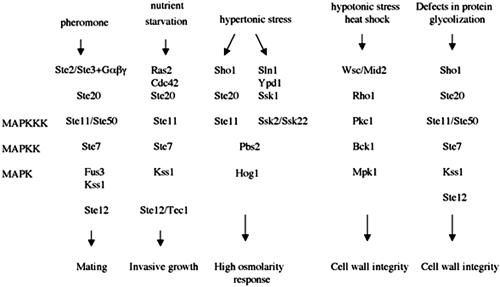
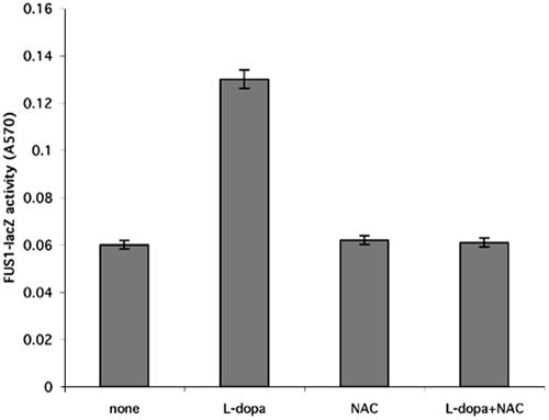
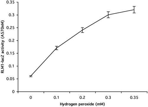
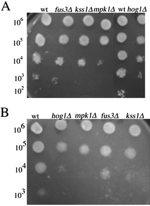
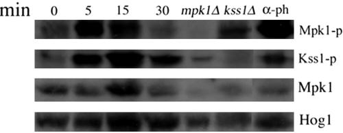
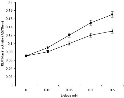
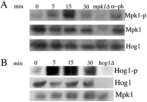
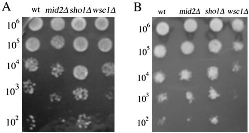
Mating in haploid Saccharomyces cerevisiae occurs after activation of the pheromone response pathway. Biochemical components of this pathway are involved in other yeast signal transduction networks. To understand more about the coordination between signaling pathways, we used a "chemical genetic" approach, searching for compounds that would activate the pheromone-responsive gene FUS1 and RLM1, a reporter for the cell integrity pathway. We found that catecholamines (l-3,4-hydroxyphenylalanine [l-dopa], dopamine, adrenaline, and noradrenaline) elevate FUS1 and RLM1 transcription. N-Acetyl-cysteine, a powerful antioxidant in yeast, completely reversed this effect, suggesting that FUS1 and RLM1 activation in response to catecholamines is a result of oxidative stress. The oxidant hydrogen peroxide also was found to activate transcription of an RLM1 reporter. Further genetic analysis combined with immunoblotting revealed that Kss1, one of the mating mitogen-activated protein kinases (MAPKs), and Mpk1, an MAPK of the cell integrity pathway, participated in l-dopa-induced stimulation of FUS1 and RLM1 transcription. We also report that Mpk1 and Hog1, the high osmolarity MAPK, were phosphorylated upon induction by hydrogen peroxide. Together, our results demonstrate that cells respond to oxidative stress via different signal transduction machinery dependent upon the nature of the oxidant.


Figures

Figure 1.
Figure 1.
MAPK cascades which regulate cellular changes in response to external stimuli in haploid S. cerevisae.
Figure 2.
Figure 2.
(A) Catecholamines activate FUS1 reporter in a dose-dependent manner. The wild-type yeast strain (EY957) was plated in 24-well plates on medium containing the β-galactosidase substrate CPRG as described in Materials and Methods. Catecholamines (adrenaline [×], noradrenaline [▴], l-dopa [▪], and dopamine [*]) were added to the media in increasing concentrations (0.01–1 mM) and β-galactosidase activity was measured at 570 nm after 48 h. Results shown are from five independent assays. (B) l-Dopa activates RLM1 reporter in a dose-dependent manner. Wild-type strain (EY957) transformed with px2RLM1 was plated in 24-well plates on media containing the β-galactosidase substrate CPRG as described in Materials and Methods. Catecholamines (adrenaline [×], noradrenaline [▴], l-dopa [▪], and dopamine ([988]) were added to the media in increasing concentrations (0.01–1 mM) and β-galactosidase activity was measured at 570 nm after 48 h. Results shown are from five independent assays.
Figure 3.
Figure 3.
N-Acetyl-cysteine reverses the l-dopa–induced activation of the reporter FUS1. The wild-type yeast strain (EY957) was plated in 24-well plates, treated with l-dopa in different concentrations and N-acetyl-cysteine. Shown are l-dopa (0.02 mM) and N-acetyl-cysteine (2 mM) alone or in combination, and β-galactosidase activity was measured as described in Materials and Methods. Results shown are from five independent assays.
Figure 4.
Figure 4.
Growth of wild-type strain (EY957) in the presence of l-dopa. Cells were grown to the early exponential phase, diluted to OD = 0.2 into fresh YEPD medium, and grown for an additional 2 h to allow the cells to adapt to the medium. The culture was split and different concentrations of l-dopa were added (wild-type [▪], 1 mM l-dopa [▴], and 2 mM l-dopa [×]). The optical density of the cultures was monitored by spectrophotometry. Results shown are from five independent assays.
Figure 5.
Figure 5.
Hydrogen peroxide activates RLM1 reporter in a dose-dependent manner. Wild-type strain (EY957) transformed with px2RLM1 was plated in 24-well plates on media containing the β-galactosidase substrate CPRG as described in Materials and Methods. Hydrogen peroxide was added to the media in increasing concentrations (0.1–1 mM), and β-galactosidase activity was measured at 570 nm after 24 h. Results shown are from five independent assays.
Figure 6.
Figure 6.
(A and B) Deletion in HOG1, KSS1, or MPK1 lead to sensitivity to l-dopa and hydrogen peroxide. Identical volumes (10 μl) of 10-fold serial dilutions of exponentially growing wild-type strain (EY957) and the isogenic kss1Δ, fus3Δ, hog1Δ strains were spotted onto YEPD plates containing various concentrations of l-dopa and hydrogen peroxide and then incubated for 48 h at 30°C. Shown are representative examples of the plates incubated with 1 mM l-dopa (A) and 1 mM hydrogen peroxide (B).
Figure 7.
Figure 7.
l-Dopa stimulates Mpk1and Kss1 MAPK. Mid-log cultures of wild-type strain cells (EY957) were treated with l-dopa at 2 mM final concentration and at 50 nM α-pheromone used as positive control. Samples were taken at the indicated times (0, 15, and 30 min). The α-pheromone was added for 60 min. The isogenic mpk1Δ and kss1Δ, used as controls, were treated with l-dopa for 15 min. Western blot analysis was performed as described in Materials and Methods by using anti-phospho-p44/p42 antibody to detect the phosphorylated Mpk1, Kss1 and Fus3, anti-Mpk1 to detect the total amount of Mpk1 protein and anti-Hog1 as a loading control.
Figure 8.
Figure 8.
l-Dopa activation of RLM1 reporter in mpk1Δ strain. Wild-type strain (DL100) (▪) and the isogenic strain mpk1Δ (▴) were transformed with px2RLM1 and plated in 24-well plates on media containing the β-galactosidase substrate CPRG as described in Materials and Methods. l-Dopa was added to the media in increasing concentrations (0.1–0.5 mM), and β-galactosidase activity was measured at 570 nm after 48 h. Results shown are from five independent assays.
Figure 9.
Figure 9.
(A) RLM1 transcription in treated with l-dopa kss1Δfus3Δ strain. Wild-type strain (EY957) (▪) and the isogenic kss1Δfus3Δ (♦) strain were transformed with px2RLM1 and then plated in 24-well plates on media containing the β-galactosidase substrate CPRG as described in Materials and Methods. l-Dopa was added to the media in increasing concentrations (0.01–0.5 mM), and β-galactosidase activity was measured at 570 nm after 24 h. Results shown are from five independent assays. (B) FUS1 transcription in mpk1Δ strain. Wild-type strain (DL100) (▪) and the isogenic mpk1Δ (▴) strain were transformed with px2RLM1 and then plated in 24-well plates on media containing the β-galactosidase substrate CPRG as described in Materials and Methods. l-Dopa was added to the media in increasing concentrations (0.01–0.5 mM), and β-galactosidase activity was measured at 570 nm after 24 h. Results shown are from five independent assays
Figure 10.
Figure 10.
(A) Mpk1 is phosphorylated on exposure of cells to hydrogen peroxide. Mid-log cultures of wild-type strain cells (EY957) were treated with hydrogen peroxide at 5 mM final concentration with 50 nM α-pheromone as a positive control. Samples were taken at the indicated times (0, 15, and 30 min). Wild-type cells were treated with α-pheromone for 60 min. Hydrogen peroxide was added to mpk1Δ strain for 15 min. Western blot analysis was performed as described in Materials and Methods by using anti-phospho-p44/p42 antibody to detect the phosphorylated Mpk1, Kss1 and Fus3, anti-Mpk1 to detect the total protein and anti-Hog1 as a loading control. (B) Hydrogen peroxide activates Hog1 MAPK. Mid-log cultures of wild-type strain cells (EY957) were treated with hydrogen peroxide at 5 mM final concentration. Samples were taken at the indicated times (0, 15, and 30 min). Strain disrupted in HOG1 gene, used as negative control, was treated with hydrogen peroxide for 15 min. Western blot analysis was performed as described in Materials and Methods by using anti-phospho-p38 antibody to detect the phosphorylated Hog1, with anti-Hog1 to detect the total protein and anti-Mpk1 as a loading control.
Figure 11.
Figure 11.
(A) Hydrogen peroxide activation of RLM1 transcription in mpk1Δ strain. Wild-type strain (Dl100) (▪) and the isogenic mpk1Δ (▴) strain were transformed with px2RLM1 and then plated in 24-well plates on media containing the β-galactosidase substrate CPRG as described in Materials and Methods. Hydrogen peroxide was added to the media in increasing concentrations (0.1–0.5 mM), and β-galactosidase activity was measured at 570 nm after 24 h. Results shown are from five independent assays. (B) RLM1 transcription induction in hog1Δ strain. Wild-type strain (EY957) (▪) and the isogenic hog1Δ strain (•) were transformed with px2RLM1 and then plated in 24-well plates on media containing the β-galactosidase substrate CPRG as described in Materials and Methods. Hydrogen peroxide was added to the media in increasing concentrations (0.1–0.5 mM), and β-galactosidase activity was measured at 570 nm after 24 h. Results shown are from five independent assays.
Figure 12.
Figure 12.
(A and B) Cells lacking WSC1 are the most sensitive to l-dopa and hydrogen peroxide. Identical volumes (10 μl) of 10-fold serial dilutions of exponentially growing wild-type strain (DL100) and the isogenic wsc1Δ, sho1Δ and mid2Δ were spotted onto YEPD plates containing various concentrations of l-dopa and hydrogen peroxide and then incubated for 48 h at 30°C. Shown are the representative examples of the plates incubated with 1 mM l-dopa (A) and 1 mM hydrogen peroxide (B).

Similar articles

Cited by

References

    1. Alonco-Monge, R., Navarro-Garcia, F., Roman, E., Negredo, A.I., Eisman, B., Nombela, C., and Pla, J. (2003). The Hog1 mitogen-activated protein kinase is essential in the oxidative stress response and chlamydospore formation in Candida albicans. Eucaryot. Cell 2, 351-361. - PMC - PubMed
    1. Bardwell, L., Cook, J.G., Zhu-Shimoni, J.X., Voora, D., and Thorner, J. (1998a). Differential regulation of transcription: repression by unactivated mitogen-activated protein kinase Kss1 requires the Dig1 and Dig2 proteins. Proc. Natl. Acad. Sci. USA 95, 15400-15405. - PMC - PubMed
    1. Bardwell, L., Cook, J.G., Voora, D., Baggott, D.M., Martinez, A.R., and Thorner, J. (1998b). Repression of yeast Ste12 transcription factor by direct binding of unphosphorylated Kss1 MAPK and its regulation by STE7 MEK. Genes Dev. 12, 2887-2898. - PMC - PubMed
    1. Basma, A.N., Nicklas, W.J., and Geller, H.M.,. (1995). L-dopa cytotoxicity to PC12 cells in culture is via its autooxidation. J. Neurochem. 64, 825-832. - PubMed
    1. Buehrer, B., and Errede, B. (1997). Coordination of the mating and cell integrity mitogen-activated protein kinase pathways in Saccharomyces cerevisiae. Mol. Cell. Biol. 17, 6517-6525. - PMC - PubMed

Publication types

MeSH terms

LinkOut - more resources