. 2004 Dec;15(12):5219-30.
doi: 10.1091/mbc.e04-03-0255. Epub 2004 Sep 22.

Spindle pole body duplication in fission yeast occurs at the G1/S boundary but maturation is blocked until exit from S by an event downstream of cdc10+

Affiliations

Spindle pole body duplication in fission yeast occurs at the G1/S boundary but maturation is blocked until exit from S by an event downstream of cdc10+

Satoru Uzawa et al. Mol Biol Cell. 2004 Dec.

Abstract

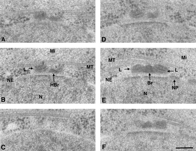
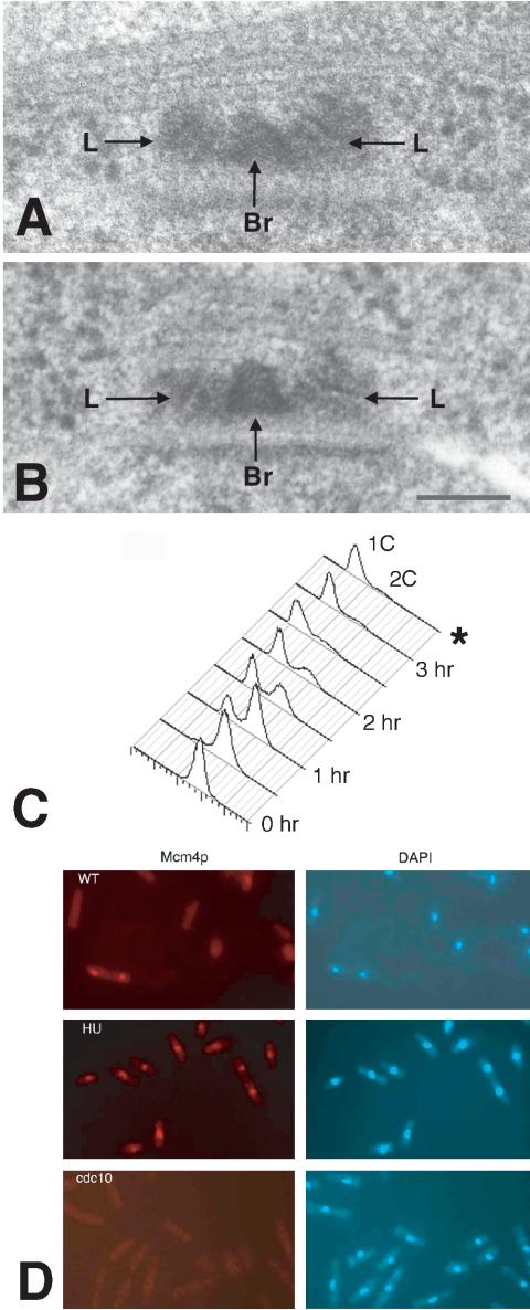
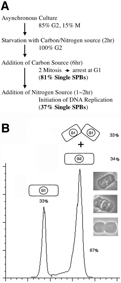
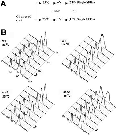
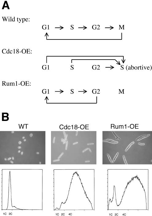
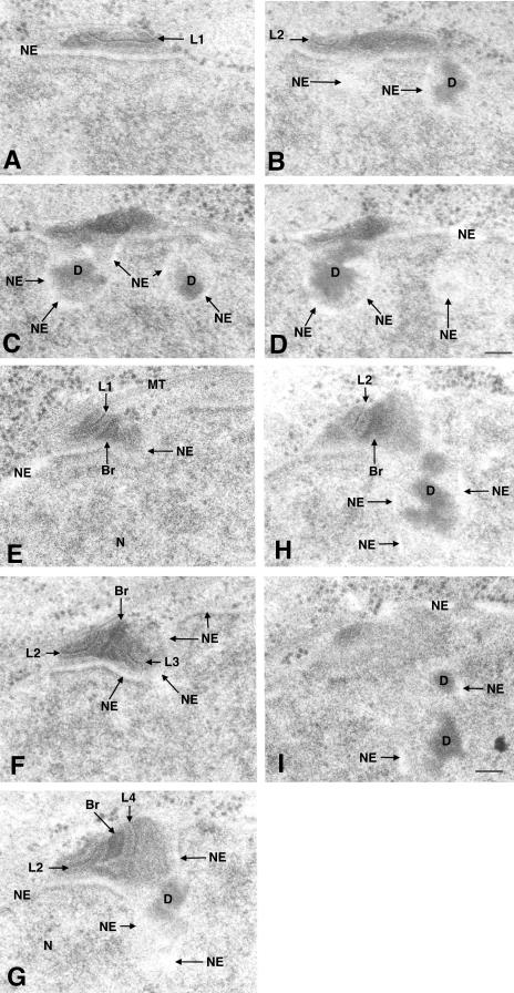
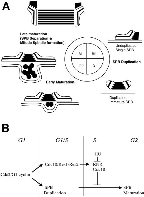
The regulation and timing of spindle pole body (SPB) duplication and maturation in fission yeast was examined by transmission electron microscopy. When cells are arrested at G1 by nitrogen starvation, the SPB is unduplicated. On release from G1, the SPBs were duplicated after 1-2 h. In cells arrested at S by hydroxyurea, SPBs are duplicated but not mature. In G1 arrest/release experiments with cdc2.33 cells at the restrictive temperature, SPBs remained single, whereas in cells at the permissive temperature, SPBs were duplicated. In cdc10 mutant cells, the SPBs seem not only to be duplicated but also to undergo partial maturation, including invagination of the nuclear envelope underneath the SPB. There may be an S-phase-specific inhibitor of SPB maturation whose expression is under control of cdc10(+). This model was examined by induction of overreplication of the genome by overexpression of rum1p or cdc18p. In cdc18p-overexpressing cells, the SPBs are duplicated but not mature, suggesting that cdc18p is one component of this feedback mechanism. In contrast, cells overexpressing rum1p have large, deformed SPBs accompanied by other features of maturation and duplication. We propose a feedback mechanism for maturation of the SPB that is coupled with exit from S to trigger morphological changes.


Figures

Figure 1.
Figure 1.
Serial sections through two SPBs comparing the structure of an unduplicated (A–C) versus a duplicated but immature (D–F) SPB. In A–C, the nitrogen-starved cell is arrested in early G1. The SPB in this cell consists of a single laminar structure (L) and a half bridge (HBr), which lies adjacent to an intact NE. In D–F, the cdc10-arrested cell at the nonpermissive temperature is arrested at the G1/S boundary. The SPB is duplicated and has two laminar structures separated by a dark staining elipsoid bridge (Br). In both cells, the nuclear envelope is continuous and unfenestrated and shows no signs of invagination, although dark material has accumulated on the nuclear but not the cytoplasmic face of the nuclear envelope adjacent to the SPB. Several microtubules (MT) are in proximity to the cytoplasmic face of the SPB in each cell, accompanied by mitochondria. A nuclear pore (NP) is always found near the SPB. Note the image of the unduplicated SPB in section B superficially resembles the image of the duplicated but immature SPB in section D. The size of each linear structure is similar (ranging from 70 to 90 nm) in unduplicated and duplicated SPBs. Bar, 100 nm.
Figure 5.
Figure 5.
(A) 3D tomographic reconstruction of a SPB in a cdc10-arrested cell. The 3D volume of the tomographic reconstruction is shown as consecutive two-dimensional projections (A–L). Each projection is 16 slices, which is the equivalent of an image of a serial section 27 nm in thickness. The SPB shows invagination of the NE, accumulation of dark material in the membrane pocket (D), microtubule (MT) in the cytoplasm associated with SPB, and expansion in overall size of the SPB (L, laminar structure). The invaginated nuclear envelope is continuous and is not fenestrated. Bar, 100 nm. A movie of the whole volume can be found in Supplemental Data. (B) Schematic DNA histograms of cells after switching cdc10 cells to the nonpermissive temperature (35°C) at time 0, as determined by flow cytometry. The first peak represents cells with 1C DNA content; the second peak cells with 2C DNA content. At zero time, almost all cells had 2C DNA content, but by 3 h almost all cells had 1C DNA content. Cells were fixed for electron microscopy after 3.5 h as indicated by the *.
Figure 2.
Figure 2.
(A and B) The images of the duplicated but immature SPBs in two hydroxurea-treated cells arrested early in S. The SPB consists of two laminar structures (L) separated by a bridge (Br). For both examples, only the center section is presented. The SPB has not undergone maturation as shown by the small size of the laminar structures (50–60 nm) and the absence of any nuclear envelope invagination or fenestration. The laminations of one of the two structures (left-hand side) lie oblique to the nuclear envelope relative to the other laminated structure. Due to the angle and the size of the smaller laminar body, it was essential to examine images of tilted specimen in series to find it reliably. Bar, 100 nm. (C) Schematic DNA histograms of cells arrested early in S after addition of hydroxyurea at time 0 as determined by flow cytometry. The first peak represents cells with 1C DNA content; the second peak cells with 2C DNA content. At zero time, almost all cells had 2C DNA content but by 3 h posthydroxyurea addition all cells had 1C DNA content. Cells were fixed for electron microscopy after 3.5 h in hydroxyurea as indicated by the *. (D) In situ chromatin binding assay for HA-tagged mcm4p proteins in cells permeabilized with 1.0% Triton X-100. HA-labeled proteins were visualized in left panels (red) by using HA antibody and a secondary conjugated to Cys3 and right (blue) panels display 4,6-diamidino-2-phenylindole staining of DNA in the same cells. In the wild-type population (top), most cells are in G2 and do not retain mcm4p in the nucleus after permeabilization. The only cell that retained mcm4p protein is a binucleate cell at the G1/S boundary that has not completed cytokinesis (lower left). After 3.5-h treatment with hydroxurea, all cells (middle) have retained mcm4p after permeabilization. No mcm4p protein was retained in the nuclei of cdc10-arrested cells grown for 3.5 h at the nonpermissive temperature (bottom).
Figure 3.
Figure 3.
(A) Flow diagram shows the method used for G1 arrest in this study (Horie et al., 1998). The cells cultured in rich medium were transferred to a synthetic medium lacking both carbon source and nitrogen source (PM-ND). The cells were cultured for 2 h to allow them to arrest in G2 phase followed by addition of carbon source (2% dextrose final). After two rapid sequential divisions, cells arrest at G1. For arrest/release experiments, nitrogen source (ammonium chloride, 1% final) was added back. The percentage of unduplicated versus duplicated SPBs is shown in bold in parentheses. (B) Schematic DNA histogram of cells arrested in G1 as determined by flow cytometry. The square represents the cells where the cell cycle phase is indicated. The highest peak represents cells with 2C DNA content, including G1 cells with incomplete separation after septation, and G2 cells. The percentage of cells that have not separated is 33%. Therefore, 66% of the cells were in G1. The lesser, left-hand peak (33%) is the cells that have 1C DNA content and have undergone septation and separation. The phase micrographs show three cells from the 2C peak; the top cell has 2C DNA, and the middle and bottom binucleate cells have 1C nuclear DNA content. The lower two cells have formed septa (arrow), and the bottom cell shows partial separation of the two daughters. The bar to the right of the histograms indicates times when cells were fixed for electron microscopy.
Figure 4.
Figure 4.
(A) Flow diagram showing the procedure used in the cdc2 arrest/release experiments. One hour after the release from G1 arrest, either at the permissive temperature or restrictive temperature, the cells were fixed and examined. At the nonpermissive temperature for cdc2 (35°C), 83% of the cells had unduplicated SPBs, but at the permissive temperature (25°C) only 13% of the cells examined had unduplicated SPBs. (B) Cell cycle arrest and release were analyzed by flow cytometry. Samples were taken of the cells after release from G1 arrest for FACS analysis at the times indicated. The bar to the right of the histograms indicates times when cells were fixed for electron microscopy. DNA histograms are shown for WT and cdc2.33 strains at permissive temperature (25°C) and nonpermissive temperature (35°C). These populations were grown in parallel. The highest peak is cells with 2C DNA content, including G1 cells that have undergone septation but not separated, and G2 cells. The percentage of cells that are incompletely separated was 25% for cdc2.33 cells and 33% for the wild-type control. The left-hand peak is the cells that have 1C DNA content and have undergone both septation and separation. In wild-type cells, the G1 peak decreases over time; the largest changes are observed at 35°C. In the cdc2.33 strain, there is no change in the G1 peak at the nonpermissive temperature (35°C) and only about a 10% decrease in the G1 peak at the permissive temperature (25°C) at 2 h. At 0 h of cell release by addition of the nitrogen source, the percentage of cdc2.33 G1 cells that have undergone both septation and separation is 35% and the percentage of G1 cells that have undergone septation but not separation is 25% (also see Figure 3B). The total percentage of cdc2.33 cells in G1 before cell release (0 h) is 60%, and the percentage of G2 cells is 40%.
Figure 6.
Figure 6.
(A) Diagram showing the impact of rum1 or cdc18p overexpression on the cell cycle (based on Moreno and Nurse, 1994; Nishitani and Nurse, 1995). When cdc18p is overexpressed, the cells start DNA replication regardless of their position in the cell cycle, and the cells remain in S. For cells overexpressing rum1p, cells in G1, S, or G2 continue to progress toward the G2/M boundary but skip M and reenter G1.(B) Phase micrographs of wt cells, cdc18p-overexpressing cells, and rum1p-overexpressing cells with schematic DNA histograms of the same cell populations as determined by flow cytometry. Wild-type cells have a 2C DNA content, whereas the other two populations of cells have the equivalent of 4C or greater DNA content.
Figure 7.
Figure 7.
Serial sections of a SPB in a cell overexpressing cdc18p. The SPB has duplicated but shows no signs of maturation. The laminar bodies (L), separated by a bridge (Br), on the cytoplasmic face of the NE adjacent to a microtubule (MT), are the same size (50 nm) as those in HU-arrested cells. Bar, 100 nm.
Figure 8.
Figure 8.
SPBs in two cells after rum1p overexpression. In A–D (the sections are not a continuous series), the SPB has undergone duplication and maturation. The nuclear envelope has invaginated, and several dense masses of dark staining material (D) have accumulated in the pockets in the NE beneath the SPB. Only one of the two pairs of laminated structures (labeled L1 and L2) is displayed, and it is accompanied by two discrete invaginations in the nuclear envelope beneath the SPB. The small one is comparable in size to ones found in wild-type, partially matured SPB (measured 80 nm) and lies at an oblique angle relative to its large partner. The larger ones were 230 nm in length. In serial sections E–I, part of another SPB in serial section is shown. In this cell, the nuclear envelope invagination is larger and the whole SPB is within it. The SPB consists of four laminated structures; two pairs, one large and one small, each labeled as L1 through L4. Careful examination of serial sections revealed that these laminated structures are not continuous but are separate structures. Two large laminated structures were connected by a bridge (Br), which has also grown in size (180 nm) compared with the ones found in other duplicated SPBs (70 nm). Bar, 100 nm.
Figure 9.
Figure 9.
Cartoon in A shows the changes in SPB morphology that occur as the cell progresses through the cell cycle. In G1, the lamellar body is associated with a half bridge outside the nuclear envelope. The lamellar bodies are duplicated at the G1/S boundary. After exiting S, the SPB undergoes early maturation, including an increase in size of the lamellar bodies, invagination of the nuclear envelope, and accumulation of material in a pocket underneath the SPB. As the cell enters M, the nuclear envelope becomes fenestrated and SPBs separate and enter the nucleus, giving rise to the spindle (late maturation). The diagram in B illustrates the regulatory mechanism that coordinates SPB duplication and maturation with the fission yeast cell cycle. Both DNA replication and SPB duplication are triggered by cdcp kinase at the G1/S boundary but follow different pathways. During S phase, early stage maturation is blocked either by cdc18p or downstream events.

Similar articles

Cited by

References

    1. Balczon, R., Bao, L., Zimmer, W.E., Brown, K., Zinkowski, R.P., and Brinkley, B.R. (1995). Dissociation of centrosome duplication events from cycles of DNA synthesis and mitotic division in hydroxyurea-arrested Chinese hamster ovary cells. J. Cell Biol. 130, 105–115. - PMC - PubMed
    1. Braunfeld, M.B., Koster, A.J., Sedat, J.W., and Agard, D.A. (1994). Cryo automated electron tomography: towards high-resolution reconstructions of plastic-embedded structures. J. Microsc. 174, 75–84. - PubMed
    1. Chen, H., Hughes, D.D., Chan, T.A., Sedat, J.W., and Agard, D.A. (1996). IVE (Image Visualization Environment): a software platform for all three-dimensional microscopy applications. J. Struct. Biol. 116, 56–60. - PubMed
    1. Ding, R., West, R.R., Morphew, D.M., Oakley, B.R., and McIntosh, J.R. (1997). The spindle pole body of Schizosaccharomyces pombe enters and leaves the nuclear envelope as the cell cycle proceeds. Mol. Biol. Cell 8, 1461–1479. - PMC - PubMed
    1. Fisher, D., and Nurse, P. (1995). Cyclins of the fission yeast Schizosaccharomyces pombe. Semin. Cell Biol. 6, 73–78. - PubMed

Publication types

MeSH terms

LinkOut - more resources