. 2004 Dec;15(12):5551-64.
doi: 10.1091/mbc.e04-07-0640. Epub 2004 Sep 22.

Requirements of fission yeast septins for complex formation, localization, and function

Affiliations

Requirements of fission yeast septins for complex formation, localization, and function

Hanbing An et al. Mol Biol Cell. 2004 Dec.

Abstract

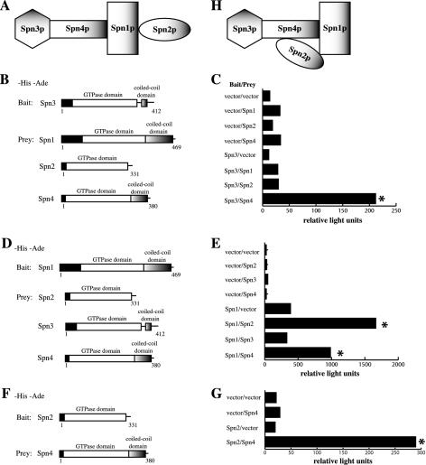
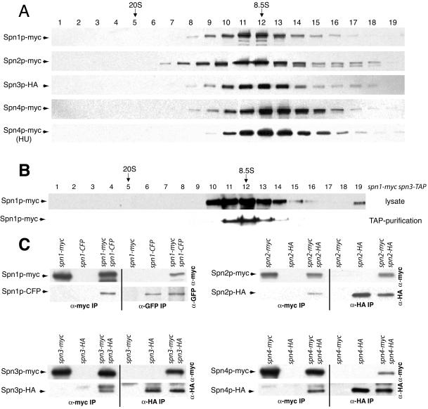
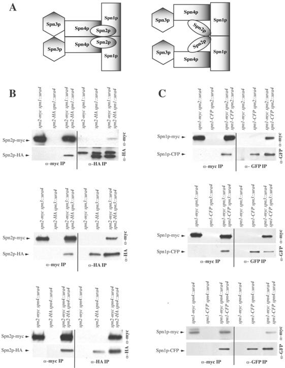
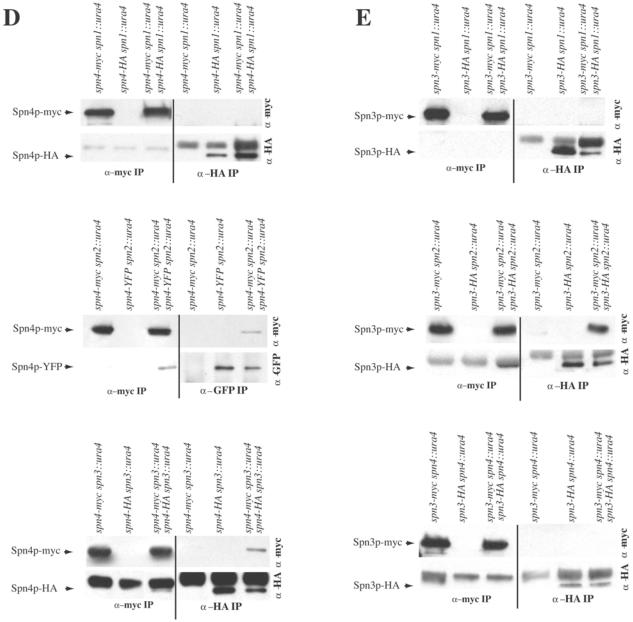
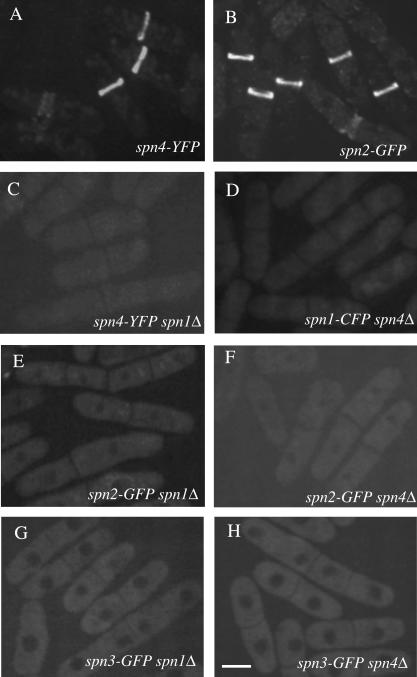
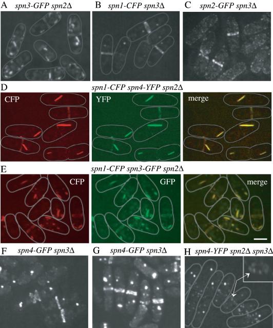
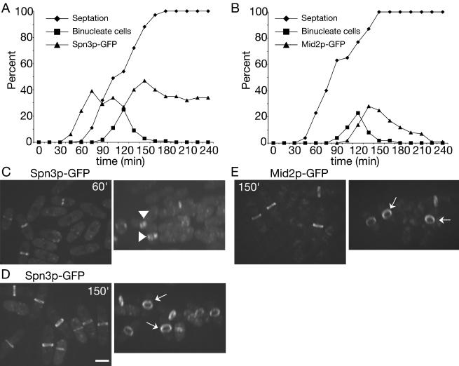
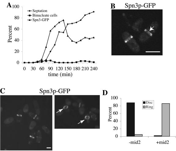
Septins are GTP binding proteins important for cytokinesis in many eukaryotes. The Schizosaccaromyces pombe genome sequence predicts orthologues of four of five Saccharomyces cerevisiae septins involved in cytokinesis and these are named Spns1-4p. That spns1-4 are not essential genes permitted the application of a combined genetic and proteomics approach to determine their functional relationships. Our findings indicate that Spns1-4p are present throughout interphase as a diffusely localized approximately 8.5S complex containing two copies of each septin linked together as a chain in the order Spn3p-Spn4p-Spn1p-Spn2p. Septin recruitment to the medial region of the cell is genetically separable from ring formation, and whereas it is normally restricted to mitosis, it can be promoted without activation of the mitotic cell cycle machinery. Coalescence into ring structures requires Spn1p and Spn4p associate with at least one other septin subunit and the expression of Mid2p that is normally restricted to mitosis. This study establishes the functional requirements for septin complex organization in vivo.


Figures

Figure 1.
Figure 1.
Composition of S. pombe septin complexes. (A–H) Silver-stained gels of a portion of Spn-TAP complexes from the indicated strains. Spn proteins are indicated with arrows. Note that the calmodulin-binding portion of the TAP tag remains after the purification and changes the mass of the proteins. Other bands are nonspecific contaminants. (I) Protein lysates prepared in denaturing conditions from the indicated strains were resolved by SDS-PAGE and blotted with anti-GFP serum. (J) anti-myc (top panel) and anti-HA (bottom panel) immunoprecipitates from the indicated strains were blotted with anti-myc antibodies.
Figure 2.
Figure 2.
Protein-protein interactions mapped between septin complex components. (A and H) Models of S. pombe septin interactions. (B) The PJ69-4A strain was transformed with pGBT9 or pGBT9spn3, and pGAD424 or pGAD424 carrying spn1, spn2, or spn4, which are shown schematically. (C) β-galactosidase activity (represented in relative light units) of the stains described in B. (D) The PJ69-4A stain was transformed with pGBT9 or pGBT9spn1 and pGAD424 or pGAD424 carrying spn2, spn3, or spn4, which are shown schematically. (E) β-galactosidase activity (represented in relative light units) of the stains described in D. (F) The PJ69-4A stain was transformed with pGBT9 or pGBT9spn2 and pGAD424 or pGAD424 carrying spn4, which are shown schematically. (G) β-galactosidase activity (represented in relative light units) of the stains described in D. Asterisk indicates positive interactions.
Figure 3.
Figure 3.
Characterization of S. pombe septin complexes. (A) Lysates prepared in NP-40 buffer from each myc-epitope– or HA-epitope–tagged septin protein from asynchronously growing cells or a hydroxyurea (HU)-arrested culture were resolved on sucrose gradients. Fractions were collected from the bottom of the gradient (1) and immunoblotted with 9E10 or 12CA5 to detect myc or HA-tagged proteins. The peaks of thyroglobulin (20S) and aldolase (8.5S) collected from gradients prepared and run in parallel are indicated. (B) A TAP and a lysate prepared in NP-40 buffer from spn1-myc spn3-TAP cells were resolved on sucrose gradients. Fractions were collected from the bottom of the gradient and immunoblotted with 9E10 to detect Spn1p-myc. The peaks of thyroglobulin (20S) and aldolase (8.5S) collected from gradients prepared and run in parallel are indicated. (C) anti-myc (left side of panels) and anti-GFP or anti-HA (right side of panels) immunoprecipitates from the indicated strains were blotted with anti-myc (top panels) and anti-GFP or anti-HA (bottom panels) antibodies.
Figure 4
Figure 4
S. pombe septins interact in cooperative manner. (A) Possible models of S. pombe septin complex organization. (B–E) anti-myc (left side of panels) and anti-GFP or anti-HA (right side of panels) immunoprecipitates from the indicated strains were blotted with anti-myc (top panels) and anti-GFP or anti-HA (bottom panels) antibodies.
Figure 4
Figure 4
S. pombe septins interact in cooperative manner. (A) Possible models of S. pombe septin complex organization. (B–E) anti-myc (left side of panels) and anti-GFP or anti-HA (right side of panels) immunoprecipitates from the indicated strains were blotted with anti-myc (top panels) and anti-GFP or anti-HA (bottom panels) antibodies.
Figure 5.
Figure 5.
Septin rings are absent in spn1Δ and spn4Δ cells. The (A) spn4-YFP (KGY496), (B) spn2-GFP (KGY4230), (C) spn4-YFP spn1Δ (KGY842), (D) spn1-CFP spn4Δ (KGY4417), (E) spn2-GFP spn1Δ (KGY4258), (F) spn2-GFP spn4Δ (KGY4259), (G) spn3-GFP spn1Δ (KGY4260), and (H) spn3-GFP spn4Δ (KGY2281) strains were grown in YE medium at 25°C, and GFP-tagged proteins were visualized in live cells. Scale bar, 5 μm.
Figure 6.
Figure 6.
Interdependence of septins for ring formation. (A) spn3-GFP spn2Δ (KGY3211), (B) spn1-CFP spn3Δ (KGY730), or (C) spn2-GFP spn3Δ (KGY3169) cells were grown at 25°C, and GFP-tagged proteins were visualized in live cells. (D) spn1-CFP spn4-YFP spn2Δ (KGY761) cells were fixed in ethanol and both Spn1p-CFP and Spn4p-YFP were photographed separately, and then the images merged. (E) spn1-CFP spn3-GFP spn2Δ (KGY887) cells were grown at 25°C, and Spn1p-CFP and Spn3p-GFP were photographed separately from live cells, and the images were merged. (F and G) spn4-GFP spn3Δ cells (KGY5000) were grown at 25°C, and either visualized live in (F) or fixed in ethanol before visualization (G). (H) Spn4p-YFP was visualized in live spn4-YFP spn2Δ spn3Δ cells (KGY1178). The inset shows the middle cell rotated on its Z-axis. Arrows point to the arrangement of Spn4p-YFP “dots” in the medial region of the cell. Gray lines represent the outlines of individual cells. Scale bar, 5 μm.
Figure 7.
Figure 7.
Septin recruitment to the cortex, but not stable ring formation, can be induced during interphase. (A) spn3-GFP cdc16-116 (KGY738) and (B) mid2-GFP cdc16-116 (KGY741) cells were grown to midlog phase at 25°C, synchronized in early G2 phase by centrifugal elutriation, and then shifted to 36°C. Samples of cells were collected every 15 min. One portion of each sample was imaged immediately to determine the percentage of septated cells and the percentage of cells with Spn3p-GFP and Mid2p-GFP medial localization, respectively. Another portion was fixed with ethanol immediately and then stained with DAPI to determine the percentage of binucleate cells. (C and D) Spn3p-GFP localization in live cells from (C) 60 min or (D) 150 min time points (left panels of each). Images in the left panels were also rotated in the Z-axis to better reveal septin ring organization (right panels). Short arrows point to disorganized septin rings at 60 min. Longer arrows point to organized rings at 150 min. (E) Mid2p-GFP localization in live cells from 150 min time point. Cells are also rotated in the Z-axis to better show Mid2p ring organization. Arrows point to organized Mid2p rings. Scale bar, 5 μm.
Figure 8.
Figure 8.
Coalescence of septin rings can be induced during interphase by mid2+ expression. (A) spn3-GFP cdc16-116 (KGY738) cells were transformed with pREP41-HA-mid2+. Transformants obtained in the presence of thiamine in the medium were then grown to midlog phase in the absence of thiamine at 25°C for 15 h. They were then synchronized in early G2 phase by centrifugal elutriation and immediately shifted to 36°C. Samples of cells were collected every 15 min. One portion of each sample was imaged immediately to determine the percentage of septated cells and the percentage of cells with Spn3p-GFP medial localization. Another portion was fixed with ethanol and stained with DAPI to determine the percentage of binucleate cells. (B and C) Spn3p-GFP localization in live cells at the 75-min time point. In B, cells imaged with a single Z-series stack (0.49 μm) are shown to provide the best visualization of split septin rings as indicated by the arrow. In C, the image in the left panel represents a 3D reconstruction of Z-series stacks. This image was rotated in the Z-axis (right panel) to illustrate the cortical restriction of the septin rings. The arrows point to organized septin rings. (C) Quantitation of disorganized septin rings (disk) and organized septin rings (ring) in interphase cdc16-116 cells in the presence or absence of Mid2p. Percentages were determined by visual inspection of Z-series stacks of live cells that contained medially placed septin structures from the experiment described in Figure 7A, 75-min time point, 93 cells examined (-mid2) and from the experiment described in part A of this figure, 75-min time point, 88 cells examined (+mid2). Scale bar, 5 μm.

References

    1. Bahler, J., Wu, J.Q., Longtine, M.S., Shah, N.G., McKenzie, A., 3rd, Steever, A.B., Wach, A., Philippsen, P., and Pringle, J.R. (1998). Heterologous modules for efficient and versatile PCR-based gene targeting in Schizosaccharomyces pombe. Yeast 14, 943-951. - PubMed
    1. Berlin, A., Paoletti, A., and Chang, F. (2003). Mid2p stabilizes septin rings during cytokinesis in fission yeast. J. Cell Biol. 160, 1083-1092. - PMC - PubMed
    1. Burns, C.G., Ohi, R., Mehta, S., O'Toole, E.T., Winey, M., Clark, T.A., Sugnet, C.W., Ares, M., Jr., and Gould, K.L. (2002). Removal of a single alpha-tubulin gene intron suppresses cell cycle arrest phenotypes of splicing factor mutations in Saccharomyces cerevisiae. Mol. Cell. Biol. 22, 801-815. - PMC - PubMed
    1. Casamayor, A. and Snyder, M. (2003). Molecular dissection of a yeast septin: distinct domains are required for septin interaction, localization, and function. Mol. Cell. Biol. 23, 2762-2777. - PMC - PubMed
    1. Caviston, J.P., Longtine, M., Pringle, J.R., and Bi, E. (2003). The Role of Cdc42p GTPase-activating proteins in assembly of the septin ring in yeast. Mol. Biol. Cell 14, 4051-4066. - PMC - PubMed

Publication types

MeSH terms

LinkOut - more resources