. 2004 Oct 13;23(20):4061-71.
doi: 10.1038/sj.emboj.7600402. Epub 2004 Sep 23.

Suz12 is essential for mouse development and for EZH2 histone methyltransferase activity

Affiliations

Suz12 is essential for mouse development and for EZH2 histone methyltransferase activity

Diego Pasini et al. EMBO J. .

Abstract

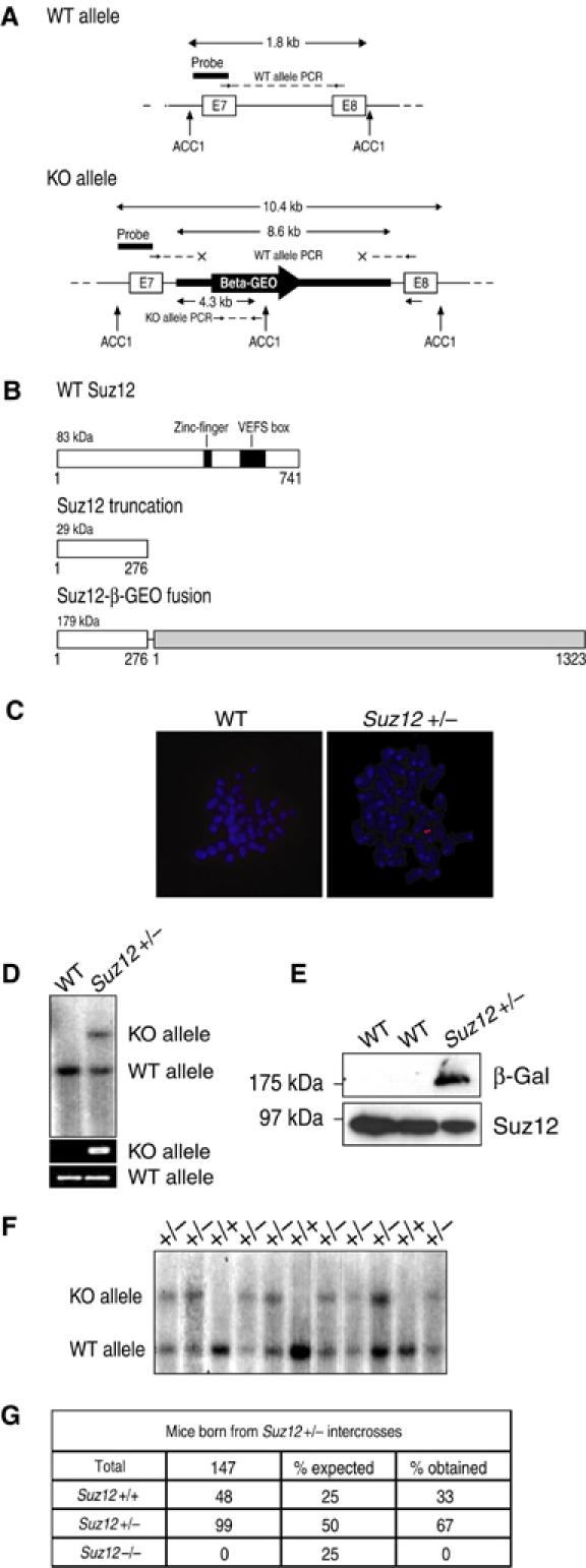
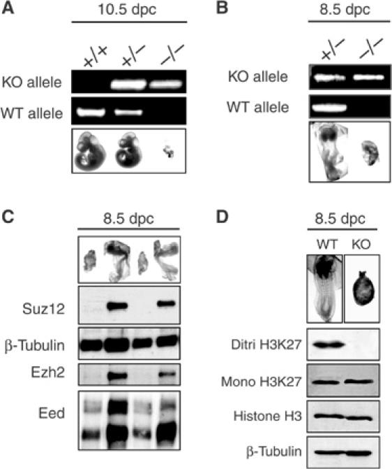
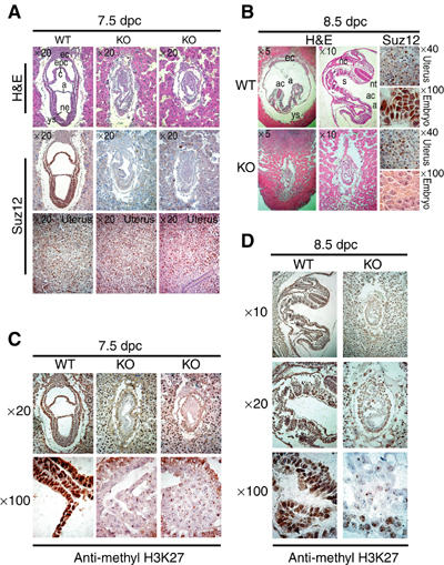
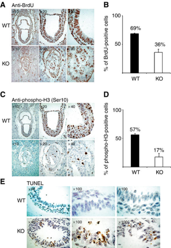
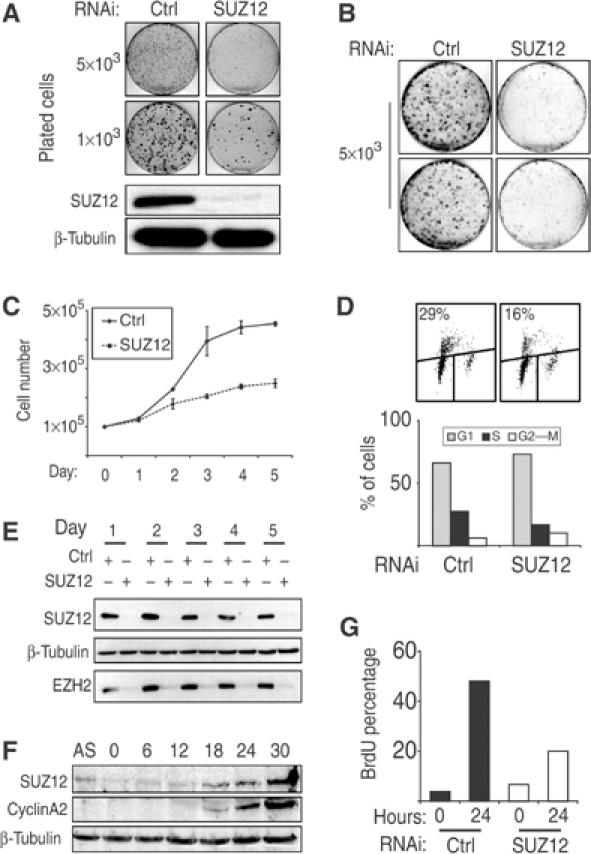
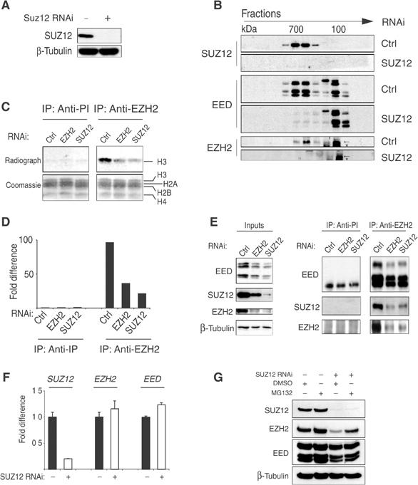
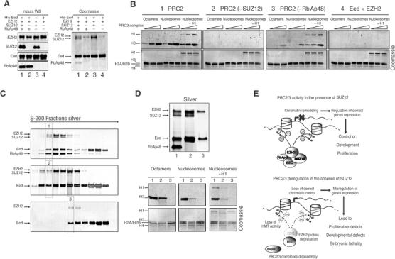
SUZ12 is a recently identified Polycomb group (PcG) protein, which together with EZH2 and EED forms different Polycomb repressive complexes (PRC2/3). These complexes contain histone H3 lysine (K) 27/9 and histone H1 K26 methyltransferase activity specified by the EZH2 SET domain. Here we show that mice lacking Suz12, like Ezh2 and Eed mutant mice, are not viable and die during early postimplantation stages displaying severe developmental and proliferative defects. Consistent with this, we demonstrate that SUZ12 is required for proliferation of cells in tissue culture. Furthermore, we demonstrate that SUZ12 is essential for the activity and stability of the PRC2/3 complexes in mouse embryos, in tissue culture cells and in vitro. Strikingly, Suz12-deficient embryos show a specific loss of di- and trimethylated H3K27, demonstrating that Suz12 is indeed essential for EZH2 activity in vivo. In conclusion, our data demonstrate an essential role of SUZ12 in regulating the activity of the PRC2/3 complexes, which are required for regulating proliferation and embryogenesis.


Figures

Figure 1
Figure 1
Suz12 is essential for mouse viability. (A) Close-up between exons 7 and 8 of the Suz12 locus is presented for both the WT (top panel) and the genetrap (KO) clones (bottom part). The strategies for genotyping the mice are presented. The probe is indicated (black horizontal bar) as well as the restriction sites (ACC1) used for Southern blot analysis. The PCR primers used are also indicated. The WT allele is detected by amplification of the entire intron 7. The presence of the KO allele was detected by PCR as part of the LacZ gene contained in the genetrap cassette. All fragments and amplified product sizes are indicated in kilobases. (B) Schematical representation of the WT Suz12 protein with the two conserved regions indicated, and the truncation products obtained in the KO mice. (C) FISH analysis of metaphase spreads of WT and Suz12 genetrap ES cells showing normal karyotype of the ES cells and a single insertion of the genetrap cassette in Suz12 targeted cells. (D) Southern blot analysis of WT and Suz12 genetrap ES cells showing the presence of the KO cassette in the Suz12 locus. (E) WB analysis showing the expression of the Suz12 WT protein in ES cells and the expression of the Suz12-β-GEO fusion only in the targeted ES cells. (F) Southern blot analysis showing the absence of Suz12 −/− mice from Suz12+/− intercrosses. (G) Summary table of the genotype of mice born from Suz12+/− intercrosses demonstrating that Suz12 KO mice are not born.
Figure 2
Figure 2
Suz12 is required for early embryonic development. (A) Picture and PCR analysis of 10.5 dpc embryos from Suz12+/− intercrosses showing nearly complete reabsorption of Suz12 KO embryos. (B) Picture and PCR analysis of 8.5 dpc embryos from Suz12+/− intercrosses showing the developmental defects of Suz12 KO embryos. (C) Picture and WB analysis of 8.5 dpc embryos from Suz12+/− intercrosses showing the complete absence of the Suz12 WT protein and the strong reduction of the Ezh2 protein levels in the KO embryos. β-Tubulin was used as loading control. (D) Picture and WB analysis of 8.5 dpc embryos from Suz12+/− intercrosses showing loss of H3K27 di/trimethylation. Each lane corresponds to a pool of four WT and four SUZ12 KO embryos. A representative picture of the pooled embryos is presented on top of each lane. Histone H3 and β-tubulin were used as loading control.
Figure 3
Figure 3
Developmental defects and lack of H3K27 methylation in Suz12 KO embryos. (A) (Top panels) H&E staining of 7.5 dpc in utero embryo sections from Suz12+/− intercrosses showing normal morphology of WT and developmental defects of Suz12 KO embryos. (Middle panels) IHC staining showing the presence or absence of the WT Suz12 protein in consecutive sections of the embryos shown in the top panels. (Lower panels) IHC staining for Suz12 in the uterus of each section is presented as a control for proper staining of the analyzed sections. ec: ectoplacental cone; epc: ectoplacental cavity; c: chorion; a: amnion; ne: neural ectoderm; ys: yolk sac. (B) H&E (left and middle panels) and Suz12 IHC staining (right panels) in the embryos and in the uterus of 8.5 dpc in utero embryo sections from Suz12+/− intercrosses. Different magnifications of the same embryos are presented to better evaluate the differences in size and morphology between WT and KO embryos. ac: amniotic cavity; s: somites; nt: neural tube; nc: notochord. (C) Different magnifications of IHC staining of H3K27 di/trimethylation in WT (left panels) and Suz12 KO (middle and right panels) 7.5 dpc embryos. (D) As in panel (C), but for 8.5 dpc embryos. WT (left panels) and Suz12 KO (right panels) embryos. Suz12 IHC staining of maternal cells in (C, D) was used as antibody staining control.
Figure 4
Figure 4
SUZ12 KO embryos are compromised in proliferation. (A) Different magnifications of IHC staining of BrdU in WT (top panels) and KO (bottom panels) 7.5 dpc embryos. The mice were injected with BrdU (150 mg/kg) for 60 min before they were killed. (B) Average of the percentage of BrdU-positive cells in both WT and KO embryos. The standard deviation (s.d.) of the mean is indicated. A total of 1086 cells in WT embryos and 936 cells in KO embryos were counted. (C) Different magnifications of IHC staining of phosphorylation of Ser10 of histone H3 in WT (top panels) and KO (bottom panels) 7.5 dpc embryos. (D) Average of the percentage of H3 Ser10 phosphorylated cells in both WT and KO embryos. s.d. is indicated. A total of 501 cells in WT embryos and 309 cells in KO embryos were counted. (E) Different magnifications of TUNEL staining in WT (top panels) and KO (bottom panels) 7.5 dpc embryos.
Figure 5
Figure 5
SUZ12 is required for proliferation of both normal and tumor cells. (A) Colony formation of U2OS cells stably interfered with empty (CTRL) and SUZ12 shRNA (SUZ12) retroviral vectors showing the proliferative impairment of SUZ12 interfered U2OS cells. In the bottom panels, WB analyses of SUZ12 levels of the same cells are presented. β-Tubulin was used as loading control. (B–D) Colony, growth curves and BrdU FACS analysis of primary human TIG3-T cells stably interfered with empty (CTRL) and SUZ12 shRNA (SUZ12) retroviral vectors, showing the requirement of SUZ12 for proliferation of diploid human cells. (E) WB analysis of TIG3-T cells at each day of the experiment presented in (C), showing the efficiency of the SUZ12 interference and the reduction of the EZH2 protein levels. β-Tubulin served as loading control. (F) Serum induction of serum-starved human diploid fibroblasts. WB analyses show the accumulation of both SUZ12 and cyclin A2 at the G1–S transition. β-Tubulin was used as loading control. (G) SUZ12 is required for S-phase entry. BrdU incorporation was measured with (24 h) and without (0 h) serum induction of serum-starved TIG3-T cells stably interfered with empty (CTRL) and SUZ12 shRNA (SUZ12) retroviral vectors.
Figure 6
Figure 6
SUZ12 is required for the formation and the HMT activity of EZH2-containing complexes. (A) WB analysis of transiently interfered HeLa cells with control (Ctrl) and SUZ12 siRNA oligos. (B) WB analysis on gel filtration fractions of transiently interfered HeLa cells presented in (A) showing the elution profile of SUZ12, EED and EZH2 in the presence or absence of SUZ12. The arrows indicate EZH2. The asterisk indicates a crossreacting protein recognized by the antibody. (C) HMT assays of IP products of preimmune (anti-PI) and anti-EZH2 serum of transiently interfered HeLa cells with Ctrl, EZH2 and SUZ12 oligos. The bottom panels show the Coomassie staining of the histones used as substrates in the reaction. In the top panel is presented the autoradiograph of the same histones showing the specific H3 HMT of the PRC2 complex and the reduction in activity upon loss of EZH2 and SUZ12. (D) Quantification of the HMT activity presented in (C) by in silico analysis. (E) WB analysis of SUZ12, EZH2 and EED proteins of the experiment presented in (C, D). (F) Expression levels of SUZ12, EZH2 and EED mRNA showing that loss of SUZ12 has no effect on EZH2 and EED expression. (G) WB analysis of transiently interfered HeLa cells with Ctrl and SUZ12 siRNA oligos treated with carrier (DMSO) or 5 μM MG132 proteosome inhibitor for 10 h before harvesting, showing that SUZ12 loss induces a proteosome-dependent degradation of EZH2.
Figure 7
Figure 7
Dual role of SUZ12 in regulating the PRC2 complex activity. (A) SUZ12 is required for the recruitment of RbAp48 to the EZH2 complex. Coomassie staining (right panel) of recombinant proteins bound and eluted from a nickel column. The indicated proteins were coexpressed in insect cells. Eed was produced as a His-tagged protein, whereas the other proteins were produced as nontagged proteins. The left panel shows the expression (input) of the various proteins before His-tag purification. (B) SUZ12 is required for PRC2 HMT activity in vitro on both histones H3 and H1. Increasing amounts (0.5, 1 and 5 μl of 0.1 μg/μl preparation) of the purified proteins shown in (A) were used in an in vitro HMT assay using octamers, oligonucleosomes and oligonucleosomes assembled with histone H1 as substrates as described in Materials and methods. (C) Purification of different PRC2 complex forms. Silver staining of S-200 gel filtration fractions of His purification: PRC2 complex (top panel), PRC2 complex in the absence of RbAp48 (middle panel) and PRC2 complex in the absence of SUZ12 (bottom panel). Highly pure complex fractions (highlighted lanes) were used in (D). (D) Silver staining (top panel) and HMT assays (bottom panels) of different forms of the PRC2 complex (labeled 1–3) showing that EZH2–SUZ12–Eed is the minimal enzymatically active PRC2 complex form. (E) Schematic model of the role of PRC2/3 complexes in transcription and a representation of the effects of SUZ12 loss on the PRC2/3 complexes highlighting the impaired recruitment of RbAp48 and the loss of HMT activity, which induce instability and degradation of EZH2.

References

    1. Birve A, Sengupta AK, Beuchle D, Larsson J, Kennison JA, Rasmuson-Lestander A, Muller J (2001) Su(z)12, a novel Drosophila Polycomb group gene that is conserved in vertebrates and plants. Development 128: 3371–3379 - PubMed
    1. Bracken AP, Pasini D, Capra M, Prosperini E, Colli E, Helin K (2003) EZH2 is downstream of the pRB–E2F pathway, essential for proliferation and amplified in cancer. EMBO J 22: 5323–5335 - PMC - PubMed
    1. Brock HW, van Lohuizen M (2001) The Polycomb group—no longer an exclusive club? Curr Opin Genet Dev 11: 175–181 - PubMed
    1. Brummelkamp TR, Bernards R, Agami R (2002) Stable suppression of tumorigenicity by virus-mediated RNA interference. Cancer Cell 2: 243–247 - PubMed
    1. Cao R, Wang L, Wang H, Xia L, Erdjument-Bromage H, Tempst P, Jones RS, Zhang Y (2002) Role of histone H3 lysine 27 methylation in Polycomb-group silencing. Science 298: 1039–1043 - PubMed

Publication types

MeSH terms