. 2004 Oct 13;23(20):3995-4006.
doi: 10.1038/sj.emboj.7600392. Epub 2004 Sep 23.

PIKE/nuclear PI 3-kinase signaling mediates the antiapoptotic actions of NGF in the nucleus

Affiliations

PIKE/nuclear PI 3-kinase signaling mediates the antiapoptotic actions of NGF in the nucleus

Jee-Yin Ahn et al. EMBO J. .

Abstract

PI 3-kinase (PI3K) occurs in the nuclei of a broad range of cell types, and various stimuli elicit PI3K nuclear translocation. However, little is known about the biological function of nuclear PI3K. Here we show that nuclear PI3K and its upstream regulator PIKE mediate the antiapoptotic activity of nerve growth factor (NGF) in the isolated nuclei. The nuclei from NGF-treated PC12 cells, EGF-treated HEK293 cells and HeLa cells are resistant to DNA fragmentation initiated by activated cell-free apoptosome. Nuclei from constitutively active PI3K adenovirus-infected cells display the same resistance as those treated by NGF, whereas PI3K inhibitors, dominant-negative PI3K or PIKE abolishes it. Knockdown of either PI3K or PIKE diminishes the antiapoptotic activity of NGF. PI (3,4,5)P3 alone mimics the antiapoptotic activity of NGF, for which nuclear Akt is required. These results demonstrate that PIKE/nuclear PI3K signaling through nuclear PI (3,4,5)P3 and Akt plays an essential role in promoting cell survival.


Figures
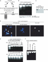
Figure 1
Figure 1
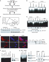
The nuclei from NGF-treated PC12 cells resist DNA fragmentation in cell-free apoptotic solution. (A) Experimental procedures employed in the DNA fragmentation assay. (B) DNA fragmentation assay. The nuclei were isolated from PC12 cells, which were treated with 50 ng/ml NGF (0, 10, 30 min and 1 h) and incubated in the activated cell-free apoptotic solution for 40 min. The fragmented DNA was extracted and resolved on 2% agarose. (C) DFF45 from the control nucleus was cleaved by active caspase-3 in HEK293 cytosolic fraction S-100. (D) Nuclear morphology assay with DAPI staining. The nuclei were isolated from PC12 cells treated with NGF and incubated in apoptotic solution for 40 min and stained with DAPI. The rat liver nuclei were employed as a positive control. (E) PC12 cells were treated with NGF in regular medium for 30 min, and the nuclear and cytosolic extracts of PC12 cells were prepared. Different amounts of the extracts were preincubated with the apoptotic solution for 10 min, then the control nuclei were introduced for DNA fragmentation assay. Nuclear, but not cytosolic, fraction inhibits active apoptosome-triggered DNA fragmentation. (F) The nuclei from HEK293 and HeLa cells, treated with 100 μg/ml EGF for 15 min, are resistant to DNA fragmentation in cell-free apoptotic solution. By contrast, evident DNA fragmentation is observed in control HEK293 cell nuclei without EGF treatment. The nuclei from PC12 cells treated with NGF were employed as positive control.
Figure 2
Figure 2
Nuclear PI3K is implicated in NGF-regulated cell survival. (A) Experimental procedures employed in the DNA fragmentation assay. (B) PI3K inhibitors abolish the antiapoptotic effect of NGF in the nucleus. PC12 cells were treated with NGF for 60 min. The isolated nuclei were pre-incubated with two specific PI3K inhibitors Wortmannin (20 nM), Ly294002 (10 μM), and vehicle solution, respectively, for 30 min. After removal of the inhibitors, the nuclei were incubated in apoptotic solution at 37°C for 40 min. The soluble DNA were extracted and analyzed. (C) Cytosolic and nuclear PI3K activity in NGF-treated PC12 cells (upper panel). The identity of cytosolic and nuclear fractions was verified by immunoblotting with anti-α-tubulin and anti-PARP (lower panels). (D) Nuclear PI3K is sufficient and necessary for the antiapoptotic effect of NGF in the nucleus. PC12 cells were infected with control adenovirus and adenovirus expressing constitutively active or dominant-negative PI3K. After 24 h, the infection efficiency was verified by GFP expression under fluorescent microscope. The isolated nuclei from NGF-treated or nontreated PC12 cells were analyzed in apoptotic solution (upper panel). In vitro PI3K activity assay of cytosolic and nuclear fractions from adenovirus-infected PC12 cells (lower panels). (E) PI3K nuclear translocation in PC12 cells infected with adenovirus expressing Myc-p110* and dominant-negative p85. The infected cells were treated with NGF for 30 min, followed by fixation and staining with anti-Myc and anti-p85 antibodies, respectively. Evident nuclear translocation occurred for p110 and p85 proteins (left panels). Similar effects were observed in biochemical fractionations (right panels). (F) Immunodepletion of PI3K from the nuclear extract abolishes its antiapoptotic effect. NGF-treated nuclear extract (10 μg) was preincubated with 2 μl anti-p110 antibody/20 μl protein-A/G conjugated beads at 4°C for 3 h, and the supernatant was supplemented with various phosphoinositol lipids or recombinant PI3K. Evident DNA fragmentation occurs when PI3K was immunodepleted (lane 1), whereas it is potently inhibited when recombinant PI3K was added back (lane 5). By contrast, rabbit IgG control failed to impair the activity (lane 6). Moreover, introduction of 10 μM PI (3,4,5)P3 but not PI (3)P or PI (3,4)P2 reconstitutes the inhibitory effect (left panel). P110 is specifically removed from nuclear extract by anti-p110 antibody (right panels). (G) Nuclear PI3K is required for the antiapoptotic effect of NGF in the nucleus. Serum-starved PC12 cells were treated with Penetratin 1-conjugated sense or antisense oligonucleotides of p110 α for 6 h, followed by 30 min NGF treatment. The isolated nuclei were analyzed in activated apoptotic solution (left panel). The protein level of p110 α and Akt phosphorylation status were verified by Western blotting. Compared to sense oligonucleotide, antisense markedly diminishes p110 α expression. By contrast, PARP protein level is not affected. Akt phosphorylation is substantially decreased in p110-knocked down cells (middle panels). Cytosolic and nuclear PI3K activity is decreased in p110-knocked down PC12 cells (right panels).
Figure 3
Figure 3
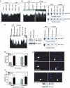
PIKE regulates the antiapoptotic effect of NGF in the nucleus. (A) Dominant-negative PIKE abrogates the antiapoptotic effect of NGF. PC12 cells were infected with control adenovirus and adenovirus expressing wild-type or dominant-negative PIKE. After 24 h, the infection efficiency was verified by GFP expression under a fluorescent microscope. The isolated nuclei from NGF-treated or nontreated PC12 cells were analyzed in apoptotic solution. (B) The stably transfected PC12 cells were induced to express wild-type or dominant-negative PIKE for 24 h, and GFP expression was verified under fluorescent microscope, then followed by NGF or vehicle solution stimulation for 1 h. The isolated nuclei were analyzed in activated apoptotic solution (left panel). Dominant-negative PIKE inhibits cytosolic and nuclear PI3K from induced PIKE stable cell lines (right panels). (C) Serum-starved PC12 cells were treated with Penetratin 1-conjugated sense or antisense oligonucleotides of PIKE for 6 h, followed by 1 h NGF treatment. The isolated nuclei were analyzed in activated cell-free apoptotic solution (left panel). Compared to sense oligonucleotide, antisense markedly diminishes both PIKE-L and -S protein expression. By contrast, PARP protein level is not affected (middle panels). In vitro PI3K activity assay of cytosolic and nuclear PI3K from oligonucleotide-treated cells. PIKE knockdown diminishes NGF-provoked PI3K activity in both the cytoplasm and the nucleus (right panels). (D) PIKE mediates the antiapoptotic effect of NGF in PC12 cells. PC12 cells were treated with Penetratin 1-conjugated sense or antisense oligonucleotides of PIKE for 6 h and induced apoptosis by 250 nM staurosporine for 24 h. (E) TUNEL assay and DAPI staining of staurosporine-treated cells. (F) PIKE mediates the antiapoptotic effect of NGF in sympathetic neurons. Sympathetic neurons were treated with Penetratin 1-conjugated sense or antisense oligonucleotides of PIKE for 6 h and treated with 250 nM staurosporine for 24 h in the presence or absence of NGF. TUNEL assay and DAPI staining of staurosporine-treated apoptotic sympathetic neurons (500 cells were counted under different fields) (right upper panels). Both PIKE-L and -S were markedly knocked down, whereas Tubulin was not changed (right lower panels). Numbers of treated cells in apoptosis were calculated as means (±s.d.) of five determinations and are representative of three experiments.
Figure 4
Figure 4
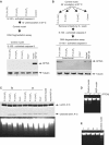
Phosphoinositol lipid PI (3,4,5)P3 mediates the antiapoptotic effect of NGF in the nucleus. (A) Phosphoinositol lipids do not directly inhibit active caspase-3. Experimental procedure scheme (upper panel). Preincubation of the lipids with the activated apoptotosome failed to prevent DFF45 cleavage in the control nuclei (lower panel). (B) PI (3,4,5)P3 pretreatment protects DFF45 in the control nuclei from apoptotic degradation. Experimental procedure scheme (upper panel). Various phosphoinositol lipids (10 μM) were preincubated with the nuclei from PC12 cells for 45 min, and removed by three times washing with buffer B, then the pretreated nuclei were analyzed in apoptotic solution. DFF45 in control nucleus pretreated with PI (3,4,5)P3 but not other lipids is intact. The nuclei from NGF-stimulated PC12 cells were employed as a positive control (lower panel). α-Tubulin was employed as loading control (bottom panel). (C) PI (3,4,5)P3 pretreatment protects the control nucleus from apoptotic degradation. PI (3,4,5)P3, but not other phosphoinositol lipids, protects lamin A/C cleavage in the pretreated control nuclei (upper panel). Consistent with DFF45 cleavage in A, none of the phosphoinositol lipids directly inhibits caspase-3-triggered lamin A/C cleavage. DNA fragmentation analysis correlates with protein cleavage assay (lower panel). (D) PTEN phosphotase abolishes the antiapoptotic effect in the nuclei from NGF-treated PC12 cells. PTEN (150 ng) preincubated with the nuclei for 15 min at 37°C before DNA fragmentation assay. (E) Cycloheximide pretreatment does not inhibit the antiapoptotic actions of NGF or PIP3. Cycloheximide (100 μg/ml) was preincubated with PC12 cells for 30 min and then treated with NGF (lane 1), or at the same time as NGF added (lane 2), cycloheximide preincubated with the nuclei from NGF-treated cells (lane 3), cycloheximide preincubated with the control nuclei from PC12 cells, followed by PIP3 treatment (lane 4), and the control nuclei (last lane) was employed as control.
Figure 5
Figure 5
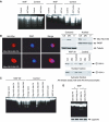
Nuclear Akt is required for the antiapoptotic action of NGF. (A) PC12 cells were infected with control adenovirus and adenovirus expressing dominant-negative Akt (left panel) or wild-type, constitutively active Akt (right panel). After 24 h, the infection efficiency was verified by GFP expression under a fluorescent microscope. The isolated nuclei from PC12 cells were analyzed in an activated cell-free apoptotic solution. (B) Characterization of stably transfected Akt in Myc-NLS-Akt cells. Stably transfected PC12 cells were cultured in tetracycline-free medium overnight, followed by fixation and staining with anti-Myc antibody. Constitutively active (CA)-NLS-Akt predominantly exists in the nucleus, whereas dominant-negative (DN)-NLS-Akt occurs in both the cytoplasm and the nucleus (left panels). Biochemical fractionation and immunoblotting analysis verify the subcellular localization of transfected Akt (right, upper panels). In vitro Akt kinase assay with anti-Akt immunocomplex from the cytosolic and nuclear fractions of PC12 cells, treated with or without NGF (right, lower panels). (C) Stably transfected PC12 cells were induced to express constitutively active or dominant-negative Akt (DN1 and DN2) for 24 h, and GFP expression was verified under a fluorescent microscope, then followed by NGF or vehicle solution stimulation for 30 min. The isolated nuclei were analyzed in the activated apoptotic solution. (D) Expression of PH domains does not interfere with the antiapoptotic actions of EGF. HEK293 cells were transfected with mammalian expression GST-Akt-PH or GST-PIKE-PH, and treated with or without EGF for 15 min. The isolated nuclei were analyzed with DNA fragmentation assay (upper panel). The expression of transfected constructs was verified (lower panel).
Figure 6
Figure 6
PI3K/PI (3,4,5)P3 signaling inhibits apoptosis both upstream and downstream of caspase-3 activation.

Similar articles

Cited by

References

    1. Andjelkovic M, Alessi DR, Meier R, Fernandez A, Lamb NJ, Frech M, Cron P, Cohen P, Lucocq JM, Hemmings BA (1997) Role of translocation in the activation and function of protein kinase B. J Biol Chem 272: 31515–31524 - PubMed
    1. Bao S, Tibbetts RS, Brumbaugh KM, Fang Y, Richardson DA, Ali A, Chen SM, Abraham RT, Wang XF (2001) ATR/ATM-mediated phosphorylation of human Rad17 is required for genotoxic stress responses. Nature 411: 969–974 - PubMed
    1. Batistatou A, Greene LA (1991) Aurintricarboxylic acid rescues PC12 cells and sympathetic neurons from cell death caused by nerve growth factor deprivation: correlation with suppression of endonuclease activity. J Cell Biol 115: 461–471 - PMC - PubMed
    1. Batistatou A, Greene LA (1993) Internucleosomal DNA cleavage and neuronal cell survival/death. J Cell Biol 122: 523–532 - PMC - PubMed
    1. Bavelloni A, Santi S, Sirri A, Riccio M, Faenza I, Zini N, Cecchi S, Ferri A, Auron P, Maraldi NM, Marmiroli S (1999) Phosphatidylinositol 3-kinase translocation to the nucleus is induced by interleukin 1 and prevented by mutation of interleukin 1 receptor in human osteosarcoma Saos-2 cells. J Cell Sci 112: 631–640 - PubMed

Publication types

MeSH terms